



PRDX3 Antibody (YA909)
(Synonyms: PRDX3; AOP1; Thioredoxin-dependent peroxide reductase; mitochondrial; Antioxidant protein 1; AOP-1; HBC189; Peroxiredoxin III; Prx-III; Peroxiredoxin-3; Protein MER5 homolog)
Based on 1 Customer Validation
PRDX3 Antibody (YA909) is a Rabbit-derived and non-conjugated IgG monoclonal antibody, targeting to PRDX3.
-
Host:
Rabbit
-
Isotype:
IgG
-
Application:
WB, ICC/IF, IHC-P, FC, IP
-
Reactivity :
Human
-
Formulation:
Supplied in 1*TBS (pH7.4), 0.05% BSA, 40% Glycerol. Preservative: 0.05% Sodium Azide.
-
Conjugation:
Non-conjugated


To place orders, for customer services and technical support, please contact: MedChemExpress USA
Tel: 609-228-6898 E-mail: [email protected] [email protected]





Applications
| Application |
WB

WB: Western Blot
|
ICC/IF
ICC/IF: Immunocytochemistry/
Immunofluorescence |
IHC-P
IHC-P: Immunohistochemistry-Paraffin
|
FC
FC: Flow Cytometry
|
IP
IP: Immunoprecipitation
|
|---|---|---|---|---|---|
| Dilution Ratio | 1:500-1:2000 | 1:50-1:200 | 1:50-1:200 | 1:50-1:100 | Use at an assay dependent concentration. |


Product Details
PRDX3 Antibody (YA909) is a Rabbit-derived and non-conjugated IgG monoclonal antibody, targeting to PRDX3.
-
Host Rabbit

-
Clonality Recombinant,Monoclonal

-
Species ReactivityHuman
-
Observed Molecular WeightObserved band size: 28 kDa
Note: Due to possible protein modifications or aggregation, the molecular weight should be confirmed by actual measurement, and the predicted value is for reference only.

-
Calculated Molecular Weight Predicted band size: 28 kDa

Synthetic peptide within Human Peroxiredoxin 3 aa 207-256 / 256.
Endogenous
Affinity Purified
Non-conjugated
Unmodified
IgG


Product Properties
-
Appearance
Solution
-
Formulation
Supplied in 1*TBS (pH7.4), 0.05% BSA, 40% Glycerol. Preservative: 0.05% Sodium Azide.
-
Concentration
Batch-dependent, Please check the COA for the concentration of each lot. Check Lot Concentration
-
Storage & Stability
Stored at -20°C for 1 year. Avoid repeated freeze / thaw cycles.
-
Shipping
Shipping with blue ice.


Verification Images
-
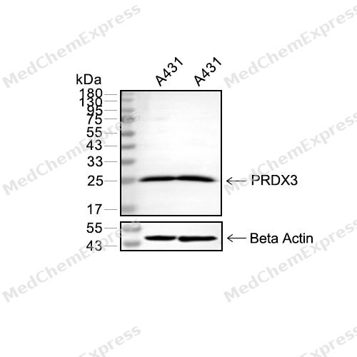 Western blot analysis of extracts from A431(lane 2(20μg) and A431(lane 3(40μg) using PRDX3(HY-P81066) Rabbit mAb. Proteins were transferred to a PVDF membrane and blocked with 5% non-fat milk in TBST for 2 hour at room temperature. The primary antibody (1/1000) and Loading control antibody (Beta Actin, HY-P80438, 1/10000) was used in 5% non-fat milk in TBST at 4°C overnight. Goat Anti-Mouse IgG-HRP Secondary Antibody (1/10000) was used for 1 hour at room temperature.
 -
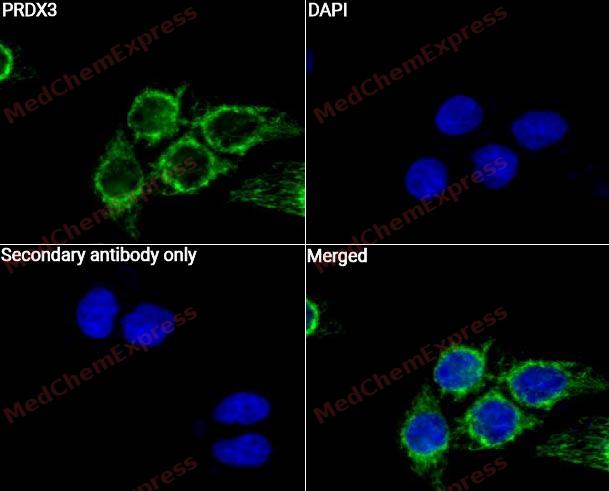 Immunocytochemistry analysis of Hela cells labeling PRDX3 with PRDX3 Antibody (HY-P81066)at 1/50 dilution. Cells were fixed in 4% paraformaldehyde for 15 minutes at room temperature, permeabilized with 0.1% Triton X-100 for 10 minutes at room temperature, then blocked with QuickBlock™ Blocking Buffer for Immunol Staining for 10 min at room temperature. Cells were then incubated with PRDX3 Antibody (HY-P81066) at 1/50 dilution in QuickBlock™ Blocking Buffer for Immunol Staining at 4 ℃. Alexa Fluor® 488-conjugated AffiniPure Goat Anti-Rabbit IgG H&L(HY-P8002, Green) was used as the secondary antibody at 1/1,000 dilution. PBS instead of the primary antibody was used as the secondary antibody only control.
 -
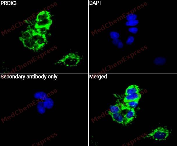 Immunocytochemistry analysis of HepG2 cells labeling PRDX3 with PRDX3 Antibody (HY-P81066) at 1/50 dilution. Cells were fixed in 4% paraformaldehyde for 15 minutes at room temperature, permeabilized with 0.1% Triton X-100 for 10 minutes at room temperature, then blocked with QuickBlock™ Blocking Buffer for Immunol Staining for 10 min at room temperature. Cells were then incubated with PRDX3 Antibody (HY-P81066) at 1/50 dilution in QuickBlock™ Blocking Buffer for Immunol Staining at 4 ℃. Alexa Fluor® 488-conjugated AffiniPure Goat Anti-Rabbit IgG H&L(HY-P8002, Green) was used as the secondary antibody at 1/1,000 dilution. PBS instead of the primary antibody was used as the secondary antibody only control. The Nuclear counterstain was DAPI (Blue).
 -
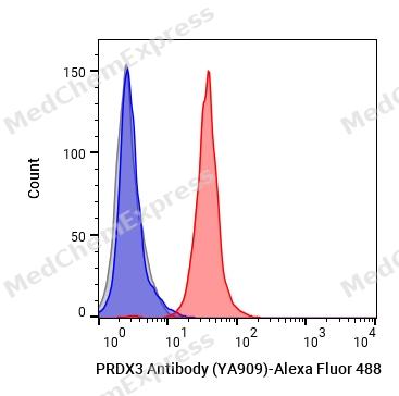 Flow cytometric analysis of 1X10^6 MCF-7 cells labeling PRDX3 Antibody (YA909) (HY-P810669, red). Cells were fixed with 4% paraformaldehyde. Then stained with the primary antibody at 1/50 dilution for an hour at 4℃. Alexa Fluor® 488-conjugated AffiniPure Goat Anti- Rabbit IgG H&L (HY-P8002) was used as the secondary antibody at 1/1,000 dilution for 30 minutes at 4℃. Rabbit IgG Isotype Control (HY-P80879, blue) was used as the isotype control, cells without incubation with primary antibody were used as the unlabeled control (black).


Background
-
Function
PRDX3 is a Thiol-specific peroxidase that catalyzes the reduction of hydrogen peroxide and organic hydroperoxides to water and alcohols, respectively. Plays a role in cell protection against oxidative stress by detoxifying peroxides. Acts synergistically with MAP3K13 to regulate the activation of NF-kappa-B in the cytosol. Required for the maintenance of physical strength[1][2][3][4][5].
-
Subcellular Localization
Mitochondrion; Cytoplasm; Early endosome
-
Isoforms & Post-Translational Modification
P30048 has 2 isomers: P30048-1: 27693 Da (predicted); P30048-2: 25839 Da (predicted).
Phosphorylated by LRRK2; phosphorylation reduces perodixase activity;The enzyme can be inactivated by further oxidation of the cysteine sulfenic acid (C(P)-SOH) to sulphinic acid (C(P)-SO2H) and sulphonic acid (C(P)-SO3H) instead of its condensation to a disulfide bond;S-palmitoylated -
Subunit
Homodimer; disulfide-linked, upon oxidation (PubMed:17707404, PubMed:27238969, PubMed:29438714). 6 homodimers assemble to form a ring-like dodecamer (PubMed:17707404, PubMed:27238969, PubMed:29438714). Interacts with NEK6 (PubMed:20873783). Interacts with LRRK2 (PubMed:21850687). Interacts with MAP3K13 (PubMed:12492477). Interacts with RPS6KC1 (via PX domain) (PubMed:15750338)
-
SwissProt ID
-
Synonyms
PRDX3; AOP1; Thioredoxin-dependent peroxide reductase; mitochondrial; Antioxidant protein 1; AOP-1; HBC189; Peroxiredoxin III; Prx-III; Peroxiredoxin-3; Protein MER5 homolog
-
Research Field
Neuroscience


Documentation
-
Data Sheet (262 KB)
-
SDS (252 KB)
- English - EN (252 KB)
- Français - FR (252 KB)
- Deutsch - DE (252 KB)
- Norwegian - NO (252 KB)
- Español - ES (252 KB)
- Swedish - SV (252 KB)
- Italian - IT (252 KB)
- Korean - KR (252 KB)
- Portuguese - PT (252 KB)
-
User Guide for Antibodies (1077 KB)
References
[1]. Cao Z, et al. Reconstitution of the mitochondrial PrxIII antioxidant defence pathway: general properties and factors affecting PrxIII activity and oligomeric state. J Mol Biol. 2007 Sep 28;372(4):1022-1033. [Content Brief]
[2]. Yewdall NA, et al. Quaternary structure influences the peroxidase activity of peroxiredoxin 3. Biochem Biophys Res Commun. 2018 Mar 4;497(2):558-563. [Content Brief]
[3]. Rebelo AP, et al. Biallelic loss-of-function variations in PRDX3 cause cerebellar ataxia. Brain. 2021 Jun 22;144(5):1467-1481. [Content Brief]
[4]. Tsuji K, et al. Mammalian antioxidant protein complements alkylhydroperoxide reductase (ahpC) mutation in Escherichia coli. Biochem J. 1995 Apr 15;307 ( Pt 2)(Pt 2):377-81. [Content Brief]
[5]. Masaki M, et al. Mixed lineage kinase LZK and antioxidant protein-1 activate NF-kappaB synergistically. Eur J Biochem. 2003 Jan;270(1):76-83. [Content Brief]

 Powered by Bioz
