



RAGE Antibody (YA5788)
(Synonyms: AGER; RAGE; Advanced glycosylation end product-specific receptor; Receptor for advanced glycosylation end products)
Based on 1 Customer Validation
RAGE Antibody (YA5788) is a Rabbit-derived and non-conjugated IgG monoclonal antibody, targeting to RAGE.
-
Host:
Rabbit
-
Isotype:
IgG/Kappa
-
Application:
WB, IHC-P, ICC/IF, ELISA
-
Reactivity :
Human, Mouse, Rat
-
Formulation:
Supplied in PBS, 50% glycerol, 0.05% Proclin 300, 0.05%BSA
-
Conjugation:
Non-conjugated


To place orders, for customer services and technical support, please contact: MedChemExpress USA
Tel: 609-228-6898 E-mail: [email protected] [email protected]





Applications
| Application |
IHC-P

IHC-P: Immunohistochemistry-Paraffin
|
WB
WB: Western Blot
|
ICC/IF
ICC/IF: Immunocytochemistry/
Immunofluorescence |
ELISA
ELISA: Enzyme Linked Immunosorbent Assay
|
|---|---|---|---|---|
| Dilution Ratio | 1:200-1:1000 | 1:2000-1:10000 | 1:200-1:1000 | 1:5000-1:20000 |


Product Details
RAGE Antibody (YA5788) is a Rabbit-derived and non-conjugated IgG monoclonal antibody, targeting to RAGE.
-
Host Rabbit

-
Clonality Monoclonal

-
Species ReactivityHuman, Mouse, Rat
-
Observed Molecular WeightObserved band size: 52,58 kDa
Note: Due to possible protein modifications or aggregation, the molecular weight should be confirmed by actual measurement, and the predicted value is for reference only.

-
Calculated Molecular Weight Predicted band size: 43 kDa

Protein A
Non-conjugated
Unmodified
IgG/Kappa


Product Properties
-
Appearance
Liquid
-
Formulation
Supplied in PBS, 50% glycerol, 0.05% Proclin 300, 0.05%BSA
-
Concentration
Batch-dependent, Please check the COA for the concentration of each lot. Check Lot Concentration
-
Storage & Stability
Stored at -20°C for 1 year. Avoid repeated freeze / thaw cycles.
-
Shipping
Shipping with blue ice.


Verification Images
-
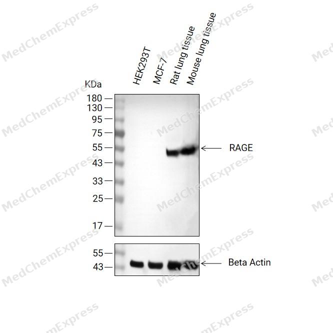 Western blot analysis of extracts from HEK293T (lane2, 20μg, Negative), MCF-7 (lane3, 20μg, Negative), Rat lung tissue (lane4, 20μg) and Mouse lung tissue (lane5, 20μg) using RAGE Antibody (HY-P86096). Proteins were transferred to a PVDF membrane and blocked with 5% non-fat milk in TBST for 2 hour at room temperature. The primary antibody (1/3000) and Loading control antibody (Beta Actin, HY-P80993, 1/10,000) was used in 5% non-fat milk in TBST at 4°C overnight. Goat Anti-Rabbit IgG-HRP Secondary Antibody (HY-P8001 ,1/10,000) was used for 1 hour at room temperature.
 -
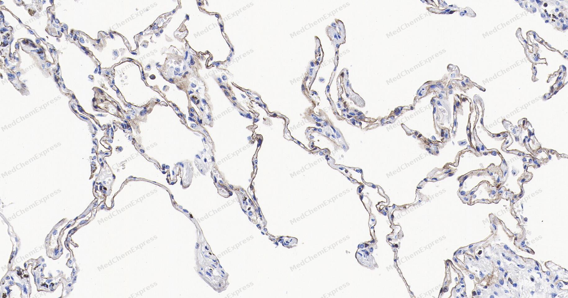 Immunohistochemical analysis of paraffin-embedded human lung tissue using RAGE Antibody (YA5788). The section was pre-treated using heat mediated antigen retrieval with Tris/EDTA buffer (pH 9.0) for 20 minutes. The tissues were probed with the primary antibody (HY-P86096, 1/200) overnight at 4℃. The detection was performed using an HRP conjugated compact polymer system. DAB was used as the chromogen. Tissues were counterstained with hematoxylin and mounted with neutral balsam.
 -
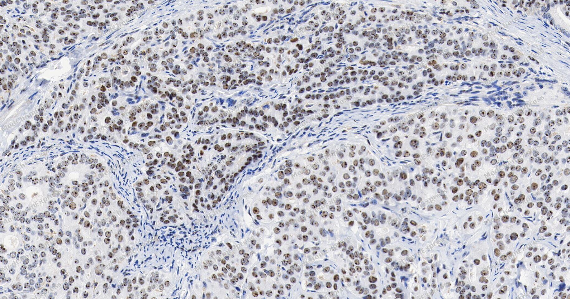 Immunohistochemical analysis of paraffin-embedded human liver cancer tissue using RAGE Antibody (YA5788). The section was pre-treated using heat mediated antigen retrieval with Tris/EDTA buffer (pH 9.0) for 20 minutes. The tissues were probed with the primary antibody (HY-P86096, 1/200) overnight at 4℃. The detection was performed using an HRP conjugated compact polymer system. DAB was used as the chromogen. Tissues were counterstained with hematoxylin and mounted with neutral balsam.
 -
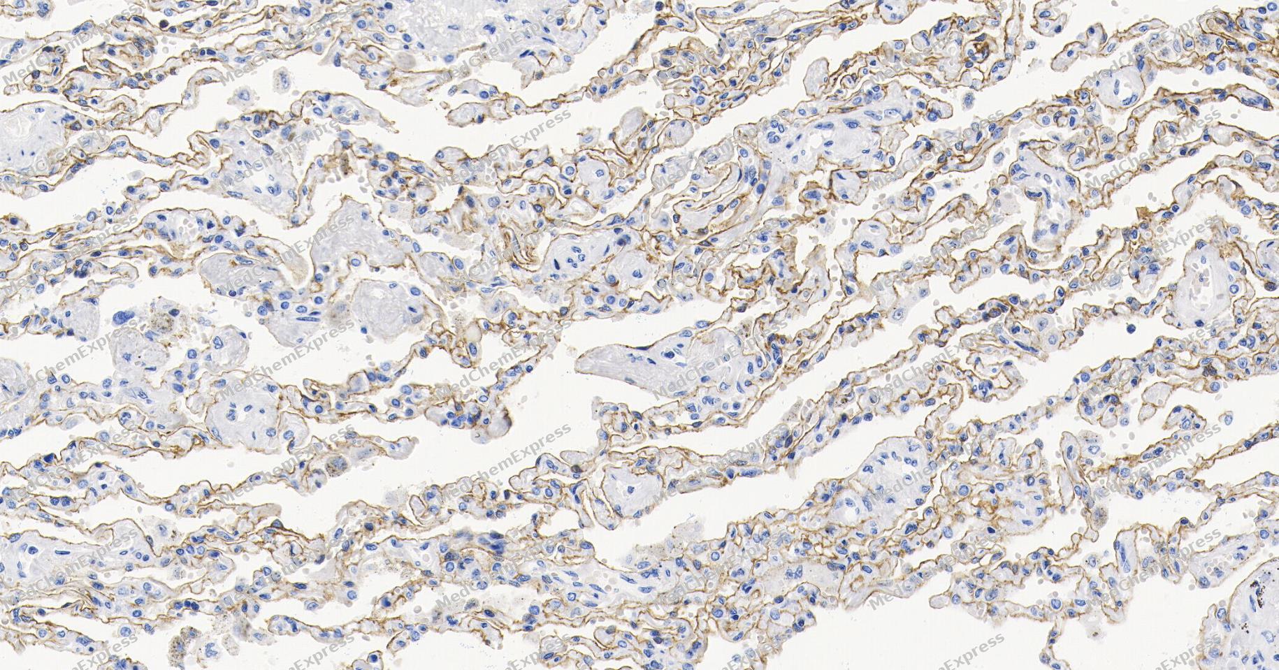 Immunohistochemical analysis of paraffin-embedded human lung tissue using RAGE Antibody (YA5788). The section was pre-treated using heat mediated antigen retrieval with Tris/EDTA buffer (pH 9.0) for 20 minutes. The tissues were probed with the primary antibody (HY-P86096, 1/200) overnight at 4℃. The detection was performed using an HRP conjugated compact polymer system. DAB was used as the chromogen. Tissues were counterstained with hematoxylin and mounted with neutral balsam.
 -
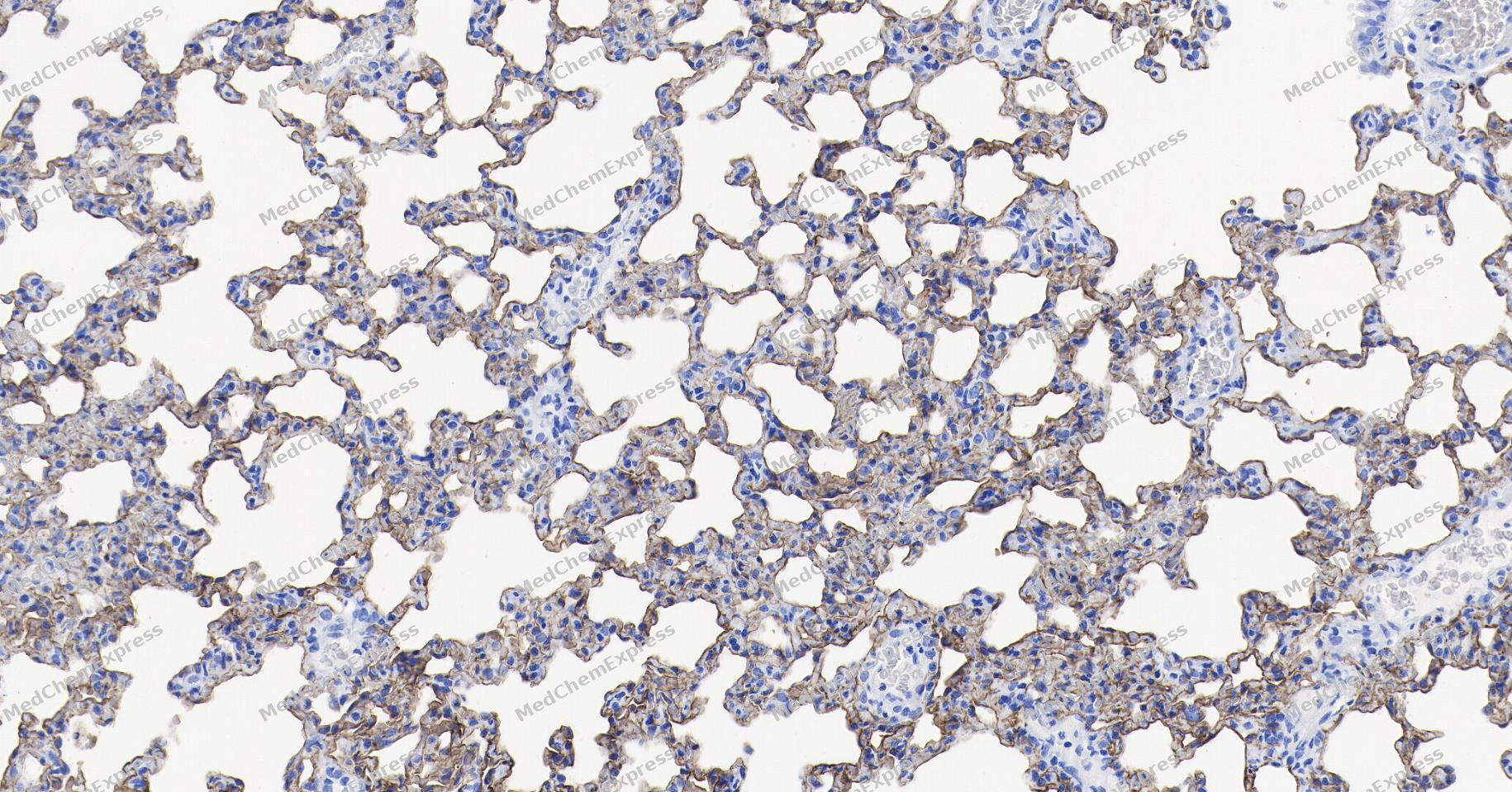 Immunohistochemical analysis of paraffin-embedded rat lung tissue using RAGE Antibody (YA5788). The section was pre-treated using heat mediated antigen retrieval with Tris/EDTA buffer (pH 9.0) for 20 minutes. The tissues were probed with the primary antibody (HY-P86096, 1/200) overnight at 4℃. The detection was performed using an HRP conjugated compact polymer system. DAB was used as the chromogen. Tissues were counterstained with hematoxylin and mounted with neutral balsam.
 -
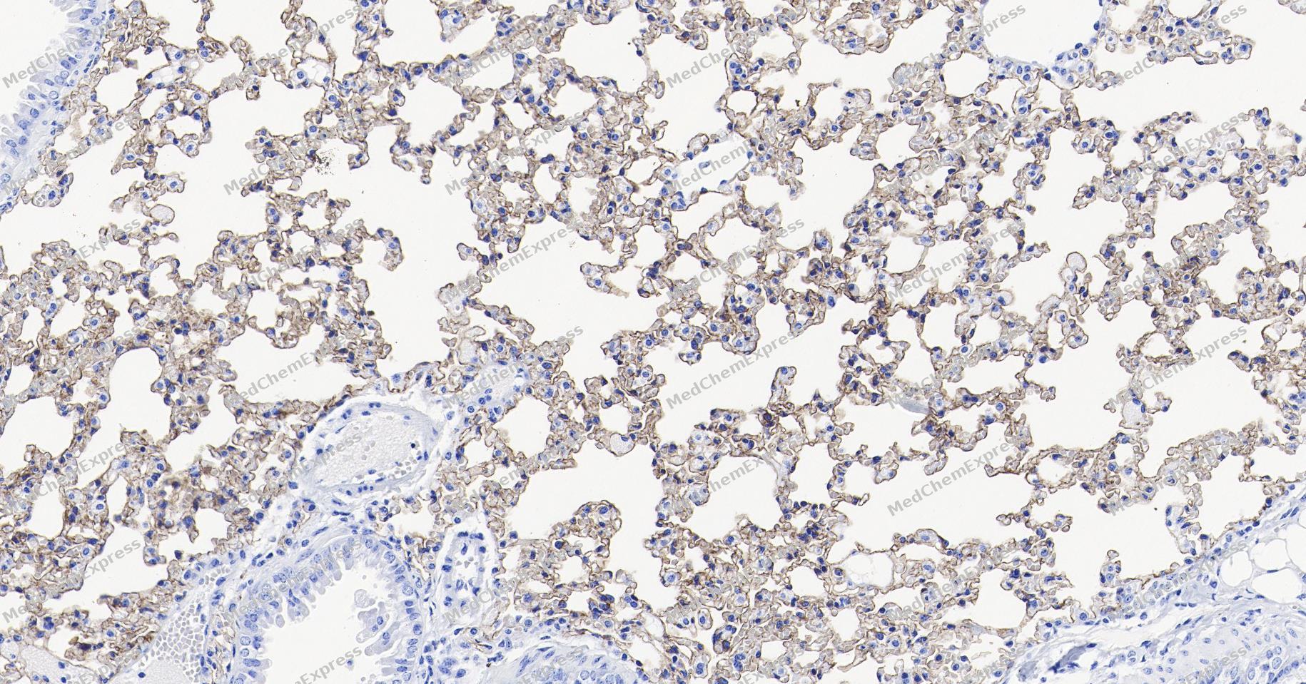 Immunohistochemical analysis of paraffin-embedded mouse lung tissue using RAGE Antibody (YA5788). The section was pre-treated using heat mediated antigen retrieval with Tris/EDTA buffer (pH 9.0) for 20 minutes. The tissues were probed with the primary antibody (HY-P86096, 1/200) overnight at 4℃. The detection was performed using an HRP conjugated compact polymer system. DAB was used as the chromogen. Tissues were counterstained with hematoxylin and mounted with neutral balsam.
 -
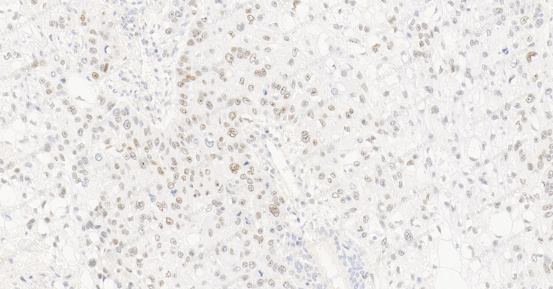 Immunohistochemical analysis of paraffin-embedded human lung squamous cell carcinoma tissue using RAGE Antibody (YA5788). The section was pre-treated using heat mediated antigen retrieval with Tris/EDTA buffer (pH 9.0) for 20 minutes. The tissues were probed with the primary antibody (HY-P86096, 1/200) overnight at 4℃. The detection was performed using an HRP conjugated compact polymer system. DAB was used as the chromogen. Tissues were counterstained with hematoxylin and mounted with neutral balsam.
 -
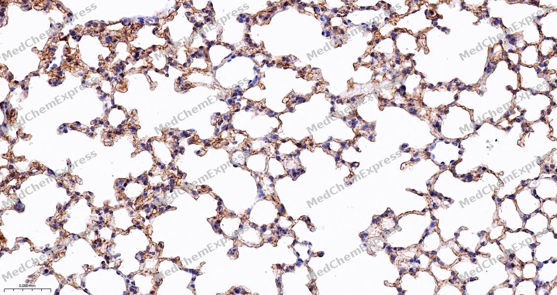 Immunohistochemical analysis of paraffin-embedded mouse lung tissue using RAGE Antibody (HY-P86096, 1/200). The section was pretreated using heat mediated antigen retrieval with Tris-EDTA buffer (pH 9.0) for 15 minutes. The tissues were blocked with quick block buffer for 0.5 hours at room temperature, washed with PBS and PBST, and then incubated with the primary antibody overnight at 4℃. The detection was performed using an HRP conjugated compact polymer system. DAB was used as the chromogen. Tissues were counterstained with hematoxylin and mounted with DPX.
 -
 Immunohistochemical analysis of paraffin-embedded human lung cancer tissue using RAGE Antibody (HY-P86096, 1/200). The section was pretreated using heat mediated antigen retrieval with Tris-EDTA buffer (pH 9.0) for 15 minutes. The tissues were blocked with quick block buffer for 0.5 hours at room temperature, washed with PBS and PBST, and then incubated with the primary antibody overnight at 4℃. The detection was performed using an HRP conjugated compact polymer system. DAB was used as the chromogen. Tissues were counterstained with hematoxylin and mounted with DPX.
 -
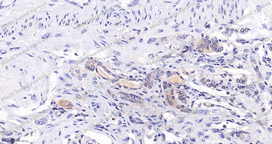 Immunohistochemical analysis of paraffin-embedded human renal cancer tissue using RAGE Antibody (HY-P86096, 1/200). The section was pretreated using heat mediated antigen retrieval with Tris-EDTA buffer (pH 9.0) for 15 minutes. The tissues were blocked with quick block buffer for 0.5 hours at room temperature, washed with PBS and PBST, and then incubated with the primary antibody overnight at 4℃. The detection was performed using an HRP conjugated compact polymer system. DAB was used as the chromogen. Tissues were counterstained with hematoxylin and mounted with DPX.
 -
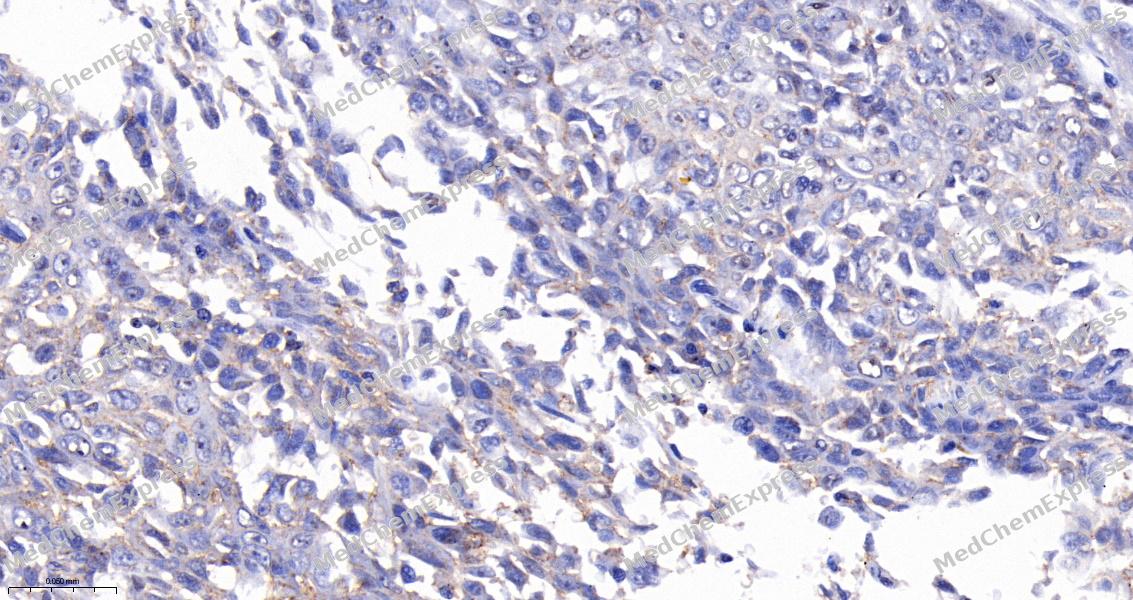 Immunohistochemical analysis of paraffin-embedded human esophagus cancer tissue using RAGE Antibody (HY-P86096, 1/200). The section was pretreated using heat mediated antigen retrieval with Tris-EDTA buffer (pH 9.0) for 15 minutes. The tissues were blocked with quick block buffer for 0.5 hours at room temperature, washed with PBS and PBST, and then incubated with the primary antibody overnight at 4℃. The detection was performed using an HRP conjugated compact polymer system. DAB was used as the chromogen. Tissues were counterstained with hematoxylin and mounted with DPX.


Background
-
Function
RAGE cell surface pattern recognition receptor that senses endogenous stress signals with a broad ligand repertoire including advanced glycation end products, S100 proteins, high-mobility group box 1 protein/HMGB1, amyloid beta/APP oligomers, nucleic acids, histones, phospholipids and glycosaminoglycans. Advanced glycosylation end products are nonenzymatically glycosylated proteins which accumulate in vascular tissue in aging and at an accelerated rate in diabetes. These ligands accumulate at inflammatory sites during the pathogenesis of various diseases including diabetes, vascular complications, neurodegenerative disorders and cancers, and RAGE transduces their binding into pro-inflammatory responses. Upon ligand binding, uses TIRAP and MYD88 as adapters to transduce the signal ultimately leading to the induction of inflammatory cytokines IL6, IL8 and TNFalpha through activation of NF-kappa-B. Interaction with S100A12 on endothelium, mononuclear phagocytes, and lymphocytes triggers cellular activation, with generation of key pro-inflammatory mediators. Interaction with S100B after myocardial infarction may play a role in myocyte apoptosis by activating ERK1/2 and p53/TP53 signaling. Contributes to the translocation of amyloid-beta peptide (ABPP) across the cell membrane from the extracellular to the intracellular space in cortical neurons. ABPP-initiated RAGE signaling, especially stimulation of p38 mitogen-activated protein kinase (MAPK), has the capacity to drive a transport system delivering ABPP as a complex with RAGE to the intraneuronal space. Participates in endothelial albumin transcytosis together with HMGB1 through the RAGE/SRC/Caveolin-1 pathway, leading to endothelial hyperpermeability. Mediates the loading of HMGB1 in extracellular vesicles (EVs) that shuttle HMGB1 to hepatocytes by transferrin-mediated endocytosis and subsequently promote hepatocyte pyroptosis by activating the NLRP3 inflammasome. Binds to DNA and promotes extracellular hypomethylated DNA (CpG DNA) uptake by cells via the endosomal route to activate inflammatory responses. Mediates phagocytosis by non-professional phagocytes (NPP) and this is enhanced by binding to ligands including RNA, DNA, HMGB1 and histones. Promotes NPP-mediated phagocytosis of Saccharomyces cerevisiae spores by binding to RNA attached to the spore wall. Also promotes NPP-mediated phagocytosis of apoptotic cells. Following DNA damage, recruited to DNA double-strand break sites where it colocalizes with the MRN repair complex via interaction with double-strand break repair protein MRE11. Enhances the endonuclease activity of MRE11, promoting the end resection of damaged DNA. Promotes DNA damage repair in trophoblasts which enhances trophoblast invasion and contributes to placental development and maintenance. Protects cells from DNA replication stress by localizing to damaged replication forks where it stabilizes the MCM2-7 complex and promotes faithful progression of the replication fork. Mediates the production of reactive oxygen species (ROS) in human endothelial cells[1][2][3][4][5][6][7][8][9][10][11][12][13].
-
Subcellular Localization
Cell membrane; Single-pass type I membrane protein; Cell projection, phagocytic cup; Early endosome; Nucleus; Cell membrane; Single-pass type I membrane protein; Secreted; Cell membrane; Single-pass type I membrane protein
-
Expression
Tissue_specificity:Endothelial cells. Endothelial cell expression was increased in preterm and preeclamptic placentas compared to the control group (PubMed: 33918759) .
Induction:Induced in T cells by antigen stimulation and by the S100B ligand (PubMed:22509345) . Induced in trophoblasts by DNA damage (PubMed:33918759) -
Isoforms & Post-Translational Modification
Q15109 has 10 isomers: Q15109-1: 42803 Da (predicted); Q15109-2: 36193 Da (predicted); Q15109-3: 37050 Da (predicted); Q15109-4: 39020 Da (predicted); Q15109-5: 13192 Da (predicted); Q15109-6: 44773 Da (predicted); Q15109-7: 41234 Da (predicted); Q15109-8: 35322 Da (predicted); Q15109-9: 38379 Da (predicted); Q15109-10: 41098 Da (predicted).
Phosphorylated on its cytoplasmic domain by PKCzeta/PRKCZ upon ligand binding (PubMed:21829704). Phosphorylated by ATM following DNA damage (By similarity);Targeted by the ubiquitin E3 ligase subunit FBXO10 to mediate its ubiquitination and degradation -
Subunit
Constitutive homodimer; disulfide-linked (PubMed:24081950). Forms homooligomers (PubMed:24081950).
-
SwissProt ID
-
Synonyms
AGER; RAGE; Advanced glycosylation end product-specific receptor; Receptor for advanced glycosylation end products


Documentation
-
Data Sheet (234 KB)
-
SDS (252 KB)
- English - EN (252 KB)
- Français - FR (252 KB)
- Deutsch - DE (252 KB)
- Norwegian - NO (252 KB)
- Español - ES (252 KB)
- Swedish - SV (252 KB)
- Italian - IT (252 KB)
- Korean - KR (252 KB)
- Portuguese - PT (252 KB)
-
User Guide for Antibodies (1077 KB)

 Powered by Bioz
