



RelB Antibody (YA5890)
(Synonyms: RELB; Transcription factor RelB; I-Rel)Based on 1 Customer Validation
RelB Antibody (YA5890) is a Rabbit-derived and non-conjugated IgG monoclonal antibody, targeting to RelB.
-
Host:
Rabbit
-
Isotype:
IgG
-
Application:
WB, IHC-P, ICC/IF, IP, ELISA
-
Reactivity :
Human, Mouse, Rat
-
Formulation:
Supplied in PBS, 50% glycerol, 0.05% Proclin 300, 0.05%BSA
-
Conjugation:
Non-conjugated


To place orders, for customer services and technical support, please contact: MedChemExpress USA
Tel: 609-228-6898 E-mail: [email protected] [email protected]





Applications
| Application |
IHC-P

IHC-P: Immunohistochemistry-Paraffin
|
WB
WB: Western Blot
|
ICC/IF
ICC/IF: Immunocytochemistry/
Immunofluorescence |
ELISA
ELISA: Enzyme Linked Immunosorbent Assay
|
IP
IP: Immunoprecipitation
|
|---|---|---|---|---|---|
| Dilution Ratio | 1:200-1:1000 | 1:2000-1:10000 | 1:200-1:1000 | 1:5000-1:20000 | 1:50-1:200 |


Product Details
RelB Antibody (YA5890) is a Rabbit-derived and non-conjugated IgG monoclonal antibody, targeting to RelB.
-
Host Rabbit

-
Clonality Monoclonal

-
Species ReactivityHuman, Mouse, Rat
-
Observed Molecular WeightObserved band size: 62 kDa
Note: Due to possible protein modifications or aggregation, the molecular weight should be confirmed by actual measurement, and the predicted value is for reference only.

-
Calculated Molecular Weight Predicted band size: 62 kDa

Protein A
Non-conjugated
Unmodified
IgG


Product Properties
-
Appearance
Solution
-
Formulation
Supplied in PBS, 50% glycerol, 0.05% Proclin 300, 0.05%BSA
-
Concentration
Batch-dependent, Please check the COA for the concentration of each lot. Check Lot Concentration
-
Storage & Stability
Stored at -20°C for 1 year. Avoid repeated freeze / thaw cycles.
-
Shipping
Shipping with blue ice.


Verification Images
-
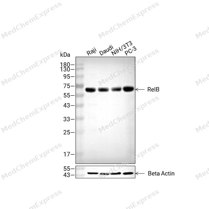 Western blot analysis of extracts from Raji (lane2(20μg), Daudi (lane3(20μg), NIH/3T3 (lane4(20μg) and PC-3 (lane5(20μg) using RelB Antibody (HY-P86198). Proteins were transferred to a PVDF membrane and blocked with 5% non-fat milk in TBST for 2 hour at room temperature. The primary antibody (1/5000) and Loading control antibody (Beta Actin, HY-P80993, 1/10,000) was used in 5% non-fat milk in TBST at 4°C overnight. Goat Anti-Rabbit IgG-HRP Secondary Antibody (HY-P8001 ,1/10,000) was used for 1 hour at room temperature.
 -
 Immunohistochemical analysis of paraffin-embedded mouse rectum tissue using RelB Antibody (HY-P86198, 1/800). The section was pretreated using heat mediated antigen retrieval with sodium citrate buffer (pH 6.0) for 8 minutes. The tissues were blocked with quick block buffer for 0.5 hours at room temperature, washed with PBS and PBST, and then incubated with the primary antibody overnight at 4℃. The detection was performed using an HRP conjugated compact polymer system. DAB was used as the chromogen. Tissues were counterstained with hematoxylin and mounted with DPX.
 -
 Immunohistochemical analysis of paraffin-embedded mouse epididymis tissue using RelB Antibody (HY-P86198, 1/800). The section was pretreated using heat mediated antigen retrieval with sodium citrate buffer (pH 6.0) for 8 minutes. The tissues were blocked with quick block buffer for 0.5 hours at room temperature, washed with PBS and PBST, and then incubated with the primary antibody overnight at 4℃. The detection was performed using an HRP conjugated compact polymer system. DAB was used as the chromogen. Tissues were counterstained with hematoxylin and mounted with DPX.
 -
 Immunohistochemical analysis of paraffin-embedded mouse colon tissue using RelB Antibody (HY-P86198, 1/800). The section was pretreated using heat mediated antigen retrieval with sodium citrate buffer (pH 6.0) for 8 minutes. The tissues were blocked with quick block buffer for 0.5 hours at room temperature, washed with PBS and PBST, and then incubated with the primary antibody overnight at 4℃. The detection was performed using an HRP conjugated compact polymer system. DAB was used as the chromogen. Tissues were counterstained with hematoxylin and mounted with DPX.
 -
 Immunohistochemical analysis of paraffin-embedded human thyroid gland tissue using RelB Antibody (HY-P86198, 1/800). The section was pretreated using heat mediated antigen retrieval with sodium citrate buffer (pH 6.0) for 8 minutes. The tissues were blocked with quick block buffer for 0.5 hours at room temperature, washed with PBS and PBST, and then incubated with the primary antibody overnight at 4℃. The detection was performed using an HRP conjugated compact polymer system. DAB was used as the chromogen. Tissues were counterstained with hematoxylin and mounted with DPX.
 -
 Immunohistochemical analysis of paraffin-embedded human adrenal gland tissue using RelB Antibody (HY-P86198, 1/800). The section was pretreated using heat mediated antigen retrieval with sodium citrate buffer (pH 6.0) for 8 minutes. The tissues were blocked with quick block buffer for 0.5 hours at room temperature, washed with PBS and PBST, and then incubated with the primary antibody overnight at 4℃. The detection was performed using an HRP conjugated compact polymer system. DAB was used as the chromogen. Tissues were counterstained with hematoxylin and mounted with DPX.
 -
 Immunohistochemical analysis of paraffin-embedded human tonsil tissue using RelB Antibody (HY-P86198, 1/800). The section was pretreated using heat mediated antigen retrieval with sodium citrate buffer (pH 6.0) for 8 minutes. The tissues were blocked with quick block buffer for 0.5 hours at room temperature, washed with PBS and PBST, and then incubated with the primary antibody overnight at 4℃. The detection was performed using an HRP conjugated compact polymer system. DAB was used as the chromogen. Tissues were counterstained with hematoxylin and mounted with DPX.
 -
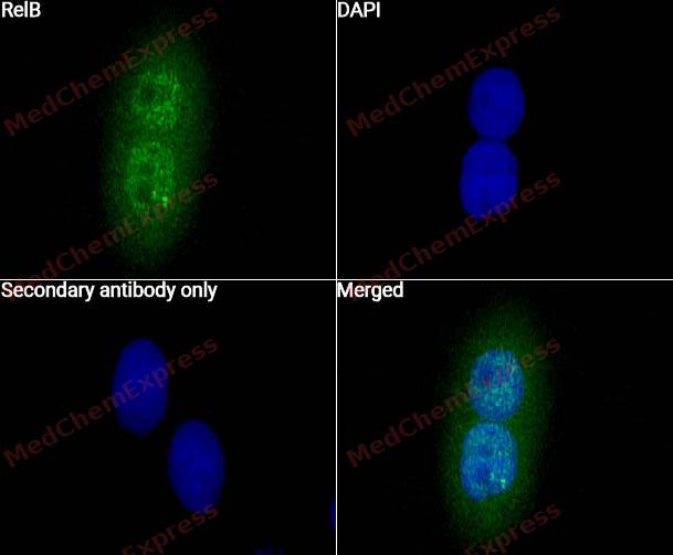 Immunocytochemistry analysis of Hela cells labeling RelB With RelB antibody (HY-P86198) at 1/300 dilution. Cells were fixed in 4% paraformaldehyde for 15 minutes at room temperature, permeabilized with 0.1% Triton X-100 in PBS for 15 minutes at room temperature, then blocked with quick block buffer for 10 minutes at room temperature. Cells were then incubated with RelB antibody (HY-P86198) at 1/300 dilution in quick block buffer overnight at 4 ℃. AF488-conjugated Goat Anti-Rabbit IgG H&L(HY-P8002, Green) was used as the secondary antibody at 1/1,000 dilution. PBS instead of the primary antibody was used as the secondary antibody only control. The Nuclear counterstain was DAPI (Blue).
 -
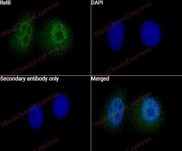 Immunocytochemistry analysis of Hela cells labeling RelB With RelB antibody (HY-P86198) at 1/500 dilution. Cells were fixed in 4% paraformaldehyde for 15 minutes at room temperature, permeabilized with 0.1% Triton X-100 in PBS for 15 minutes at room temperature, then blocked with quick block buffer for 10 minutes at room temperature. Cells were then incubated with RelB antibody (HY-P86198) at 1/500 dilution in quick block buffer overnight at 4 ℃. AF488-conjugated Goat Anti-Rabbit IgG H&L(HY-P8002, Green) was used as the secondary antibody at 1/1,000 dilution. PBS instead of the primary antibody was used as the secondary antibody only control. The Nuclear counterstain was DAPI (Blue).


Background
-
Function
RelB is a member of the NF-κB transcription factor family and functions as a key regulator of immune homeostasis, antigen-presenting cell activity, and lymphoid tissue organization[4][1]. Mechanistically, RelB is the principal transcriptional component of the non-canonical NF-κB pathway, where NF-κB-inducing kinase (NIK) and IKKα promote processing of p100 into p52, leading to formation and nuclear translocation of RelB:p52 heterodimers that regulate distinct target genes involved in lymphoid organogenesis, lymphocyte trafficking, and immune regulation[1][5]. In dendritic cells, RelB is highly expressed and is required for dendritic cell maturation, antigen presentation, and T-cell activation, highlighting its central role in adaptive immune responses[2]. Disease relevance arises from the broad involvement of non-canonical NF-κB signaling in inflammatory, autoimmune, and malignant disorders, while RelB-dependent transcriptional programs contribute to the regulation of immune response genes and maintenance of immune homeostasis[1][3][6]. Compared with the closely related NF-κB subunit RelA (p65), which predominantly mediates canonical NF-κB signaling through p50:RelA dimers, RelB preferentially forms transcriptionally active complexes with p52 and controls a distinct set of genes associated with developmental and homeostatic immune functions[1][5]. Experimental studies further indicate that RelB can modulate inflammatory gene expression by competing with RelA for κB-binding sites, providing a mechanistic basis for functional divergence among NF-κB family members[6]. For research applications, pharmacological strategies targeting NF-κB signaling, including inhibitors of upstream kinases and nuclear NF-κB activity, are widely used to investigate RelB-regulated pathways in immunity, inflammation, and cancer models[3].
-
Subcellular Localization
Nucleus; Cytoplasm, cytoskeleton, microtubule organizing center, centrosome
-
Expression
Induction:Up-regulated by mitogens and NUPR1 -
Subunit
Component of the NF-kappa-B RelB-p50 complex. Component of the NF-kappa-B RelB-p52 complex. Self-associates; the interaction seems to be transient and may prevent degradation allowing for heterodimer formation with p50 or p52. Interacts with NFKB1/p50, NFKB2/p52 and NFKB2/p100. Interacts with NFKBID. Interacts with BMAL1 and the interaction is enhanced in the presence of CLOCK (By similarity)
-
SwissProt ID
-
Synonyms
RELB; Transcription factor RelB; I-Rel


Documentation
-
Data Sheet (262 KB)
-
SDS (251 KB)
- English - EN (251 KB)
- Français - FR (251 KB)
- Deutsch - DE (251 KB)
- Norwegian - NO (251 KB)
- Español - ES (251 KB)
- Swedish - SV (251 KB)
- Italian - IT (251 KB)
- Korean - KR (251 KB)
- Portuguese - PT (251 KB)
-
User Guide for Antibodies (1077 KB)
References
[1]. Sun SC. The non-canonical NF-kappaB pathway in immunity and inflammation. Nat Rev Immunol. 2017;17(9):545-558. [Content Brief]
[2]. Shih VFS, et al. Control of RelB during dendritic cell activation integrates canonical and noncanonical NF-kappaB pathways. Nat Immunol. 2012;13(12):1162-1170. [Content Brief]
[3]. Guo Q, et al. NF-κB in biology and targeted therapy: new insights and translational implications. Signal Transduct Target Ther. 2024 Mar 4;9(1):53. [Content Brief]

 Powered by Bioz
