



SNX1 Antibody (YA2044)
(Synonyms: SNX1; Sorting nexin-1)
Based on 1 Customer Validation
SNX1 Antibody (YA2044) is a Rabbit-derived and non-conjugated IgG monoclonal antibody, targeting to SNX1.
-
Host:
Rabbit
-
Isotype:
IgG
-
Application:
WB, IHC-P, ICC/IF, FC
-
Reactivity :
Human, Mouse, Rat
-
Formulation:
Supplied in 50mM Tris-Glycine(pH 7.4), 0.15M NaCl, 40%Glycerol, 0.01% sodium azide and 0.05% BSA.
-
Conjugation:
Non-conjugated


To place orders, for customer services and technical support, please contact: MedChemExpress USA
Tel: 609-228-6898 E-mail: [email protected] [email protected]





Applications
| Application |
WB

WB: Western Blot
|
IHC-P
IHC-P: Immunohistochemistry-Paraffin
|
ICC/IF
ICC/IF: Immunocytochemistry/
Immunofluorescence |
FC
FC: Flow Cytometry
|
|---|---|---|---|---|
| Dilution Ratio | 1:500-1:1000 | 1:50-1:100 | 1:50-1:200 | 1:50-1:100 |


Product Details
SNX1 Antibody (YA2044) is a Rabbit-derived and non-conjugated IgG monoclonal antibody, targeting to SNX1.
-
Host Rabbit

-
Clonality Recombinant,Monoclonal

-
Species ReactivityHuman, Mouse, Rat
-
Observed Molecular WeightObserved band size: 74 kDa
Note: Due to possible protein modifications or aggregation, the molecular weight should be confirmed by actual measurement, and the predicted value is for reference only.

-
Calculated Molecular Weight Predicted band size: 59 kDa

Entrez Gene: 6642 Human ; 56440 Mouse ; 84471 Rat
SwissProt: Q13596 Human ; Q9WV80 Mouse ; Q99N27 Rat
OMIM: 601272 Human
A synthesized peptide derived from human SNX1 aa50-100/522.
Endogenous
Affinity Chromatography
Non-conjugated
Unmodified
IgG


Product Properties
-
Appearance
Solution
-
Formulation
Supplied in 50mM Tris-Glycine(pH 7.4), 0.15M NaCl, 40%Glycerol, 0.01% sodium azide and 0.05% BSA.
-
Concentration
Batch-dependent, Please check the COA for the concentration of each lot. Check Lot Concentration
-
Storage & Stability
Stored at -20°C for 1 year. Avoid repeated freeze / thaw cycles.
-
Shipping
Shipping with blue ice.


Verification Images
-
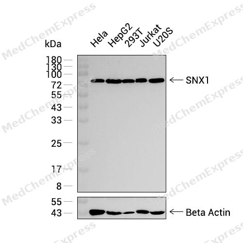 Western blot analysis was performed on extracts from Hela (lane 1, 20 μg), HepG2 (lane 2, 20 μg), 293T (lane 3, 20 μg), Jurkat (lane 4, 20 μg), and U2OS (lane 5, 20 μg) using SNX1 Rabbit mAb.Proteins were transferred to a PVDF membrane and blocked with 5% non - fat milk in TBST at 4°C overnight.The primary antibody (1:1000 dilution) and the loading control antibody (beta-Actin, HY-P83730, 1:5000 dilution) were incubated in 5% non-fat milk in TBST for 1 hour at 37°C.Goat Anti - Rabbit IgG - HRP Secondary Antibody (1:20000 dilution) was then applied for 40 minutes at 37°C.
 -
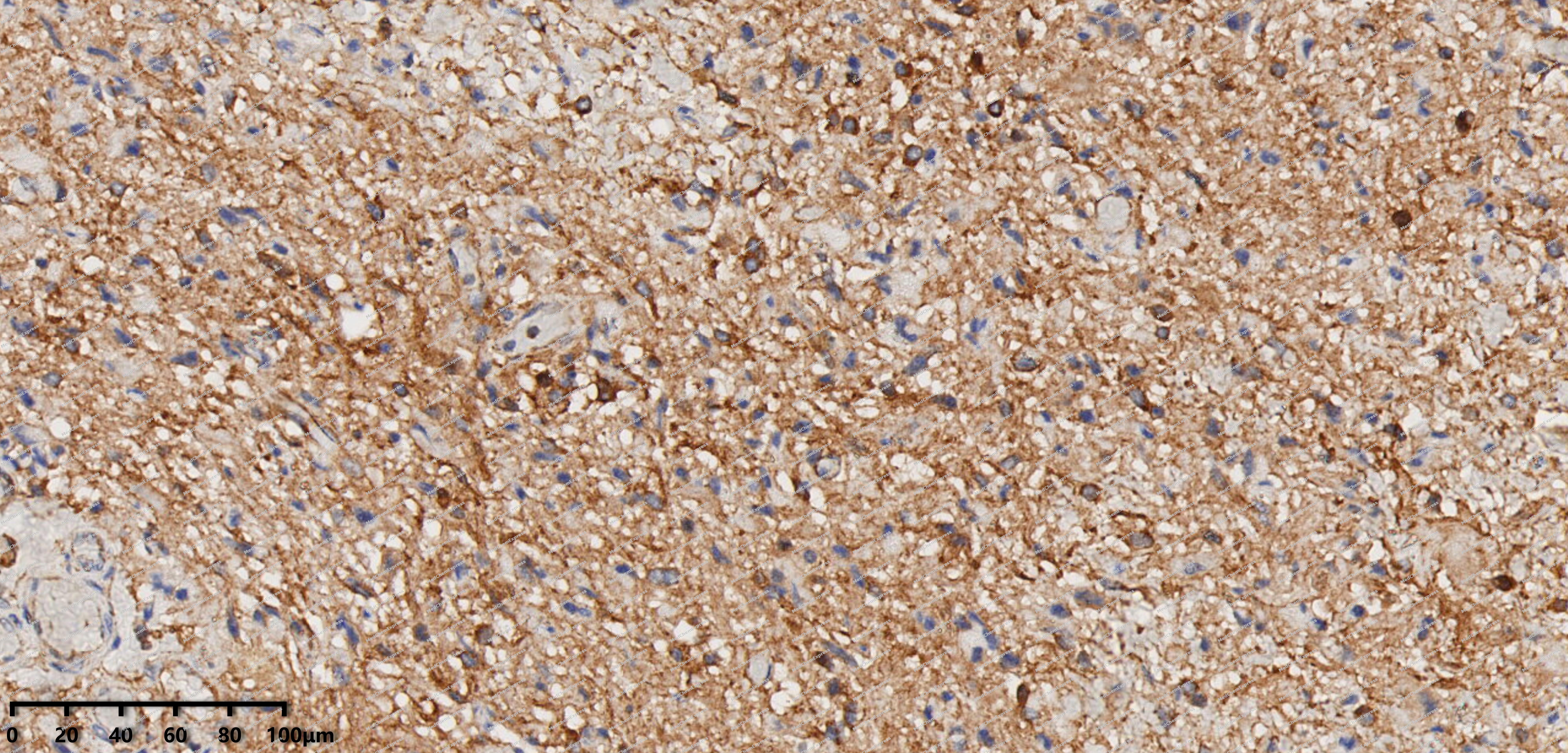 Immunohistochemical analysis of paraffin-embedded human Glioma tissue using SNX1 antibody was performed. The section was pretreated using high-temperature mediated EDTA antigen retrieval buffer (pH 9.0), for 20 minutes. The tissues were incubated with primary antibody (HY-P82299, 1:200 dilution) at room temperature for 20 minutes. Detection was performed using an HRP conjugated compact polymer system. DAB was used as the chromogen. The tissues were counterstained with hematoxylin and mounted with neutral balsam mounting medium.
 -
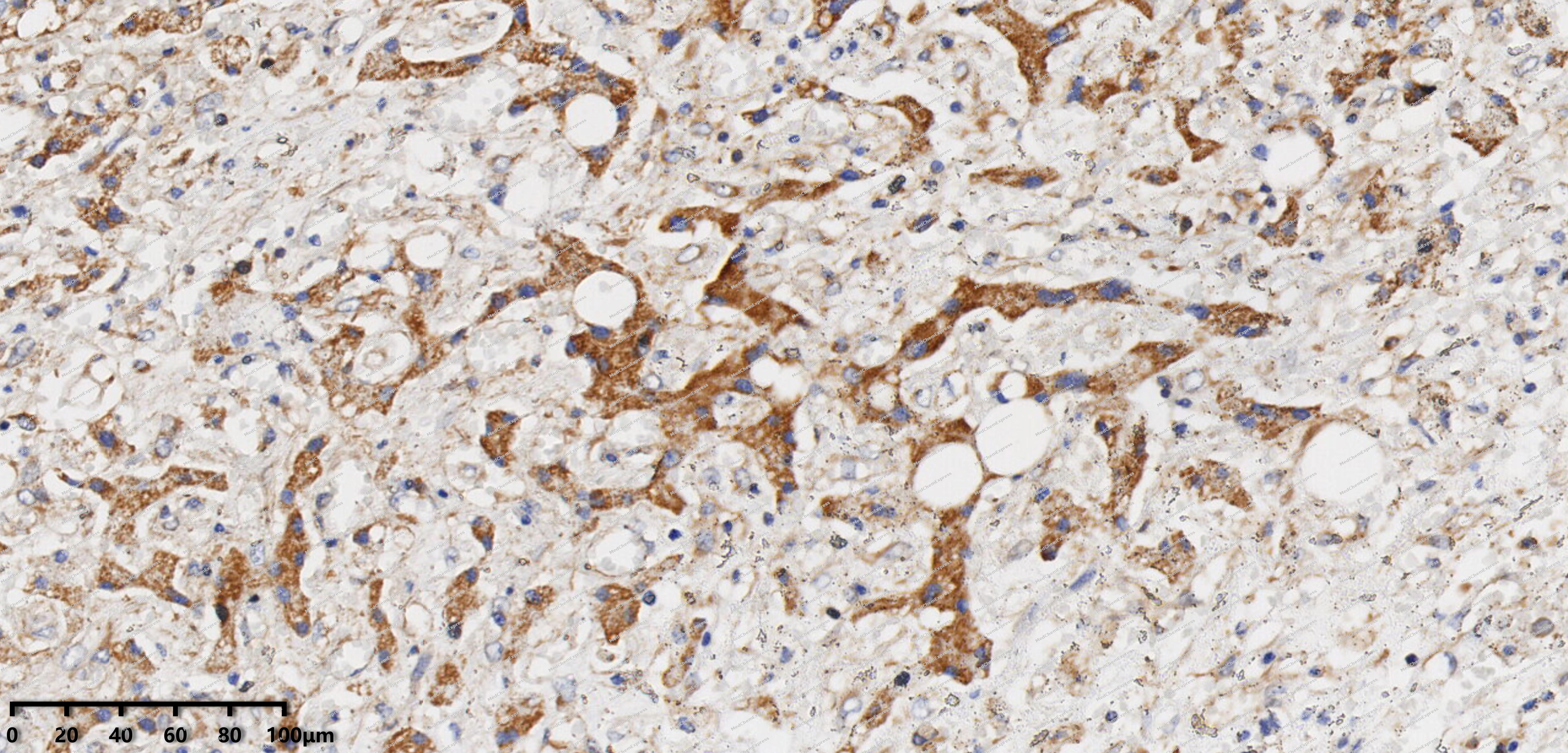 Immunohistochemical analysis of paraffin-embedded human Liver cyst tissue using SNX1 antibody was performed. The section was pretreated using high-temperature mediated EDTA antigen retrieval buffer (pH 9.0), for 20 minutes. The tissues were incubated with primary antibody (HY-P82299, 1:200 dilution) at room temperature for 20 minutes. Detection was performed using an HRP conjugated compact polymer system. DAB was used as the chromogen. The tissues were counterstained with hematoxylin and mounted with neutral balsam mounting medium.
 -
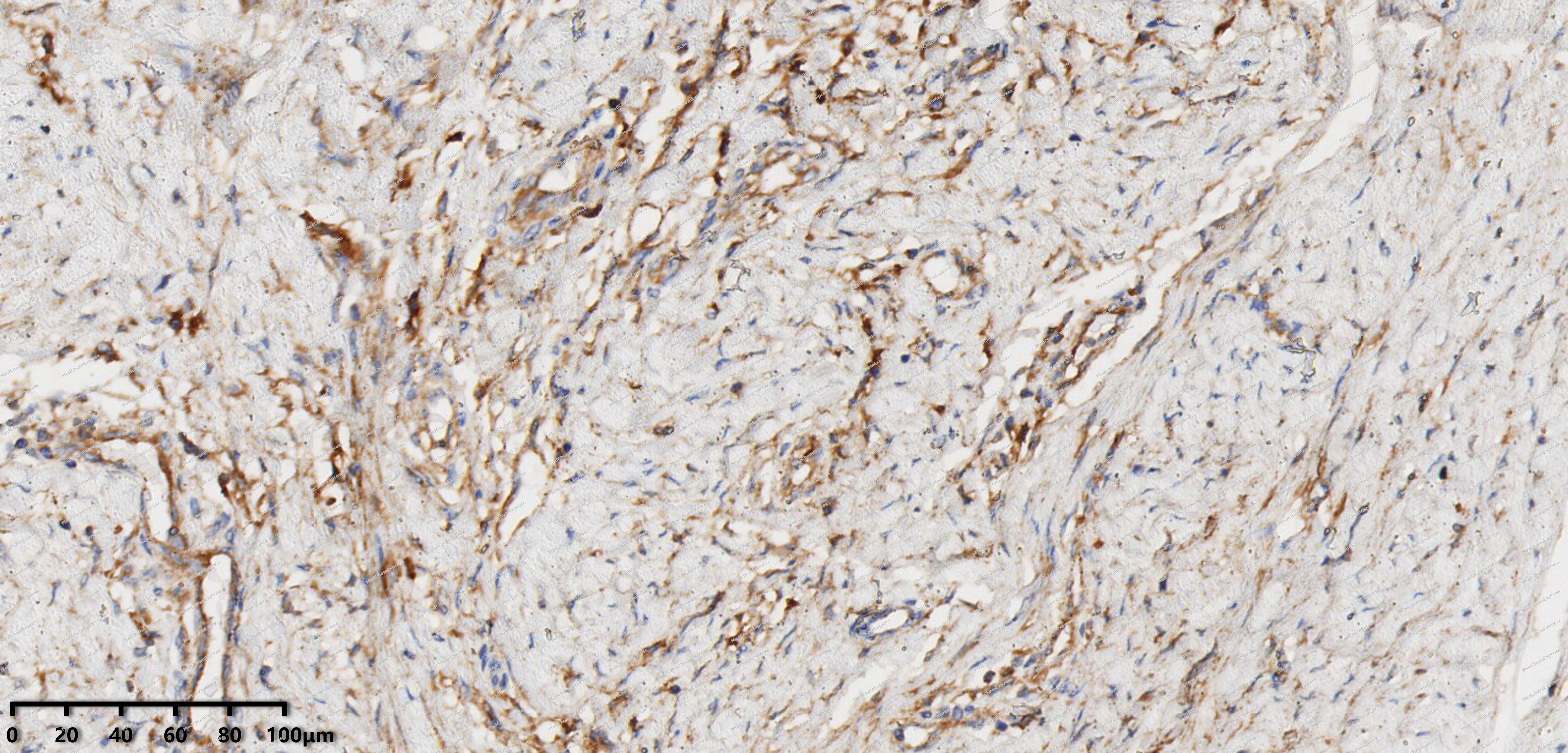 Immunohistochemical analysis of paraffin-embedded human pancreatic serous cystadenoma tissue using SNX1 antibody was performed. The section was pretreated using high-temperature mediated EDTA antigen retrieval buffer (pH 9.0), for 20 minutes. The tissues were incubated with primary antibody (HY-P82299, 1:200 dilution) at room temperature for 20 minutes. Detection was performed using an HRP conjugated compact polymer system. DAB was used as the chromogen. The tissues were counterstained with hematoxylin and mounted with neutral balsam mounting medium.
 -
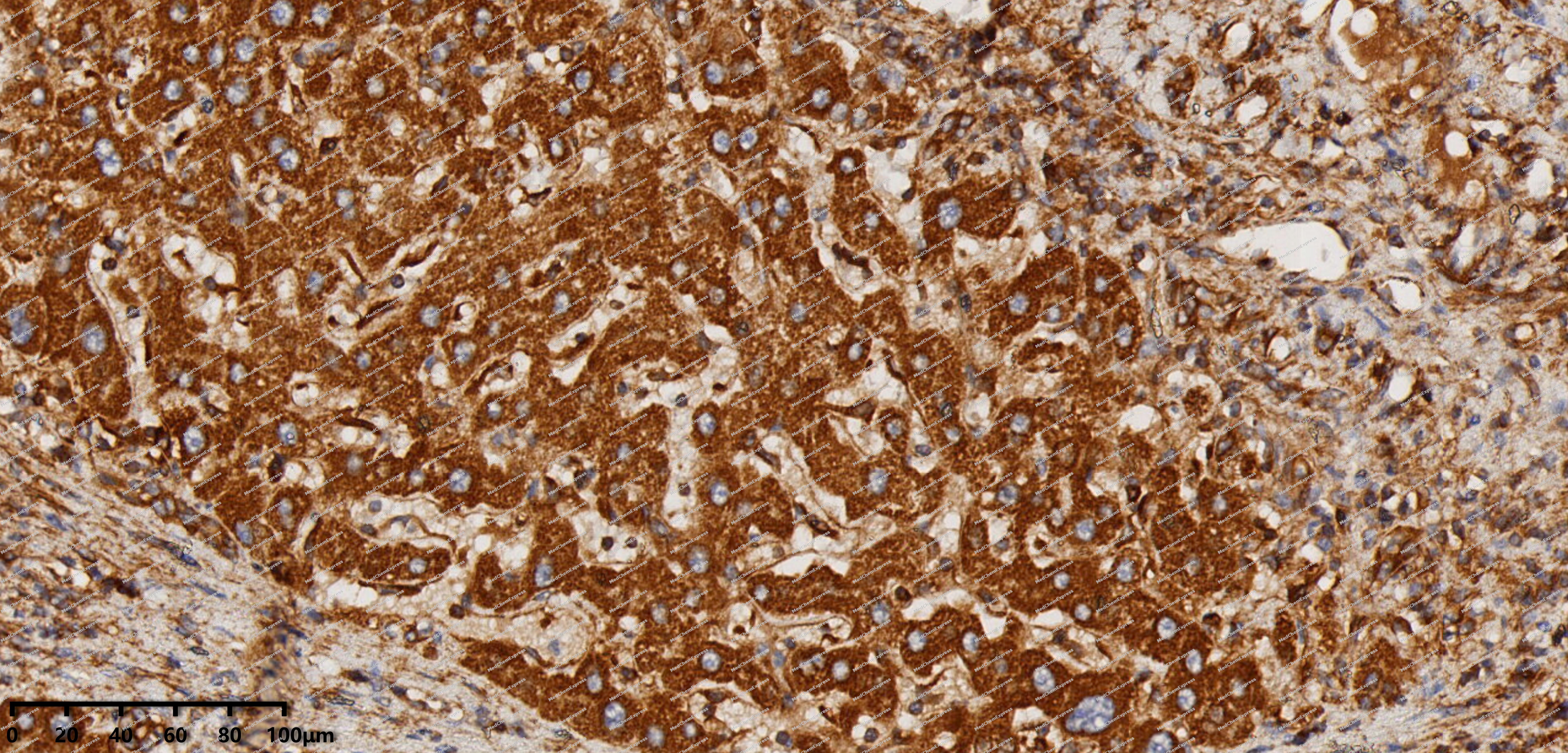 Immunohistochemical analysis of paraffin-embedded human Hepatocellular Carcinoma tissue using SNX1 antibody was performed. The section was pretreated using high-temperature mediated EDTA antigen retrieval buffer (pH 9.0), for 20 minutes. The tissues were incubated with primary antibody (HY-P82299, 1:200 dilution) at room temperature for 20 minutes. Detection was performed using an HRP conjugated compact polymer system. DAB was used as the chromogen. The tissues were counterstained with hematoxylin and mounted with neutral balsam mounting medium.
 -
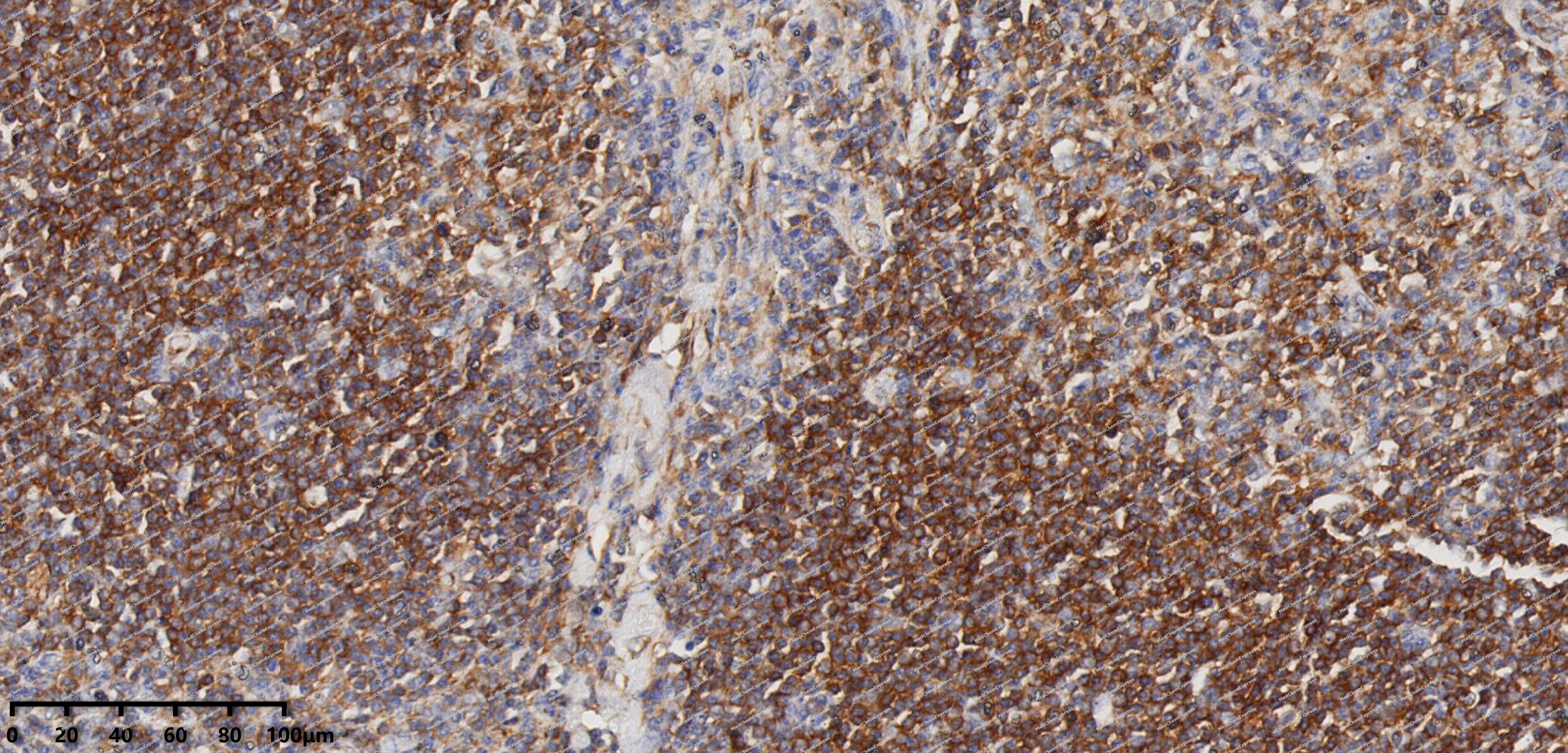 Immunohistochemical analysis of paraffin-embedded human Kimura's disease tissue using SNX1 antibody was performed. The section was pretreated using high-temperature mediated EDTA antigen retrieval buffer (pH 9.0), for 20 minutes. The tissues were incubated with primary antibody (HY-P82299, 1:200 dilution) at room temperature for 20 minutes. Detection was performed using an HRP conjugated compact polymer system. DAB was used as the chromogen. The tissues were counterstained with hematoxylin and mounted with neutral balsam mounting medium.
 -
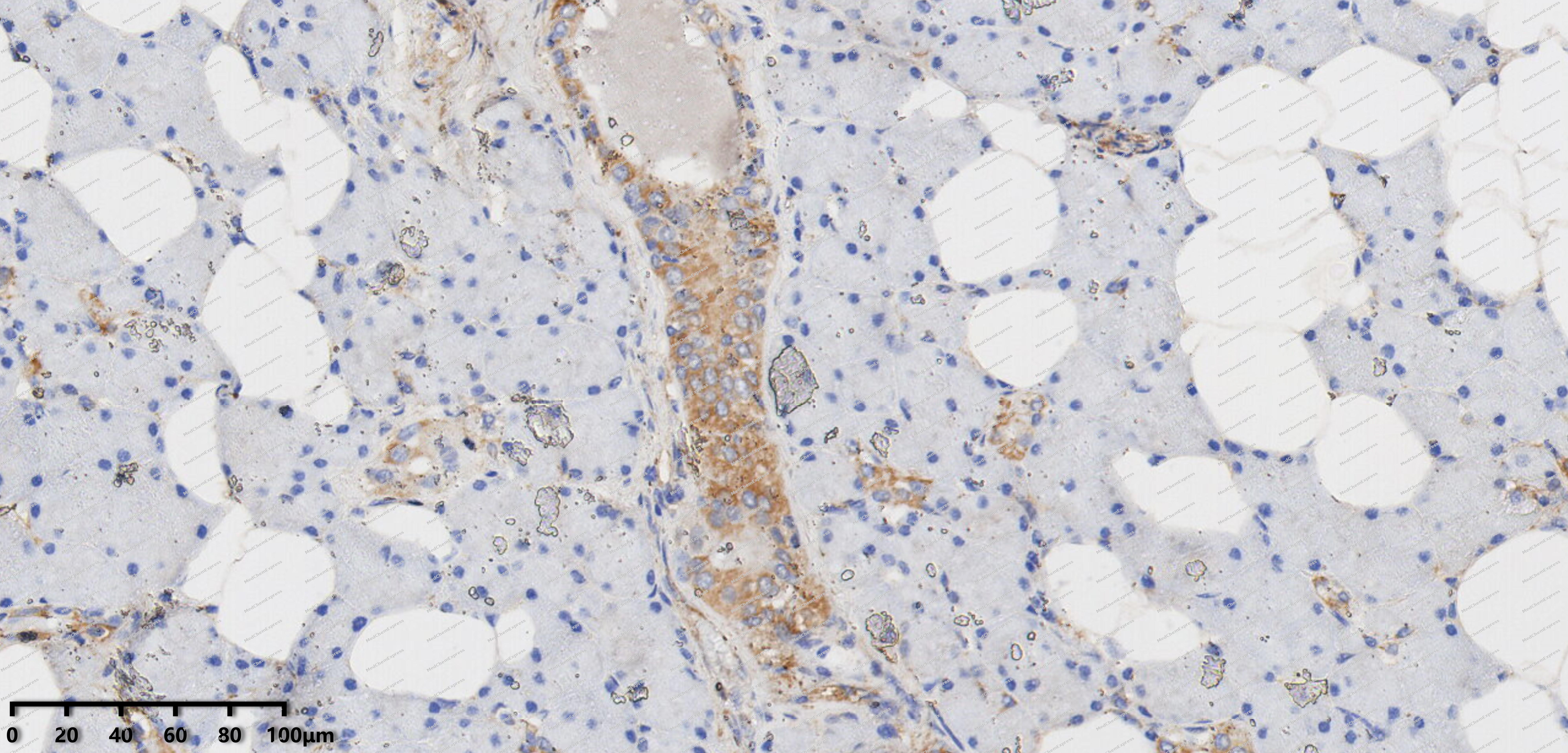 Immunohistochemical analysis of paraffin-embedded human adenolymphoma tissue using SNX1 antibody was performed. The section was pretreated using high-temperature mediated EDTA antigen retrieval buffer (pH 9.0), for 20 minutes. The tissues were incubated with primary antibody (HY-P82299, 1:200 dilution) at room temperature for 20 minutes. Detection was performed using an HRP conjugated compact polymer system. DAB was used as the chromogen. The tissues were counterstained with hematoxylin and mounted with neutral balsam mounting medium.
 -
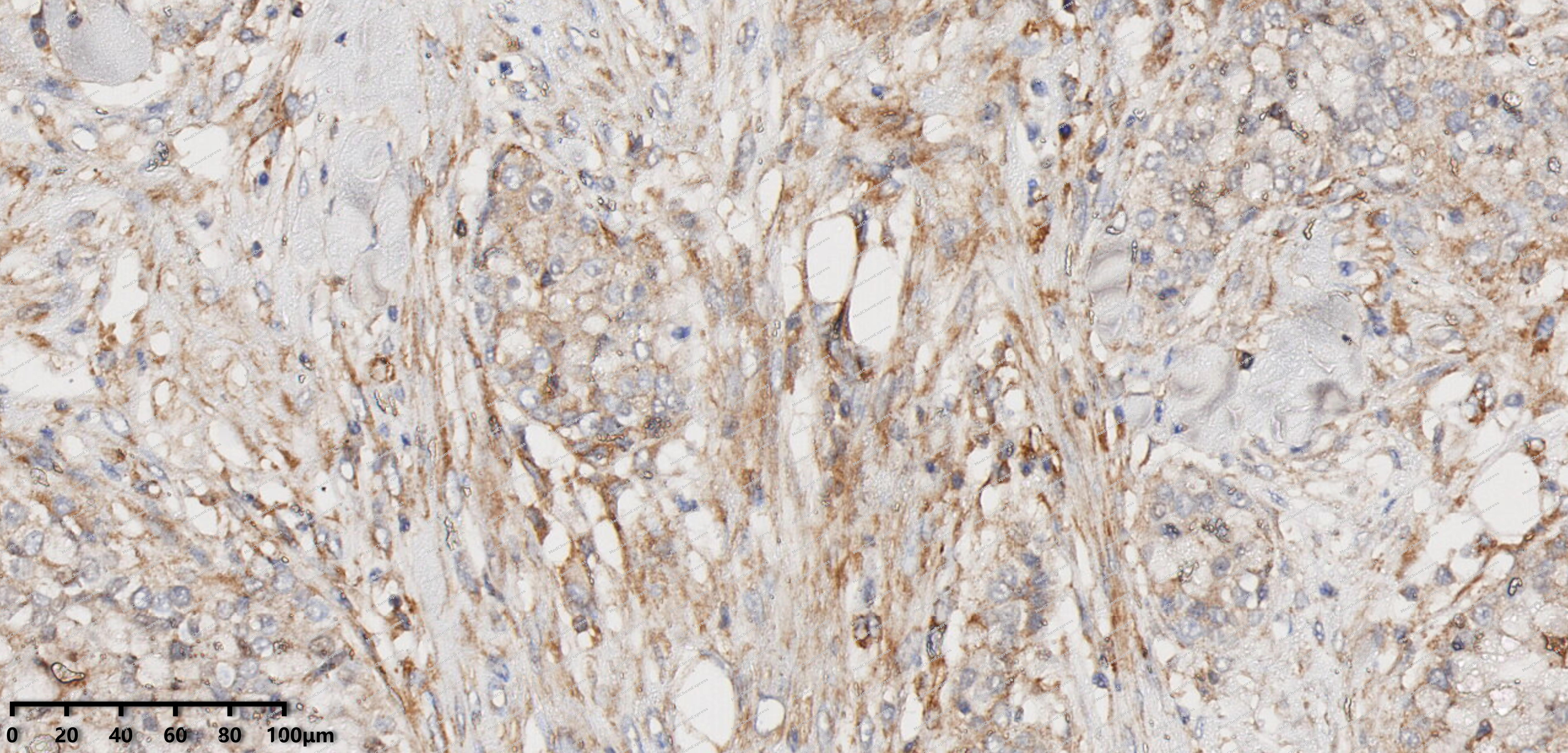 Immunohistochemical analysis of paraffin-embedded human Mucoepidermoid carcinoma tissue using SNX1 antibody was performed. The section was pretreated using high-temperature mediated EDTA antigen retrieval buffer (pH 9.0), for 20 minutes. The tissues were incubated with primary antibody (HY-P82299, 1:200 dilution) at room temperature for 20 minutes. Detection was performed using an HRP conjugated compact polymer system. DAB was used as the chromogen. The tissues were counterstained with hematoxylin and mounted with neutral balsam mounting medium.
 -
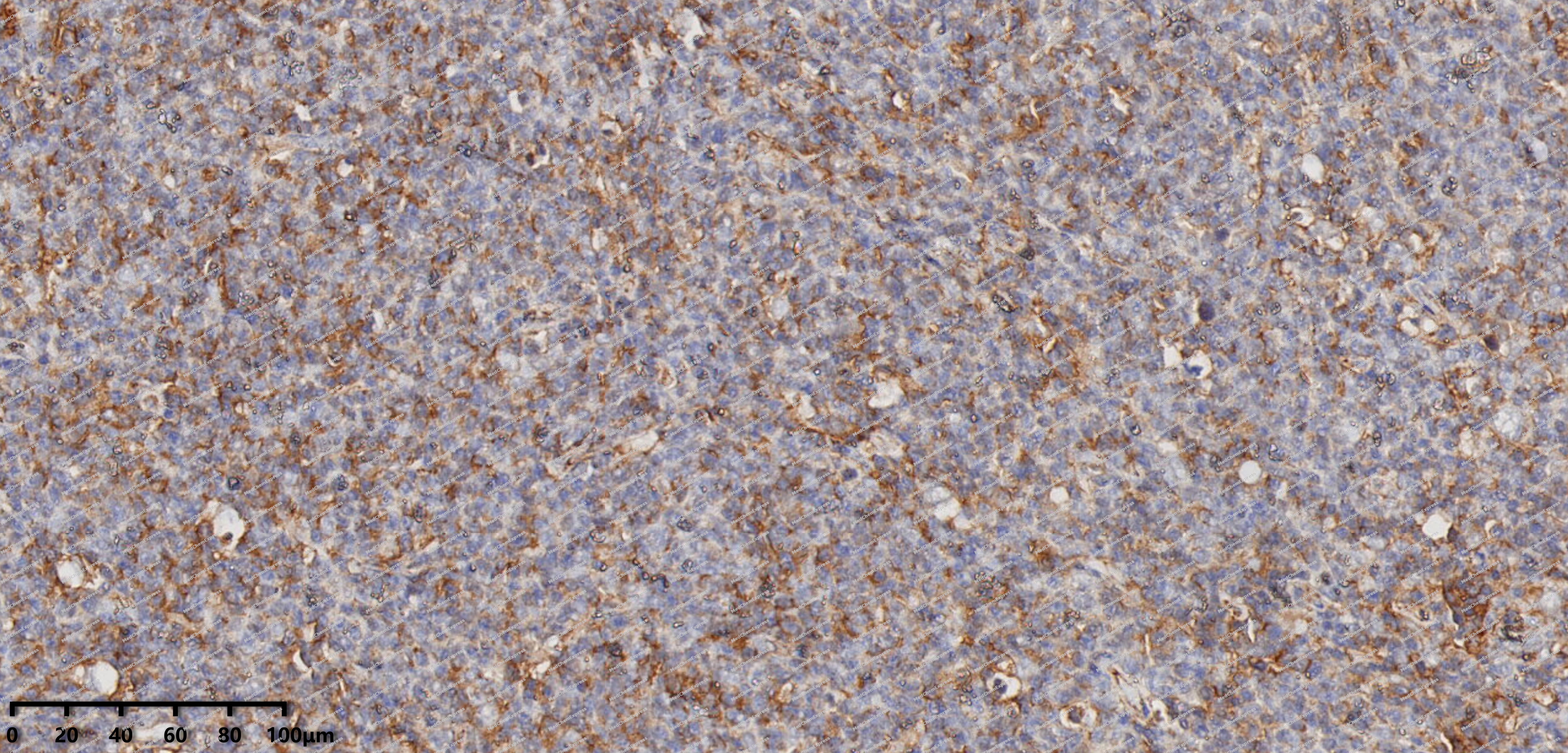 Immunohistochemical analysis of paraffin-embedded human Follicular Lymphoma tissue using SNX1 antibody was performed. The section was pretreated using high-temperature mediated EDTA antigen retrieval buffer (pH 9.0), for 20 minutes. The tissues were incubated with primary antibody (HY-P82299, 1:200 dilution) at room temperature for 20 minutes. Detection was performed using an HRP conjugated compact polymer system. DAB was used as the chromogen. The tissues were counterstained with hematoxylin and mounted with neutral balsam mounting medium.
 -
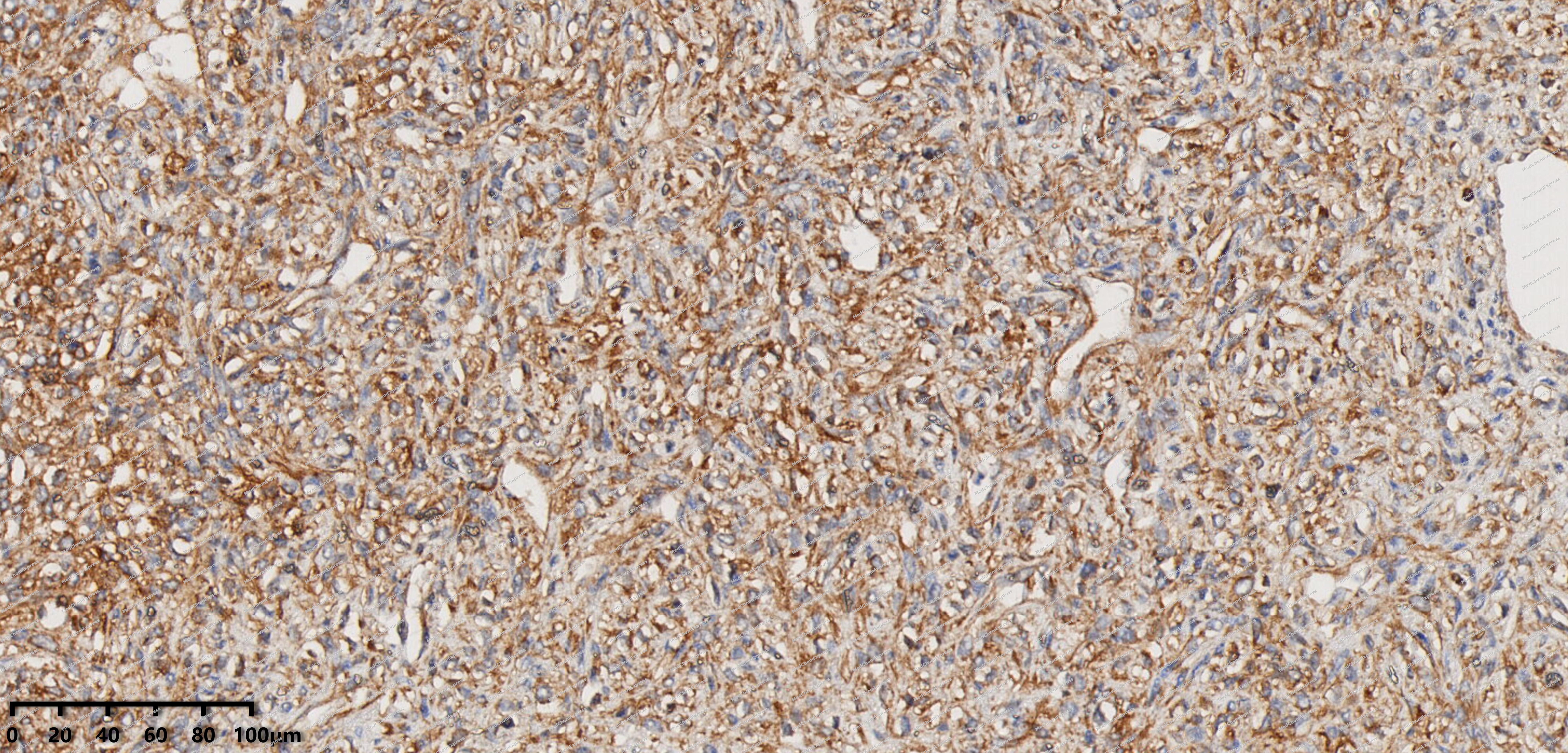 Immunohistochemical analysis of paraffin-embedded human solitary fibrous tumor tissue using SNX1 antibody was performed. The section was pretreated using high-temperature mediated EDTA antigen retrieval buffer (pH 9.0), for 20 minutes. The tissues were incubated with primary antibody (HY-P82299, 1:200 dilution) at room temperature for 20 minutes. Detection was performed using an HRP conjugated compact polymer system. DAB was used as the chromogen. The tissues were counterstained with hematoxylin and mounted with neutral balsam mounting medium.
 -
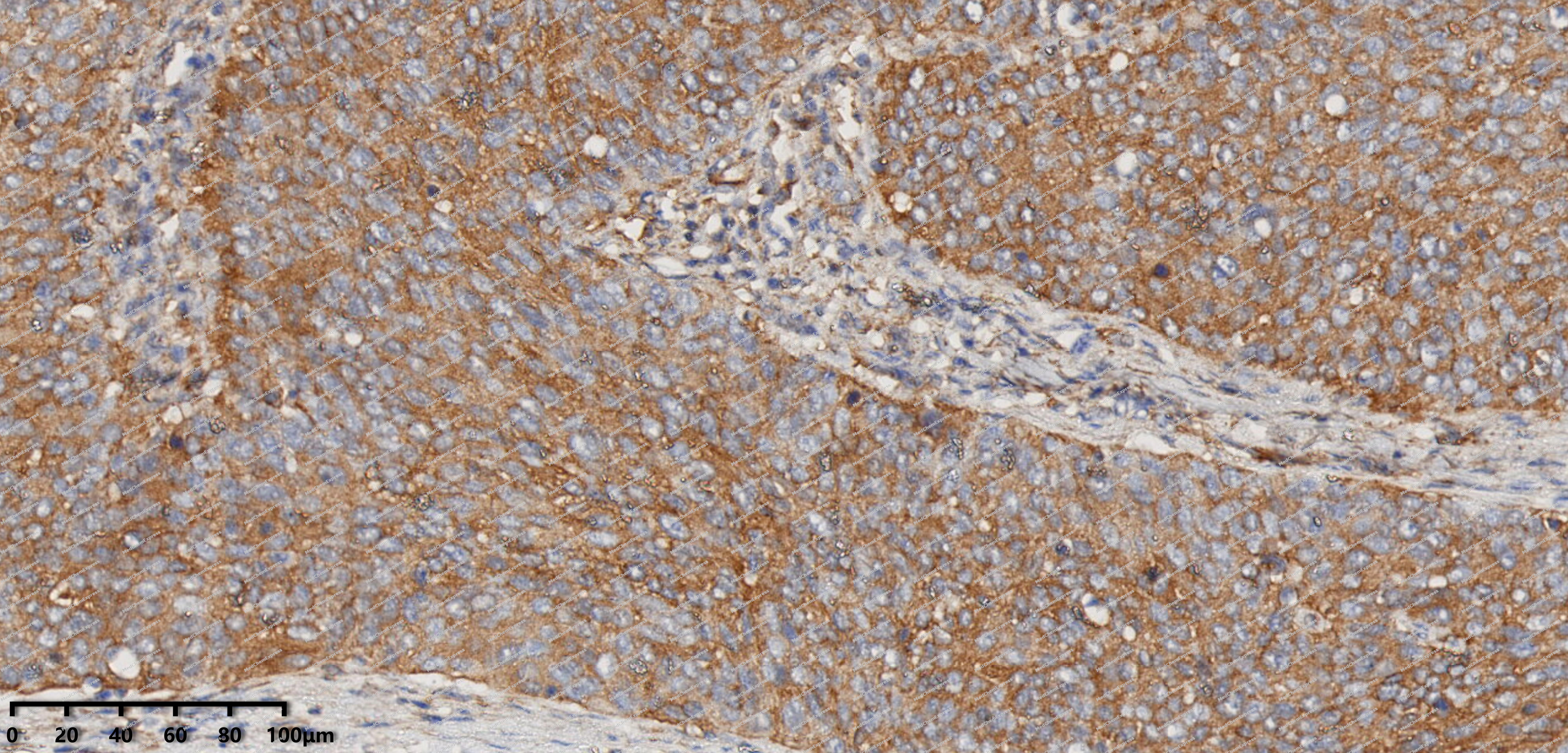 Immunohistochemical analysis of paraffin-embedded human cervical squamous cell carcinoma tissue using SNX1 antibody was performed. The section was pretreated using high-temperature mediated EDTA antigen retrieval buffer (pH 9.0), for 20 minutes. The tissues were incubated with primary antibody (HY-P82299, 1:200 dilution) at room temperature for 20 minutes. Detection was performed using an HRP conjugated compact polymer system. DAB was used as the chromogen. The tissues were counterstained with hematoxylin and mounted with neutral balsam mounting medium.
 -
 Immunocytochemistry analysis of HepG2 cells labeling SNX1 with SNX1 Antibody (HY-P82299) at 1/100 dilution. Cells were fixed in 4% paraformaldehyde for 15 minutes at room temperature, permeabilized with 0.1% Triton X-100 in PBS for 15 minutes at room temperature, then blocked with quick block buffer for 10 minutes at room temperature. Cells were then incubated with SNX1 Antibody (HY-P82299) at 1/100 dilution in quick block buffer overnight at 4 ℃. AF488-conjugated Goat Anti-Rabbit IgG H&L (HY-P8002, Green) was used as the secondary antibody at 1/1,000 dilution. PBS instead of the primary antibody was used as the secondary antibody only control. The Nuclear counterstain was DAPI (Blue).
 -
 Immunocytochemistry analysis of NIH-3T3 cells labeling SNX1 with SNX1 Antibody (HY-P82299) at 1/100 dilution. Cells were fixed in 4% paraformaldehyde for 15 minutes at room temperature, permeabilized with 0.1% Triton X-100 in PBS for 15 minutes at room temperature, then blocked with quick block buffer for 10 minutes at room temperature. Cells were then incubated with SNX1 Antibody (HY-P82299) at 1/100 dilution in quick block buffer overnight at 4 ℃. AF488-conjugated Goat Anti-Rabbit IgG H&L (HY-P8002, Green) was used as the secondary antibody at 1/1,000 dilution. PBS instead of the primary antibody was used as the secondary antibody only control. The Nuclear counterstain was DAPI (Blue).
 -
 Flow cytometric analysis of 1X10⁶ HeLa cells labeling SNX1 Antibody (HY-P82299, red). Cells were fixed with 4% paraformaldehyde and permeabilised with 90% methanol. Then stained with the primary antibody at 1/100 dilution for an hour at 4℃. AF488-conjugated Goat Anti-Rabbit IgG H&L (HY-P8002) was used as the secondary antibody at 1/1,000 dilution for 30 minutes at 4℃. Rabbit IgG Isotype Control (HY-P80879, blue) was used as the isotype control, cells without incubation with primary antibody were used as the unlabeled control (black).


Background
-
Function
SNX1 is involved in several stages of intracellular trafficking. Interacts with membranes containing phosphatidylinositol 3-phosphate (PtdIns(3P)) or phosphatidylinositol 3,5-bisphosphate (PtdIns(3,5)P2). Acts in part as component of the retromer membrane-deforming SNX-BAR subcomplex. The SNX-BAR retromer mediates retrograde transport of cargo proteins from endosomes to the trans-Golgi network (TGN) and is involved in endosome-to-plasma membrane transport for cargo protein recycling. The SNX-BAR subcomplex functions to deform the donor membrane into a tubular profile called endosome-to-TGN transport carrier (ETC) (Probable). Can sense membrane curvature and has in vitro vesicle-to-membrane remodeling activity. Involved in retrograde endosome-to-TGN transport of lysosomal enzyme receptors (IGF2R, M6PR and SORT1) and Shiginella dysenteria toxin stxB. Plays a role in targeting ligand-activated EGFR to the lysosomes for degradation after endocytosis from the cell surface and release from the Golgi. Involvement in retromer-independent endocytic trafficking of P2RY1 and lysosomal degradation of protease-activated receptor-1/F2R. Promotes KALRN- and RHOG-dependent but retromer-independent membrane remodeling such as lamellipodium formation; the function is dependent on GEF activity of KALRN. Required for endocytosis of DRD5 upon agonist stimulation but not for basal receptor trafficking[1][2][3][4][5][6][7][8][9][10][11][12].
-
Subcellular Localization
Endosome membrane; Peripheral membrane protein; Cytoplasmic side; Golgi apparatus, trans-Golgi network membrane; Peripheral membrane protein; Cytoplasmic side; Early endosome membrane; Peripheral membrane protein; Cytoplasmic side; Cell projection, lamellipodium
-
Isoforms & Post-Translational Modification
Q13596 has 3 isomers: Q13596-1: 59070 Da (predicted); Q13596-2: 51813 Da (predicted); Q13596-3: 63082 Da (predicted).
-
Subunit
Predominantly forms heterodimers with BAR domain-containing sorting nexins SNX5, SNX6 and SNX32; can self-associate to form homodimers (PubMed:23085988). The heterodimers are proposed to self-assemble into helical arrays on the membrane to stabilize and expand local membrane curvature underlying endosomal tubule formation. Thought to be a component of the originally described retromer complex (also called SNX-BAR retromer) which is a pentamer containing the heterotrimeric retromer cargo-selective complex (CSC), also described as vacuolar protein sorting subcomplex (VPS) and a heterodimeric membrane-deforming subcomplex formed between SNX1 or SNX2 and SNX5 or SNX6 (also called SNX-BAR subcomplex); the respective CSC and SNX-BAR subcomplexes associate with low affinity (Probable). Interacts with SNX5, SNX6, SNX32, VPS26A, VPS29, VPS35, DRD5, DENND5A, KALRN, RHOG (GDP-bound form) (PubMed:11102511, PubMed:19619496, PubMed:19935774, PubMed:20604901, PubMed:23085988, PubMed:23152498, PubMed:9819414). The interaction with SNX2 is reported controversially (PubMed:11997453, PubMed:19619496, PubMed:23085988, PubMed:9819414). Interacts with DNAJC13; prevented by presence of HGS (PubMed:19874558). Interacts with HGS (By similarity)
-
SwissProt ID
-
Synonyms
SNX1; Sorting nexin-1
-
Research Field
Signal Transduction


Documentation
-
Data Sheet (234 KB)
-
SDS (251 KB)
- English - EN (251 KB)
- Français - FR (251 KB)
- Deutsch - DE (251 KB)
- Norwegian - NO (251 KB)
- Español - ES (251 KB)
- Swedish - SV (251 KB)
- Italian - IT (251 KB)
- Korean - KR (251 KB)
- Portuguese - PT (251 KB)
-
User Guide for Antibodies (1077 KB)

 Powered by Bioz
