



TSG101 Antibody (YA025)
(Synonyms: TSG101; Tumor susceptibility gene 101 protein; ESCRT-I complex subunit TSG101)Based on 3 publication(s) in Google Scholar
TSG101 Antibody (YA025) is a Rabbit-derived and non-conjugated IgG monoclonal antibody, targeting to TSG101.
-
Host:
Rabbit
-
Isotype:
IgG
-
Application:
WB, IHC-P, ICC/IF, FC
-
Reactivity :
Human, Mouse, Rat
-
Formulation:
Supplied in PBS(pH 7.4), 150mM NaCl, 50% Glycerol, 0.02% Sodium azide and 0.05% BSA.
-
Conjugation:
Non-conjugated


To place orders, for customer services and technical support, please contact: MedChemExpress USA
Tel: 609-228-6898 E-mail: [email protected] [email protected]



Publications Citing Use of MedChemExpress (MCE) TSG101 Antibody (YA025)
More
 3 Publications Citing Use of MCE TSG101 Antibody (YA025) (1)
-
WB


Applications
| Application |
WB

WB: Western Blot
|
IHC-P
IHC-P: Immunohistochemistry-Paraffin
|
ICC/IF
ICC/IF: Immunocytochemistry/
Immunofluorescence |
FC
FC: Flow Cytometry
|
|---|---|---|---|---|
| Dilution Ratio | 1:2000-20000 | 1:100-1000 | 1:200-500 | 1:20-50 |


Product Details
TSG101 Antibody (YA025) is a Rabbit-derived and non-conjugated IgG monoclonal antibody, targeting to TSG101.
-
Host Rabbit

-
Clonality Recombinant,Monoclonal

-
Species ReactivityHuman, Mouse, Rat
-
Observed Molecular WeightObserved band size: 47, 52 kDa
Note: Due to possible protein modifications or aggregation, the molecular weight should be confirmed by actual measurement, and the predicted value is for reference only.

-
Calculated Molecular Weight Predicted band size: 44 kDa

Entrez Gene: 7251 Human ; 22088 Mouse ; 292925 Rat
SwissProt: Q99816 Human ; Q61187 Mouse ; Q6IRE4 Rat
OMIM: 601387 Human
Synthetic peptide corresponding to Human TSG101.The exact sequence is proprietary to MCE.
Endogenous
affinity purified
Non-conjugated
Unmodified
IgG


Product Properties
-
Appearance
Solution
-
Formulation
Supplied in PBS(pH 7.4), 150mM NaCl, 50% Glycerol, 0.02% Sodium azide and 0.05% BSA.
-
Concentration
Batch-dependent, Please check the COA for the concentration of each lot. Check Lot Concentration
-
Storage & Stability
Stored at -20°C for 1 year. Avoid repeated freeze / thaw cycles.
-
Shipping
Shipping with blue ice.


Publications (3)
-
Journal Impact Factor
-
Most Recent
-
J Transl Med
Tumor-derived exosomal lncRNA-MIR193BHG promotes bone metastasis of breast cancer by targeting the miR-489-3p/DNMT3A signaling axis in osteoclasts. [Abstract]2025 Jan 31;23(1):142. PMID: 39891171
TSG101 Antibody (YA025) purchased from MedChemExpress. Usage Cited in: J Transl Med. 2025 Jan 31;23(1):142. [Abstract]
Western blot analysis detecting the expression of exosome marker proteins CD63 and TSG101.
-
Int J Nanomedicine
Comparison of the Cytotoxicity, Internalization and Anti-Cancer Drug Delivery Efficacy of Nature Killer Cell Derived Nanovesicles and Extracellular Vesicles. [Abstract]2025 Sep 2:20:10683-10700. PMID: 40918949


Verification Images
-
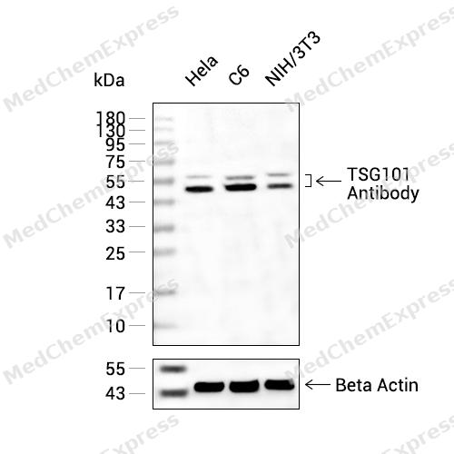 Western blot analysis of extracts from Hela(lane 2(20ug) , C6(lane 3(20ug) and NIH/3T3(lane 4(20ug) using TSG101 Antibody (HY-P80924) Rabbit mAb. Proteins were transferred to a PVDF membrane and blocked with 5% non-fat milk in TBST for 2 hour at room temperature. The primary antibody (1/1000) and Loading control antibody (Beta Actin, HY-P80438, 1/10000) was used in 5% non-fat milk in TBST at 4°C overnight. Goat Anti-Mouse/Rabbit IgG-HRP Secondary Antibody (1/10000) was used for 1 hour at room temperature.
 -
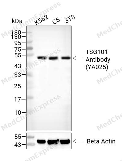 Western blot analysis of extracts from K562 (lane 2(40μg),C6 (lane 3(40μg), 3T3 (lane 4(40μg)using TSG101 Antibody (YA025). Proteins were transferred to a PVDF membrane and blocked with 5% BSA in TBST for 2 hour at room temperature. The primary antibody and Loading control antibody (Beta Actin, HY-P80438, 1/3000) was used in 5% BSA in TBST at 4°C overnight. Goat Anti-Mouse/Rabbit IgG-HRP Secondary Antibody (HY-P8004/HY-P8001, 1/10,000) was used for 1 hour at room temperature.
 -
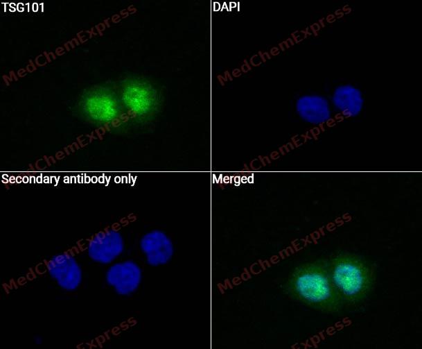 Immunocytochemistry analysis of Hela cells labeling TSG101 with TSG101 Antibody (HY-P80924)at 1/50 dilution. Cells were fixed in 4% paraformaldehyde for 15 minutes at room temperature, permeabilized with 0.1% Triton X-100 for 10 minutes at room temperature, then blocked with QuickBlock™ Blocking Buffer for Immunol Staining for 10 min at room temperature. Cells were then incubated with TSG101 Antibody (HY-P80924) at 1/50 dilution in QuickBlock™ Blocking Buffer for Immunol Staining at 4 ℃. Alexa Fluor® 488-conjugated AffiniPure Goat Anti-Rabbit IgG H&L(HY-P8002, Green) was used as the secondary antibody at 1/1,000 dilution. PBS instead of the primary antibody was used as the secondary antibody only control. The Nuclear counterstain was DAPI (Blue).
 -
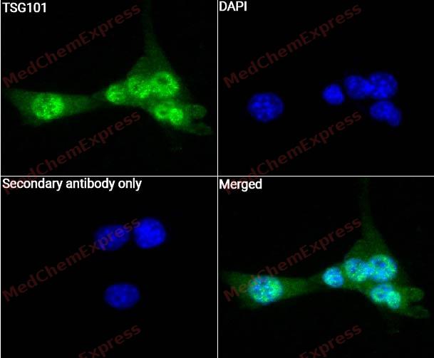 Immunocytochemistry analysis of NIH3T3 cells labeling TSG101 with TSG101 Antibody (HY-P80924) at 1/50 dilution. Cells were fixed in 4% paraformaldehyde for 15 minutes at room temperature, permeabilized with 0.1% Triton X-100 for 10 minutes at room temperature, then blocked with QuickBlock™ Blocking Buffer for Immunol Staining for 10 min at room temperature. Cells were then incubated with TSG101 Antibody (HY-P80924) at 1/50 dilution in QuickBlock™ Blocking Buffer for Immunol Staining at 4 ℃. Alexa Fluor® 488-conjugated AffiniPure Goat Anti-Rabbit IgG H&L(HY-P8002, Green) was used as the secondary antibody at 1/1,000 dilution. PBS instead of the primary antibody was used as the secondary antibody only control. The Nuclear counterstain was DAPI (Blue).
 -
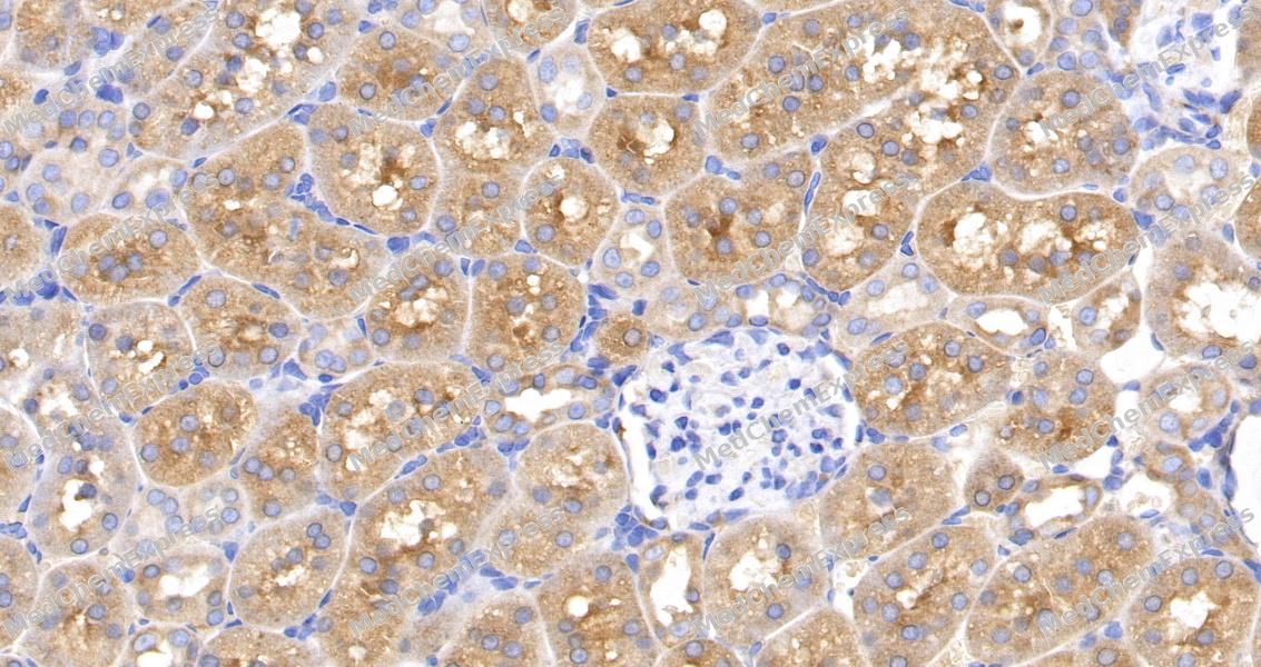 Immunohistochemical analysis of paraffin-embedded rat kidney tissue using TSG101 Antibody. The section was pre-treated using heat mediated antigen retrieval with Tris-EDTA buffer (pH 9.0) for 8 minutes. The tissues were blocked in QuickBlock for 20 minutes at room temperature, washed with ddH2O and PBS, and then probed with the primary antibody at 1/100 dilution in 4℃ overnight. The detection was performed using an HRP conjugated compact polymer system. DAB was used as the chromogen. Tissues were counterstained with hematoxylin and mounted with DPX.
 -
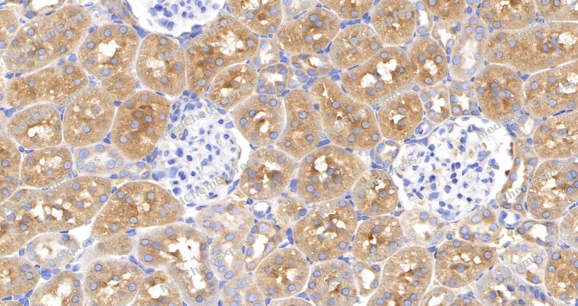 Immunohistochemical analysis of paraffin-embedded rat kidney tissue using TSG101 Antibody. The section was pre-treated using heat mediated antigen retrieval with Tris-EDTA buffer (pH 9.0) for 8 minutes. The tissues were blocked in QuickBlock for 20 minutes at room temperature, washed with ddH2O and PBS, and then probed with the primary antibody at 1/100 dilution in 4℃ overnight. The detection was performed using an HRP conjugated compact polymer system. DAB was used as the chromogen. Tissues were counterstained with hematoxylin and mounted with DPX.


Background
-
Function
TSG101 component of the ESCRT-I complex, a regulator of vesicular trafficking process. Binds to ubiquitinated cargo proteins and is required for the sorting of endocytic ubiquitinated cargos into multivesicular bodies (MVBs). Mediates the association between the ESCRT-0 and ESCRT-I complex. Required for completion of cytokinesis; the function requires CEP55. May be involved in cell growth and differentiation. Acts as a negative growth regulator. Involved in the budding of many viruses through an interaction with viral proteins that contain a late-budding motif P-[ST]-A-P. This interaction is essential for viral particle budding of numerous retroviruses. Required for the exosomal release of SDCBP, CD63 and syndecan. It may also play a role in the extracellular release of microvesicles that differ from the exosomes[1][2].
-
Subcellular Localization
Cytoplasm; Early endosome membrane; Peripheral membrane protein; Cytoplasmic side; Late endosome membrane; Peripheral membrane protein; Cytoplasm, cytoskeleton, microtubule organizing center, centrosome; Midbody, Midbody ring; Nucleus
-
Expression
Tissue_specificity:Heart, brain, placenta, lungs, liver, skeletal muscles, kidneys, and pancreas -
Isoforms & Post-Translational Modification
Q99816 has 2 isomers: Q99816-1: 43944 Da (predicted); Q99816-2: 31732 Da (predicted).
Monoubiquitinated at multiple sites by LRSAM1 and by MGRN1. Ubiquitination inactivates it, possibly by regulating its shuttling between an active membrane-bound protein and an inactive soluble form. Ubiquitination by MGRN1 requires the presence of UBE2D1 -
Subunit
Component of the ESCRT-I complex (endosomal sorting complex required for transport I) which consists of TSG101, VPS28, a VPS37 protein (VPS37A to -D) and MVB12A or MVB12B in a 1:1:1:1 stoichiometry (PubMed:18005716). Interacts with VPS37A, VPS37B and VPS37C (PubMed:15218037, PubMed:15509564). Interacts with DMAP1 (PubMed:10888872). Interacts with ubiquitin (PubMed:11595185). Interacts with stathmin, GMCL and AATF (By similarity). Component of an ESCRT-I complex (endosomal sorting complex required for transport I) which consists of TSG101, VPS28, VPS37A and UBAP1 in a 1:1:1:1 stoichiometry (PubMed:21757351). Interacts with HGS; the interaction mediates the association with the ESCRT-0 complex. Interacts with GGA1 and GGA3 (PubMed:15039775, PubMed:15143060). Interacts (via UEV domain) with PDCD6IP/AIP1 (PubMed:14505570, PubMed:14519844). Interacts with VPS28, SNF8 and VPS36 (PubMed:14505570). Self-associates (PubMed:14505570, PubMed:14519844). Interacts with MVB12A; the association appears to be mediated by the TSG101-VPS37 binary subcomplex. Interacts with VPS37D. Interacts with LRSAM1. Interacts with CEP55; the interaction is required for cytokinesis but not for viral budding (PubMed:17853893). Interacts with PDCD6 (PubMed:18256029). Interacts with LITAF (PubMed:23166352). Interacts with MGRN1 (PubMed:17229889). Interacts with ARRDC1; recruits TSG101 to the plasma membrane (PubMed:21191027, PubMed:22315426)
-
SwissProt ID
-
Synonyms
TSG101; Tumor susceptibility gene 101 protein; ESCRT-I complex subunit TSG101
-
Research Field
Cell Biology
Documentation
-
Data Sheet (261 KB)
-
SDS (252 KB)
- English - EN (252 KB)
- Français - FR (252 KB)
- Deutsch - DE (252 KB)
- Norwegian - NO (252 KB)
- Español - ES (252 KB)
- Swedish - SV (252 KB)
- Italian - IT (252 KB)
- Korean - KR (252 KB)
- Portuguese - PT (252 KB)
-
User Guide for Antibodies (1077 KB)
References
[1]. Baietti MF, et al. Syndecan-syntenin-ALIX regulates the biogenesis of exosomes. Nat Cell Biol. 2012 Jun 3;14(7):677-85. [Content Brief]
[2]. Nabhan JF, et al. Formation and release of arrestin domain-containing protein 1-mediated microvesicles (ARMMs) at plasma membrane by recruitment of TSG101 protein. Proc Natl Acad Sci U S A. 2012 Mar 13;109(11):4146-51. [Content Brief]

 Powered by Bioz
