



Wilms Tumor Protein Antibody (YA2277)
(Synonyms: GUD; AWT1; WAGR; WT33; NPHS4; WIT-2; EWS-WT1; WIT 2 antibody; WT 1 antibody; FWT1 antibody; GUD antibody; NPHS4 antibody; WAGR antibody; Wilms tumor 1 antibody; Wilms Tumor antibody; Wilms tumor protein antibody; Wilms' tumor gene antibody; WIT2 antibody; WT antibody; WT1 antibody; WT1_HUMAN antibody; WT33 antibody)
Based on 1 Customer Validation
Wilms Tumor Protein Antibody (YA2277) is a Rabbit-derived and non-conjugated IgG monoclonal antibody, targeting to Wilms Tumor Protein.
-
Host:
Rabbit
-
Isotype:
IgG
-
Application:
WB, IHC-P, ICC/IF, FC
-
Reactivity :
Human, Mouse
-
Formulation:
Supplied in 50mM Tris-Glycine(pH 7.4), 0.15M NaCl, 40%Glycerol, 0.01% sodium azide and 0.05% BSA.
-
Conjugation:
Non-conjugated


To place orders, for customer services and technical support, please contact: MedChemExpress USA
Tel: 609-228-6898 E-mail: [email protected] [email protected]





Applications
| Application |
WB

WB: Western Blot
|
IHC-P
IHC-P: Immunohistochemistry-Paraffin
|
ICC/IF
ICC/IF: Immunocytochemistry/
Immunofluorescence |
FC
FC: Flow Cytometry
|
|---|---|---|---|---|
| Dilution Ratio | 1:500-1:1000 | 1:50-1:100 | 1:50-1:200 | 1:50-1:100 |


Product Details
Wilms Tumor Protein Antibody (YA2277) is a Rabbit-derived and non-conjugated IgG monoclonal antibody, targeting to Wilms Tumor Protein.
-
Host Rabbit

-
Clonality Recombinant,Monoclonal

-
Species ReactivityHuman, Mouse
-
Observed Molecular WeightObserved band size: 55 kDa
Note: Due to possible protein modifications or aggregation, the molecular weight should be confirmed by actual measurement, and the predicted value is for reference only.

-
Calculated Molecular Weight Predicted band size: 49 kDa

A synthesized peptide derived from human Wilms Tumor Protein
Endogenous
Affinity Chromatography
Non-conjugated
Unmodified
IgG


Product Properties
-
Appearance
Solution
-
Formulation
Supplied in 50mM Tris-Glycine(pH 7.4), 0.15M NaCl, 40%Glycerol, 0.01% sodium azide and 0.05% BSA.
-
Concentration
Batch-dependent, Please check the COA for the concentration of each lot. Check Lot Concentration
-
Storage & Stability
Stored at -20°C for 1 year. Avoid repeated freeze / thaw cycles.
-
Shipping
Shipping with blue ice.


Verification Images
-
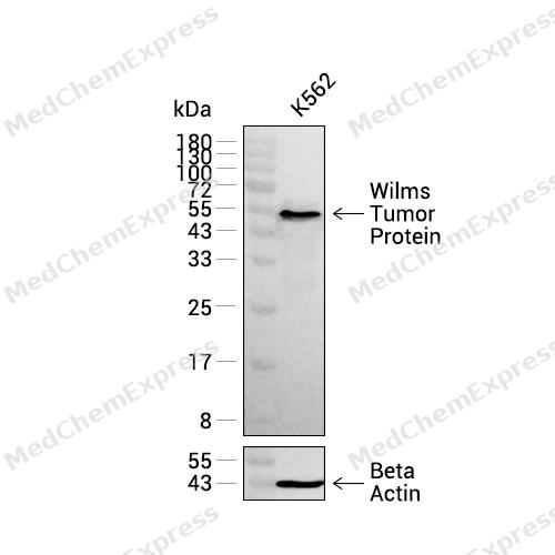 Western blot analysis was performed on extracts from K562 (lane 1, 20 μg) using Wilms Tumor Protein Rabbit mAb.Proteins were transferred to a PVDF membrane and blocked with 5% non - fat milk in TBST at 4°C overnight.The primary antibody (1:1000 dilution) and the loading control antibody (beta-Actin, HY-P83730, 1:5000 dilution) were incubated in 5% non-fat milk in TBST for 1 hour at 37°C.Goat Anti - Rabbit IgG - HRP Secondary Antibody (1:20000 dilution) was then applied for 40 minutes at 37°C.
 -
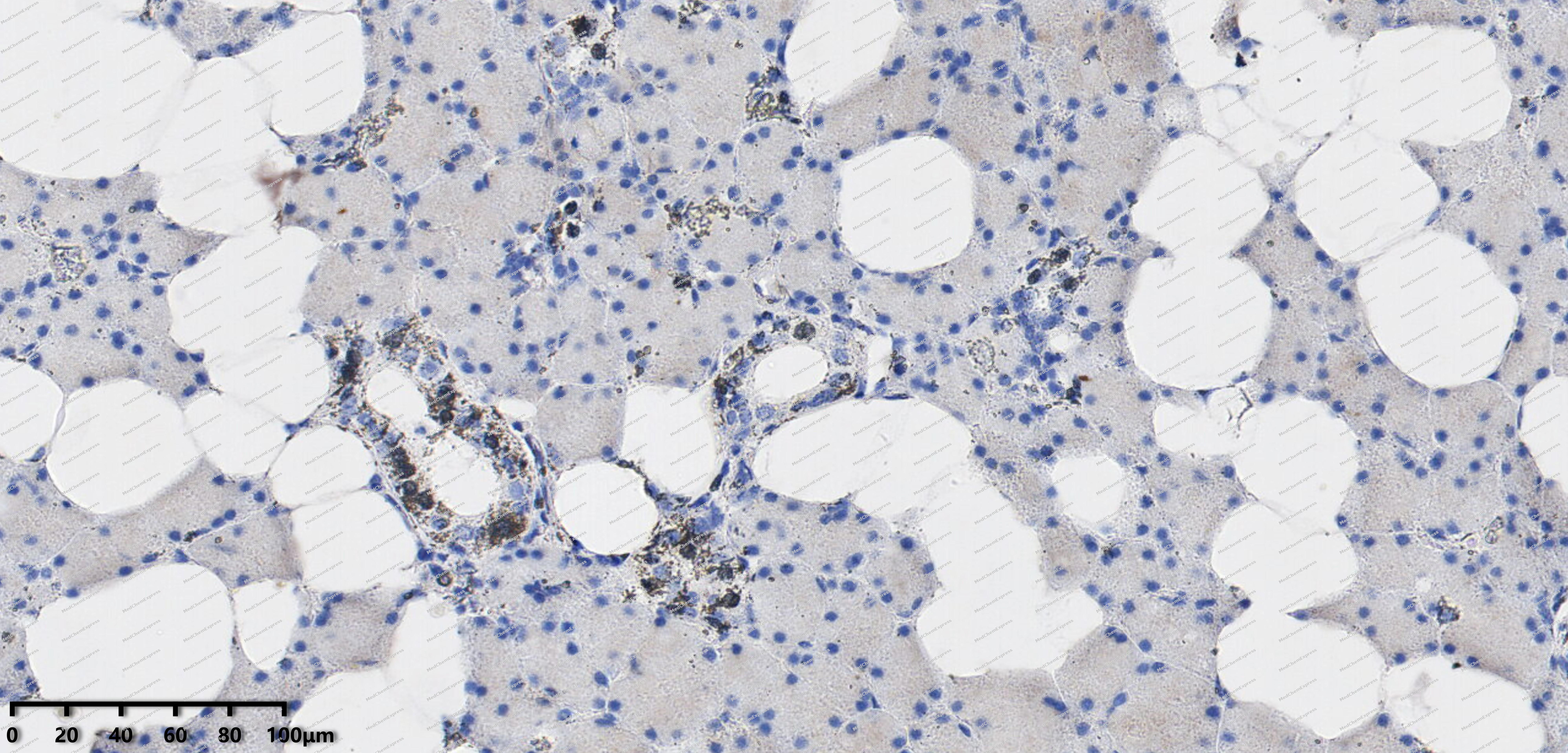 Immunohistochemical analysis of paraffin-embedded human adenolymphoma tissue using Wilms Tumor Protein antibody was performed. The section was pretreated using high-temperature mediated EDTA antigen retrieval buffer (pH 9.0), for 20 minutes. The tissues were incubated with primary antibody (HY-P82532, 1:100 dilution) at room temperature for 20 minutes. Detection was performed using an HRP conjugated compact polymer system. DAB was used as the chromogen. The tissues were counterstained with hematoxylin and mounted with neutral balsam mounting medium.
 -
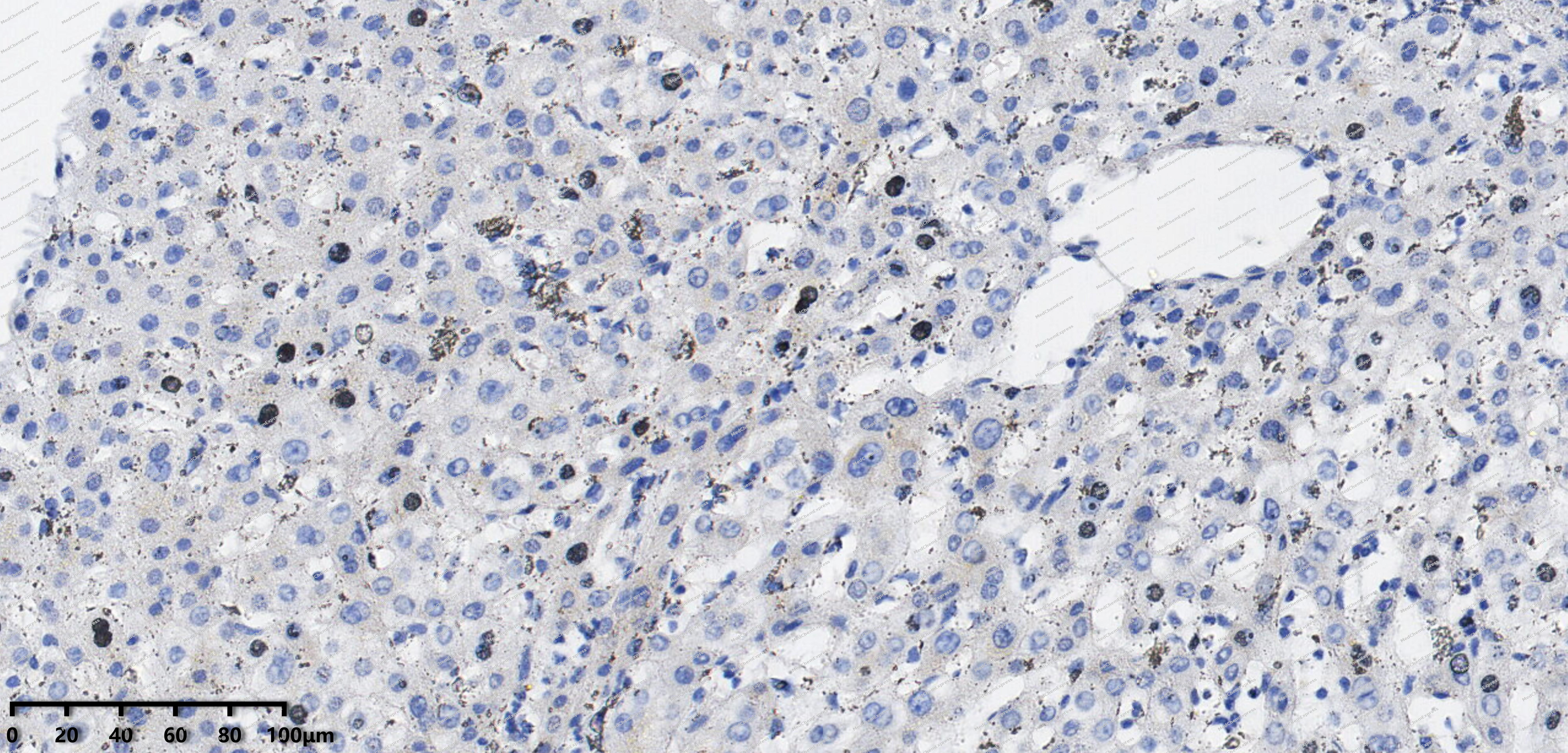 Immunohistochemical analysis of paraffin-embedded human Hepatocellular Carcinoma tissue using Wilms Tumor Protein antibody was performed. The section was pretreated using high-temperature mediated EDTA antigen retrieval buffer (pH 9.0), for 20 minutes. The tissues were incubated with primary antibody (HY-P82532, 1:100 dilution) at room temperature for 20 minutes. Detection was performed using an HRP conjugated compact polymer system. DAB was used as the chromogen. The tissues were counterstained with hematoxylin and mounted with neutral balsam mounting medium.
 -
 Flow cytometric analysis of 1X10⁶ K562 cells labeling Wilms Tumor Protein Antibody (HY-P82532, red). Cells were fixed with 4% paraformaldehyde and permeabilised with 90% methanol. Then stained with the primary antibody at 1/50 dilution for an hour at 4℃. AF488-conjugated Goat Anti-Rabbit IgG H&L (HY-P8002) was used as the secondary antibody at 1/1,000 dilution for 30 minutes at 4℃. Rabbit IgG Isotype Control (HY-P80879, blue) was used as the isotype control, cells without incubation with primary antibody were used as the unlabeled control (black).


Background
-
Function
Wilms Tumor Protein transcription factor that plays an important role in cellular development and cell survival. Recognizes and binds to the DNA sequence 5'-GCG(T/G)GGGCG-3'. Regulates the expression of numerous target genes, including EPO. Plays an essential role for development of the urogenital system. It has a tumor suppressor as well as an oncogenic role in tumor formation. Function may be isoform-specific: isoforms lacking the KTS motif may act as transcription factors. Isoforms containing the KTS motif may bind mRNA and play a role in mRNA metabolism or splicing. Isoform 1 has lower affinity for DNA, and can bind RNA[1][2][3][4][5][6].
-
Subcellular Localization
Nucleus; Nucleus, nucleolus; Cytoplasm; Nucleus speckle; Nucleus, nucleoplasm
-
Expression
Tissue_specificity:It is expressed in the kidneys and some hematopoietic cells. -
Isoforms & Post-Translational Modification
P19544 has 8 isomers: P19544-1: 49188 Da (predicted); P19544-2: 47195 Da (predicted); P19544-3: 47511 Da (predicted); P19544-4: 48872 Da (predicted); P19544-6: 34447 Da (predicted); P19544-7: 56883 Da (predicted); P19544-8: 55206 Da (predicted); P19544-9: 33086 Da (predicted).
-
Subunit
Homodimer. Interacts with WTIP. Interacts with actively translating polysomes. Detected in nuclear ribonucleoprotein (mRNP) particles. Interacts with HNRNPU via the zinc-finger region. Interacts with U2AF2. Interacts with CITED2 (By similarity). Interacts with ZNF224 via the zinc-finger region. Interacts with WTAP and SRY. Interacts with AMER1. Interacts with RBM4
-
SwissProt ID
-
Synonyms
GUD; AWT1; WAGR; WT33; NPHS4; WIT-2; EWS-WT1; WIT 2 antibody; WT 1 antibody; FWT1 antibody; GUD antibody; NPHS4 antibody; WAGR antibody; Wilms tumor 1 antibody; Wilms Tumor antibody; Wilms tumor protein antibody; Wilms' tumor gene antibody; WIT2 antibody; WT antibody; WT1 antibody; WT1_HUMAN antibody; WT33 antibody
-
Research Field
Tags & Cell Markers


Documentation
-
Data Sheet (235 KB)
-
SDS (252 KB)
- English - EN (252 KB)
- Français - FR (252 KB)
- Deutsch - DE (252 KB)
- Norwegian - NO (252 KB)
- Español - ES (252 KB)
- Swedish - SV (252 KB)
- Italian - IT (252 KB)
- Korean - KR (252 KB)
- Portuguese - PT (252 KB)
-
User Guide for Antibodies (1077 KB)

 Powered by Bioz
