



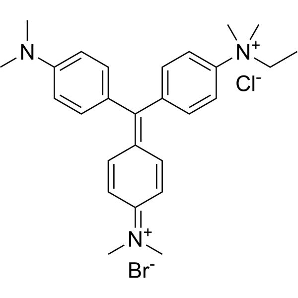
Methyl Green
Based on 1 publication(s) in Google Scholar
Methyl Green is a non-intercalating fluorescent labeling agent that selectively binds to the major groove of DNA. Methyl Green electrostatically interacts with the major groove of DNA through positively charged groups, exhibiting key activities such as high affinity, resistance to photobleaching, and stable fluorescence emission. Methyl Green can be directly measured by microscopy and flow cytometry, with peaks at 633 and 677 nm. Methyl Green can be used for fluorescent labeling of the nuclei of embryonic tissues or cells, or DNA staining and cell activity detection in gel electrophoresis[1][2][3].
Nur für Forschungszwecke. Wir verkaufen nicht an Patienten.
- Assay : 82.5%
- CAS. Nr.: 14855-76-6
- Formel: C27H35BrClN3
- Molecular Weight:516.94
-
Speicherung:
4°C, sealed storage, away from moisture and light
* In solvent : -80°C, 6 months; -20°C, 1 month (sealed storage, away from moisture and light)

To place orders, for customer services and technical support, please contact: MedChemExpress USA
Tel: 609-228-6898 E-mail: [email protected] [email protected]


-
 Biologische Aktivität
-
Chemical Information
-
Lösungsmittel & Löslichkeit
- Protokoll
- Reinheit & Dokumentation
- Verweise
-
 Help & FAQs


Publications Citing Use of MedChemExpress (MCE) Methyl Green
More

Biologische Aktivität
Beschreibung
In Vitro
Guide (The following is our recommended experimental protocol. This protocol is for reference only and should be modified according to your specific needs).
I. Zebrafish/chicken embryo whole embryo fluorescence staining reference[1][2]
Steps:
1. Sample fixation: 48 hpf zebrafish embryos or target stage chicken embryos were fixed with 4% paraformaldehyde (HY-Y0333) (prepared with PBS) at 4°C overnight. The fixation time was adjusted according to the thickness of the embryo.
2. Permeabilization: The embryos were washed 3 times with PBS (PBS-T) (HY-K1022) containing 1% Triton X-100 for 5 minutes each time to destroy the egg membrane/tissue barrier.
3. Methyl green staining: dilute 2% methyl green stock solution to 1:5,000-1:10,000 with PBS-T (final concentration of about 2-4 μg/mL), incubate embryos at 4°C for at least 6 hours (overnight for thick embryos), with gentle shaking.
4. Washing and removal of impurities: wash 3 times with PBS, 30 minutes each time, to remove unbound dye.
5. Mounting and imaging: mount the slides with 75% glycerol + 0.1 M Tris·HCl (pH 8.0), use laser confocal microscopy, excitation wavelength 633 nm, emission wavelength 650-750 nm (peak 677 nm).
II. Agarose gel DNA staining[2]
Steps:
1. Gel preparation: Add 2% Methyl Green (HY-D0163) stock solution to 1% agarose (prepared with TAE buffer) at a ratio of 1:5,000-1:10,000, and cool after gel preparation.
2. Sample loading: Load DNA/RNA ladder (such as 100 bp DNA ladder), conventional electrophoresis (voltage/time adjusted according to the experiment).
3. Fluorescence detection: Use 635 nm red light excitation, 705 nm bandpass filter, exposure for 30-60 seconds, or observe with UV transilluminator (only DNA color development).
III. Flow cytometry to detect cell viability[2][3]
Steps:
1. Cell treatment: Cultured Raji cells (or PBMCs) were treated with drugs (e.g. 50-100 μg/mL Fludarabine (HY-B0069)) for 24 hours, and then the cell suspension (5×10⁵ cells) was collected.
2. Staining: Add 4 μg/mL Methyl Green (HY-D0163) + 10 μg/mL Propidium Iodide (HY-D0815) and incubate at room temperature in the dark for 15 minutes.
3. Flow cytometry: Use a CyAn flow cytometer, set the excitation wavelength to 633 nm (MG)/488 nm (PI), and the emission wavelength to 677 nm (MG)/609 nm (PI), and analyze the ratio of live and dead cells.
IV. Fluorescence spectroscopy (DNA binding analysis)[2]
Chinese steps:
1. Solution preparation: Prepare a 100 μg/mL calf thymus DNA (HY-109517) solution in PBS and add methyl green to a final concentration of 10 μg/mL (or 125 ng/mL free state).
2. Spectral detection: Use a fluorescence spectrophotometer, excite at 633 nm, scan the emission spectrum from 645-800 nm, and record the peak at 677 nm (DNA binding state).
MedChemExpress (MCE) has not independently confirmed the accuracy of these methods. They are for reference only.


Chemical Information
-
CAS. Nr. 14855-76-6
-
Appearance Solid
-
Molecular Weight 516.94
-
Formel C27H35BrClN3
-
Color Green to dark green
-
SMILES
C[N+](C)(CC)C1=CC=C(/C(C2=CC=C(N(C)C)C=C2)=C3C=C/C(C=C\3)=[N+](C)\C)C=C1.[Br-].[Cl-]
-
Versand
Room temperature in continental US; may vary elsewhere.
-
Speicherung
4°C, sealed storage, away from moisture and light
* In solvent : -80°C, 6 months; -20°C, 1 month (sealed storage, away from moisture and light)


Publications (1)
-
Journal Impact Factor
-
Most Recent


Lösungsmittel & Löslichkeit
In Vitro:
DMSO : 125 mg/mL (241.81 mM; Need ultrasonic; Hygroscopic DMSO has a significant impact on the solubility of product, please use newly opened DMSO)
H2O : 5.83 mg/mL (11.28 mM; Need ultrasonic)
Please refer to the solubility information to select the appropriate solvent. Once prepared, please aliquot and store the solution to prevent product inactivation from repeated freeze-thaw cycles.
Storage method and period of stock solution: -80°C, 6 months; -20°C, 1 month (sealed storage, away from moisture and light). When stored at -80°C, please use it within 6 months. When stored at -20°C, please use it within 1 month.
* Note: If you choose water as the stock solution, please dilute it to the working solution, then filter and sterilize it with a 0.22 μm filter before use.
Please refer to the solubility information to select the appropriate solvent. Once prepared, please aliquot and store the solution to prevent product inactivation from repeated freeze-thaw cycles.
Storage method and period of stock solution: -80°C, 6 months; -20°C, 1 month (sealed storage, away from moisture and light). When stored at -80°C, please use it within 6 months. When stored at -20°C, please use it within 1 month.
* Note: If you choose water as the stock solution, please dilute it to the working solution, then filter and sterilize it with a 0.22 μm filter before use.
Konzentration (Stammlösung) × Volumen (Stammlösung) = Konzentration (Ziellösung) × Volumen (Ziellösung)


Reinheit & Dokumentation
-
Data Sheet (277 KB)
-
SDS (923 KB)
- English - EN (923 KB)
- Français - FR (923 KB)
- Deutsch - DE (923 KB)
- Norwegian - NO (923 KB)
- Español - ES (923 KB)
- Swedish - SV (923 KB)
- Italian - IT (923 KB)
- Korean - KR (923 KB)
- Portuguese - PT (923 KB)
-
Handling Instructions (2659 KB)
Verweise
[1]. Prieto D, et al. Application of the DNA-specific stain methyl green in the fluorescent labeling of embryos. J Vis Exp. 2015 May 2;(99):e52769. [Content Brief]
[2]. Prieto D, et, al. A fast, low cost, and highly efficient fluorescent DNA labeling method using methyl green. Histochem Cell Biol. 2014 Sep;142(3):335-45. [Content Brief]
[3]. Kim SK, et al. Methyl green. A DNA major-groove binding drug. FEBS Lett. 1993 Jan 2;315(1):61-4. [Content Brief]


Complete Stock Solution Preparation Table
Please refer to the solubility information to select the appropriate solvent. Once prepared, please aliquot and store the solution to prevent product inactivation from repeated freeze-thaw cycles.
Storage method and period of stock solution: -80°C, 6 months; -20°C, 1 month (sealed storage, away from moisture and light). When stored at -80°C, please use it within 6 months. When stored at -20°C, please use it within 1 month.
| Optional Solvent | Concentration Solvent Mass | 1 mg | 5 mg | 10 mg | 25 mg |
|---|---|---|---|---|---|
| H2O / DMSO | 1 mM | 1.9345 mL | 9.6723 mL | 19.3446 mL | 48.3615 mL |
| 5 mM | 0.3869 mL | 1.9345 mL | 3.8689 mL | 9.6723 mL | |
| 10 mM | 0.1934 mL | 0.9672 mL | 1.9345 mL | 4.8362 mL | |
| DMSO | 15 mM | 0.1290 mL | 0.6448 mL | 1.2896 mL | 3.2241 mL |
| 20 mM | 0.0967 mL | 0.4836 mL | 0.9672 mL | 2.4181 mL | |
| 25 mM | 0.0774 mL | 0.3869 mL | 0.7738 mL | 1.9345 mL | |
| 30 mM | 0.0645 mL | 0.3224 mL | 0.6448 mL | 1.6121 mL | |
| 40 mM | 0.0484 mL | 0.2418 mL | 0.4836 mL | 1.2090 mL | |
| 50 mM | 0.0387 mL | 0.1934 mL | 0.3869 mL | 0.9672 mL | |
| 60 mM | 0.0322 mL | 0.1612 mL | 0.3224 mL | 0.8060 mL | |
| 80 mM | 0.0242 mL | 0.1209 mL | 0.2418 mL | 0.6045 mL | |
| 100 mM | 0.0193 mL | 0.0967 mL | 0.1934 mL | 0.4836 mL |
* Note: If you choose water as the stock solution, please dilute it to the working solution, then filter and sterilize it with a 0.22 μm filter before use.


Methyl Green Related Classifications
HY-D0163 Related Classifications
 Powered by Bioz