



McMMAF
Based on 8 publication(s) in Google Scholar
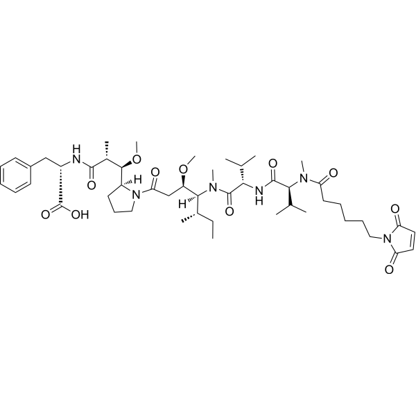
McMMAF (Maleimidocaproyl monomethylauristatin F) is a drug-linker conjugate for ADCs, formed by coupling the potent microtubule inhibitor Monomethyl auristatin F (MMAF) (HY-15579) with a maleimidocaproyl (mc) linker. McMMAF can be conjugated with anti-BCMA antibodies to form J6M0-mcMMAF, promoting apoptosis and inhibiting tumor growth.
Nos produits utilisent uniquement pour la recherche. Nous ne vendons pas aux patients.
- Pureté : 99.97%
- CAS No.: 863971-19-1
- Formule: C49H76N6O11
- Masse moléculaire:925.16
-
Stockage:
-20°C, stored under nitrogen
* The compound is unstable in solutions, freshly prepared is recommended.

To place orders, for customer services and technical support, please contact: MedChemExpress USA
Tel: 609-228-6898 E-mail: [email protected] [email protected]


-
 Activité biologique
-
Chemical Information
-
Solvant et solubilité
- Protocole
- Pureté et documentation
- Références
-
 Help & FAQs


-
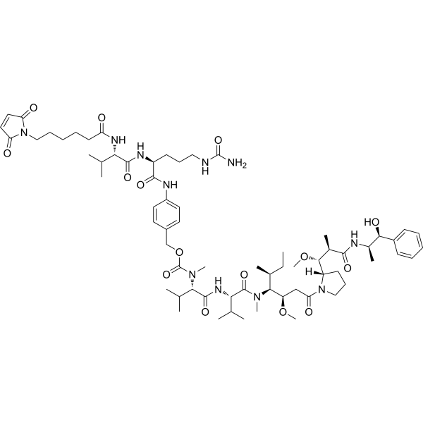
HY-15575
VcMMAEDrug-Linker Conjugate for ADC -
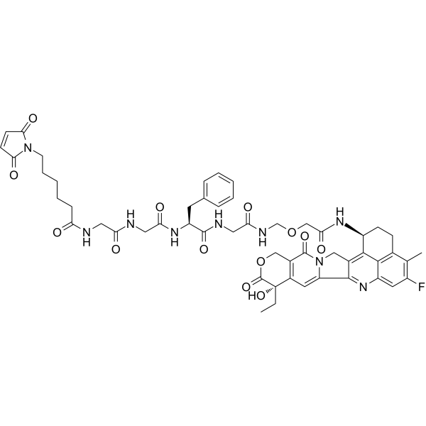
HY-13631E
DeruxtecanDrug-Linker Conjugates for ADC -
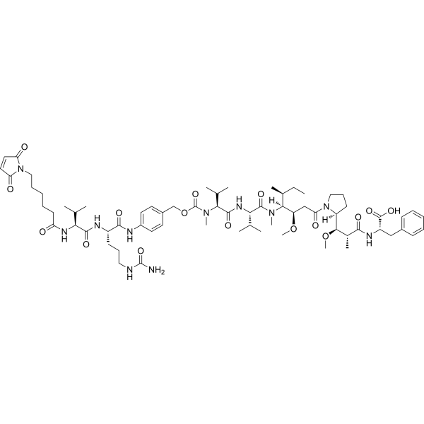
HY-112786
MC-Val-Cit-PAB-MMAFDrug-Linker Conjugate for ADC -
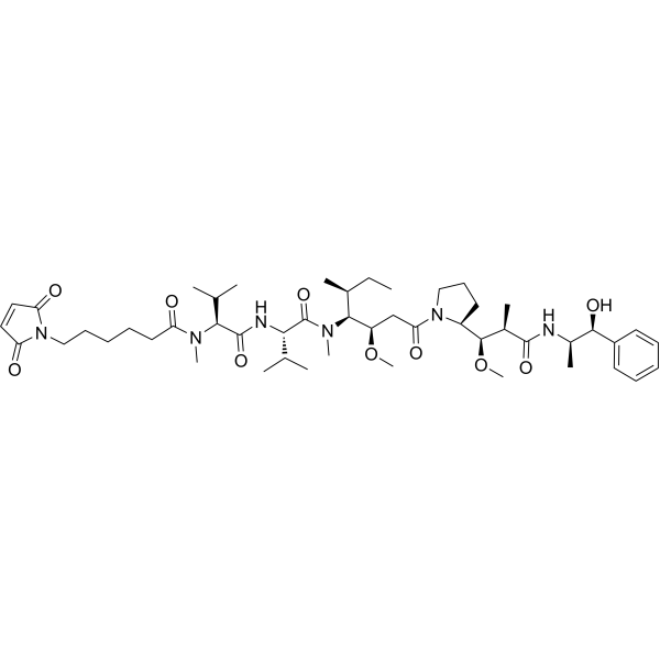
HY-15741
Mc-MMAEDrug-Linker Conjugate for ADC -
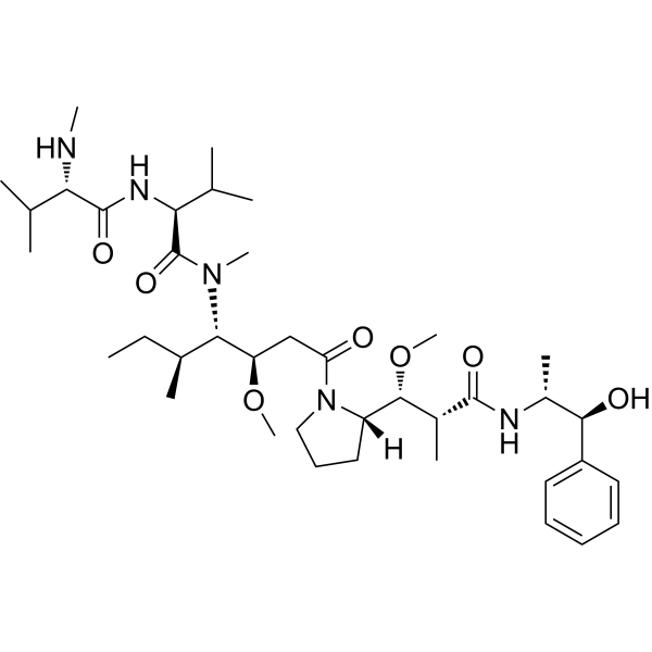
HY-15162
Monomethyl auristatin EAntimitotic Agent -
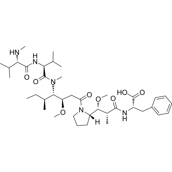
HY-15579
MMAFTubulin Inhibitor -
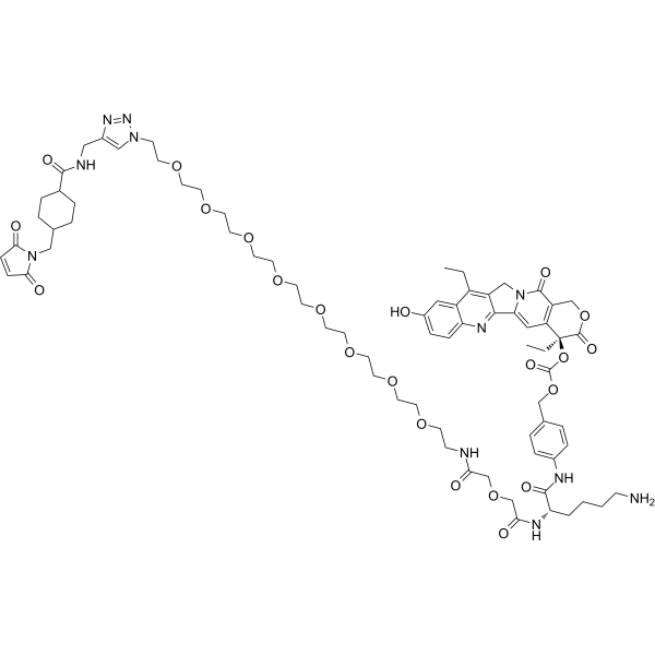
HY-128946
CL2A-SN-38DNA Topoisomerase I Inhibitor -
HY-101070
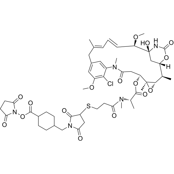
SMCC-DM1Drug-Linker Conjugate for ADC -
HY-100374
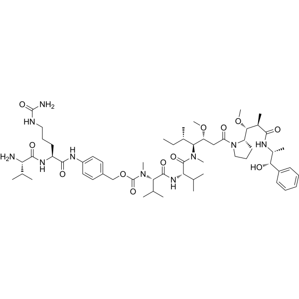
Val-Cit-PAB-MMAEDrug-Linker Conjugate For ADC -
HY-19792
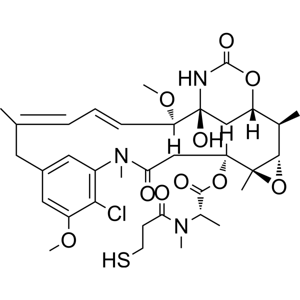
MertansineMicrotubule-Targeted Cytotoxin -
HY-15575G
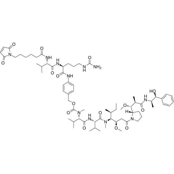
VcMMAE (GMP)Drug-Linker Conjugate for ADC -

HY-P9921
Trastuzumab emtansineAntibody-drug Conjugate
Publications Citing Use of MedChemExpress (MCE) McMMAF
More- J Control Release. 2020 Nov 10:327:186-197. [Abstract]
- J Pharm Anal. 2025 May;15(5):101100. [Abstract]
- Anal Chem. 2022 Feb 15;94(6):2772-2778. [Abstract]
- Pharmaceutics. 2025 Jul 25;17(8):967. [Abstract]
- Neoplasia. 2026 Mar 12:74:101295. [Abstract]
- Patent. US20210393733A1.
- Patent. US10087260B2.
- Patent. US20160304621A1.
Voir tous les produits spécifiques à Isoform Drug-Linker Conjugates for ADC
More

Activité biologique
Description
IC50 & Target
|
Auristatin |
In Vitro
ADCs are comprised of an antibody to which is attached an ADC cytotoxin through an ADC linker.
MedChemExpress (MCE) has not independently confirmed the accuracy of these methods. They are for reference only.


Chemical Information
-
CAS No. 863971-19-1
-
Appearance Solid
-
Masse moléculaire 925.16
-
Formule C49H76N6O11
-
Color White to off-white
-
SMILES
O=C([C@H](C)[C@H]([C@]1([H])CCCN1C(C[C@@H](OC)[C@@]([H])(N(C([C@H](C(C)C)NC([C@H](C(C)C)N(C)C(CCCCCN2C(C=CC2=O)=O)=O)=O)=O)C)[C@@H](C)CC)=O)OC)N[C@H](C(O)=O)CC3=CC=CC=C3
-
Synonyms
Maleimidocaproyl monomethylauristatin F
-
Livraison
Room temperature in continental US; may vary elsewhere.
-
Stockage
-20°C, stored under nitrogen
* The compound is unstable in solutions, freshly prepared is recommended.


Publications (8)
-
Journal Impact Factor
-
Most Recent
-
J Control Release
Optimizing the anti-tumor efficacy of protein-drug conjugates by engineering the molecular size and half-life. [Abstract]2020 Nov 10:327:186-197. PMID: 32768630 -
J Pharm Anal
Synergistic approach to combating triple-negative breast cancer: DDR1-targeted antibody-drug conjugate combined with pembrolizumab. [Abstract]2025 May;15(5):101100. PMID: 40521369 -
Anal Chem
Site-Specific Conjugation Quantitation of a Cysteine-Conjugated Antibody-Drug Conjugate Using Stable Isotope Labeling Peptide Mapping LC-MS/MS Analysis. [Abstract]2022 Feb 15;94(6):2772-2778. PMID: 35100801 -
Pharmaceutics
A Dual-Payload Bispecific ADC Improved Potency and Efficacy over Single-Payload Bispecific ADCs. [Abstract]2025 Jul 25;17(8):967. PMID: 40870990 -
Neoplasia
Rational payload selection enables high antitumoral efficacy of an anti-EGFR antibody-drug conjugate against ovarian tumors. [Abstract]2026 Mar 12:74:101295. PMID: 41825107 -
-
-


Solvant et solubilité
In Vitro:
DMSO : ≥ 100 mg/mL (108.09 mM; Hygroscopic DMSO has a significant impact on the solubility of product, please use newly opened DMSO)
* "≥" means soluble, but saturation unknown.
Please refer to the solubility information to select the appropriate solvent. The compound is unstable in solutions, freshly prepared is recommended.
Please refer to the solubility information to select the appropriate solvent. The compound is unstable in solutions, freshly prepared is recommended.
Concentration (start) × Volume (start) = Concentration (final) × Volume (final)
In Vivo:
Select the appropriate dissolution method based on your experimental animal and administration route.
- For the following dissolution methods, please ensure to first prepare a clear stock solution using an In Vitro approach and then sequentially add co-solvents:
- To ensure reliable experimental results, the clarified stock solution can be appropriately stored based on storage conditions. As for the working solution for In Vivo experiments, it is recommended to prepare freshly and use it on the same day.
- The percentages shown for the solvents indicate their volumetric ratio in the final prepared solution. If precipitation or phase separation occurs during preparation, heat and/or sonication can be used to aid dissolution.
Add each solvent one by one: 10% DMSO 40% PEG300 5% Tween-80 45% Saline
Solubility: ≥ 5 mg/mL (5.40 mM); Clear solution
This protocol yields a clear solution of ≥ 5 mg/mL (saturation unknown).
Taking 1 mL working solution as an example, add 100 μL DMSO stock solution (50.0 mg/mL) to 400 μL PEG300, and mix evenly; then add 50 μL Tween-80 and mix evenly; then add 450 μL Saline to adjust the volume to 1 mL.
Preparation of Saline: Dissolve 0.9 g sodium chloride in ddH₂O and dilute to 100 mL to obtain a clear Saline solution.
Add each solvent one by one: 1% DMSO 99% Saline
Solubility: ≥ 0.5 mg/mL (0.54 mM); Clear solution
In Vivo Dissolution Calculator
Please enter the basic information of animal experiments:
-
-
-
-
Recommended: Prepare an additional quantity of animals to account for potential losses during experiments.
Please enter your animal formula composition:
-
%DMSO +
Recommended: Keep the proportion of DMSO in working solution below 2% if your animal is weak.
-
%+
-
+%Tween-80 + +
-
%Saline +
The co-solvents required include: DMSO, . All of co-solvents are available by MedChemExpress (MCE). , Tween 80. All of co-solvents are available by MedChemExpress (MCE).
Working solution concentration: 0.22 mg/mL
Method for preparing stock solution: mg drug dissolved in μL DMSO. Stock solution concentration: mg/mL. * The compound is unstable in solutions, freshly prepared is recommended.
1. Take μL DMSO stock solution;
2. Add μL .
μL , mix evenly;
3. Then add μL Tween 80, mix evenly;
4. Then add μL
Please ensure that the stock solution in the first step is dissolved to a clear state, and add co-solvents in sequence. You can use ultrasonic heating (ultrasonic cleaner, recommended frequency 20-40 kHz), vortexing, etc. to assist dissolution.


Pureté et documentation
-
Fiche technique (292 KB)
-
SDS (480 KB)
- English - EN (480 KB)
- Français - FR (480 KB)
- Deutsch - DE (480 KB)
- Norwegian - NO (480 KB)
- Español - ES (480 KB)
- Swedish - SV (480 KB)
- Italian - IT (480 KB)
- Korean - KR (480 KB)
- Portuguese - PT (480 KB)
-
Instruction de manipulation (2659 KB)
Références
[1]. Polson AG, et al. Antibody-drug conjugates for the treatment of non-Hodgkin's lymphoma: target and linker-drug selection. Cancer Res. 2009 Mar 15;69(6):2358-2364. [Content Brief]
[2]. Jianmin Fang, et al. Anti-her2 antibody and conjugate thereof. US 20160304621 A1.
[3]. Yu-Tzu Tai, et al. Novel anti-B-cell maturation antigen antibody-drug conjugate (GSK2857916) selectively induces killing of multiple myeloma. Blood. 2014 May 15;123(20):3128-38. [Content Brief]


Complete Stock Solution Preparation Table
Please refer to the solubility information to select the appropriate solvent. The compound is unstable in solutions, freshly prepared is recommended.
| Optional Solvent | Concentration Solvent Mass | 1 mg | 5 mg | 10 mg | 25 mg |
|---|---|---|---|---|---|
| DMSO | 1 mM | 1.0809 mL | 5.4045 mL | 10.8089 mL | 27.0224 mL |
| 5 mM | 0.2162 mL | 1.0809 mL | 2.1618 mL | 5.4045 mL | |
| 10 mM | 0.1081 mL | 0.5404 mL | 1.0809 mL | 2.7022 mL | |
| 15 mM | 0.0721 mL | 0.3603 mL | 0.7206 mL | 1.8015 mL | |
| 20 mM | 0.0540 mL | 0.2702 mL | 0.5404 mL | 1.3511 mL | |
| 25 mM | 0.0432 mL | 0.2162 mL | 0.4324 mL | 1.0809 mL | |
| 30 mM | 0.0360 mL | 0.1801 mL | 0.3603 mL | 0.9007 mL | |
| 40 mM | 0.0270 mL | 0.1351 mL | 0.2702 mL | 0.6756 mL | |
| 50 mM | 0.0216 mL | 0.1081 mL | 0.2162 mL | 0.5404 mL | |
| 60 mM | 0.0180 mL | 0.0901 mL | 0.1801 mL | 0.4504 mL | |
| 80 mM | 0.0135 mL | 0.0676 mL | 0.1351 mL | 0.3378 mL | |
| 100 mM | 0.0108 mL | 0.0540 mL | 0.1081 mL | 0.2702 mL |


McMMAF Related Classifications
HY-15578 Related Classifications
 Powered by Bioz