



Pterin

Based on 1 Customer Validation
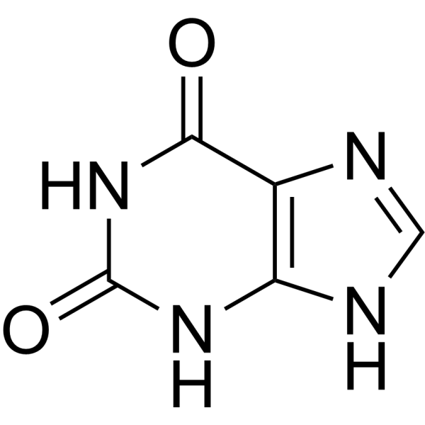
Pterin is a pteridine with antibacterial activity. Pterin binds to the Fe2+ active site via its carbonyl group, reducing the oxygen activation energy barrier in hydroxylases. Pterin participates in metabolic redox processes. Pterin can be used in research related to cancer, vitiligo, bacterial infections, phenylketonuria, Parkinson's disease, schizophrenia and depression.
Nos produits utilisent uniquement pour la recherche. Nous ne vendons pas aux patients.
- Pureté : 99.75%
- CAS No.: 2236-60-4
- Formule: C6H5N5O
- Masse moléculaire:163.14
-
Stockage:
4°C, protect from light
* In solvent : -80°C, 6 months; -20°C, 1 month (protect from light)


To place orders, for customer services and technical support, please contact: MedChemExpress USA
Tel: 609-228-6898 E-mail: [email protected] [email protected]


-
 Activité biologique
-
Chemical Information
-
Solvant et solubilité
- Protocole
- Pureté et documentation
- Références
-
 Help & FAQs




Activité biologique
Description
In Vitro
MedChemExpress (MCE) has not independently confirmed the accuracy of these methods. They are for reference only.


Chemical Information
-
CAS No. 2236-60-4
-
Appearance Solid
-
Masse moléculaire 163.14
-
Formule C6H5N5O
-
Color Brown to reddish brown
-
SMILES
O=C1NC(N)=NC2=NC=CN=C12
-
Livraison
Room temperature in continental US; may vary elsewhere.
-
Stockage
4°C, protect from light
* In solvent : -80°C, 6 months; -20°C, 1 month (protect from light)


Solvant et solubilité
In Vitro:
TFA : 10 mg/mL (61.30 mM; Need ultrasonic)
Please refer to the solubility information to select the appropriate solvent. Once prepared, please aliquot and store the solution to prevent product inactivation from repeated freeze-thaw cycles.
Storage method and period of stock solution: -80°C, 6 months; -20°C, 1 month (protect from light). When stored at -80°C, please use it within 6 months. When stored at -20°C, please use it within 1 month.
Please refer to the solubility information to select the appropriate solvent. Once prepared, please aliquot and store the solution to prevent product inactivation from repeated freeze-thaw cycles.
Storage method and period of stock solution: -80°C, 6 months; -20°C, 1 month (protect from light). When stored at -80°C, please use it within 6 months. When stored at -20°C, please use it within 1 month.
Concentration (start) × Volume (start) = Concentration (final) × Volume (final)


Pureté et documentation
-
Fiche technique (274 KB)
-
SDS (394 KB)
- English - EN (394 KB)
- Français - FR (394 KB)
- Deutsch - DE (394 KB)
- Norwegian - NO (394 KB)
- Español - ES (394 KB)
- Swedish - SV (394 KB)
- Italian - IT (394 KB)
- Korean - KR (394 KB)
- Portuguese - PT (394 KB)
-
Instruction de manipulation (2659 KB)
Références
[2]. Mahendran R, et al. Evaluation of Pterin, a Promising Drug Candidate from Cyanide Degrading Bacteria. Current microbiology. 2018 Jun;75(6):684-693. [Content Brief]
[8]. Rutowski RL, et al. Pterin pigments amplify iridescent ultraviolet signal in males of the orange sulphur butterfly, Colias eurytheme. Proceedings. Biological sciences. 2005 Nov 07;272(1578):2329-35. [Content Brief]


Complete Stock Solution Preparation Table
Please refer to the solubility information to select the appropriate solvent. Once prepared, please aliquot and store the solution to prevent product inactivation from repeated freeze-thaw cycles.
Storage method and period of stock solution: -80°C, 6 months; -20°C, 1 month (protect from light). When stored at -80°C, please use it within 6 months. When stored at -20°C, please use it within 1 month.
| Optional Solvent | Concentration Solvent Mass | 1 mg | 5 mg | 10 mg | 25 mg |
|---|---|---|---|---|---|
| TFA | 1 mM | 6.1297 mL | 30.6485 mL | 61.2970 mL | 153.2426 mL |
| 5 mM | 1.2259 mL | 6.1297 mL | 12.2594 mL | 30.6485 mL | |
| 10 mM | 0.6130 mL | 3.0649 mL | 6.1297 mL | 15.3243 mL | |
| 15 mM | 0.4086 mL | 2.0432 mL | 4.0865 mL | 10.2162 mL | |
| 20 mM | 0.3065 mL | 1.5324 mL | 3.0649 mL | 7.6621 mL | |
| 25 mM | 0.2452 mL | 1.2259 mL | 2.4519 mL | 6.1297 mL | |
| 30 mM | 0.2043 mL | 1.0216 mL | 2.0432 mL | 5.1081 mL | |
| 40 mM | 0.1532 mL | 0.7662 mL | 1.5324 mL | 3.8311 mL | |
| 50 mM | 0.1226 mL | 0.6130 mL | 1.2259 mL | 3.0649 mL | |
| 60 mM | 0.1022 mL | 0.5108 mL | 1.0216 mL | 2.5540 mL |


Pterin Related Classifications
HY-W037825 Related Classifications
 Powered by Bioz