



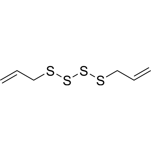
Diallyl tetrasulfide
Diallyl tetrasulfide is an orally active diallyl tetrasulfide. Diallyl tetrasulfide ameliorates cadmium-induced changes in acetylcholinesterase and adenosine triphosphatase activities as well as oxidative stress injury in the brain of rats. Diallyl tetrasulfide inhibits lipid peroxidation in rat liver microsomes. Diallyl tetrasulfide ameliorates cadmium-induced oxidative liver injury in rats. Diallyl tetrasulfide protects cells against cadmium-induced loss of cell viability, reduces apoptosis rate and ROS production. Diallyl tetrasulfide is applicable to research related to cadmium-induced neurotoxicity and cadmium-induced oxidative liver injury.
Nur für Forschungszwecke. Wir verkaufen nicht an Patienten.
- CAS. Nr.: 2444-49-7
- Formel: C6H10S4
- Molecular Weight:210.40
-
Speicherung:
Please store the product under the recommended conditions in the Certificate of Analysis.

To place orders, for customer services and technical support, please contact: MedChemExpress USA
Tel: 609-228-6898 E-mail: [email protected] [email protected]


-
 Biologische Aktivität
-
Chemical Information
- Protokoll
- Reinheit & Dokumentation
- Verweise
-
 Help & FAQs


-
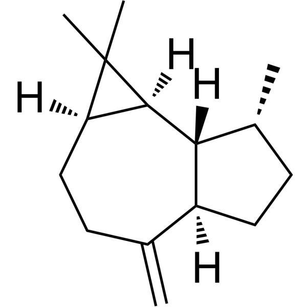
HY-N8411
AromadendreneAntibacterial Agent -
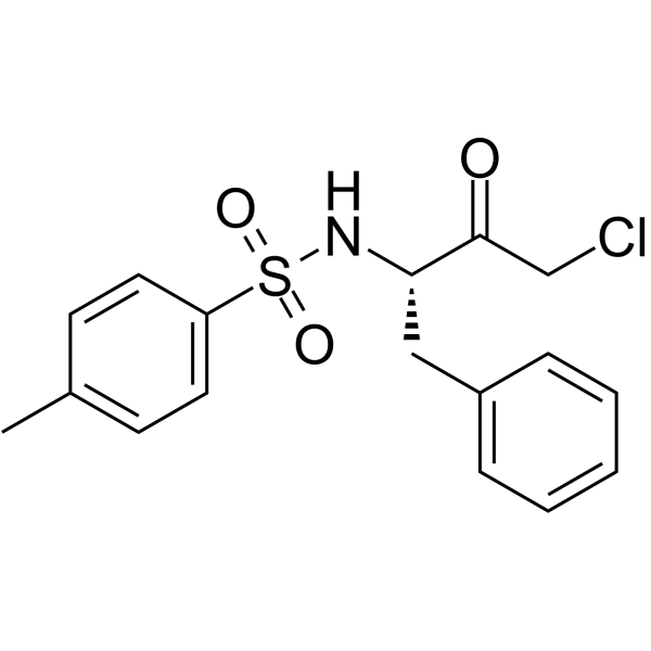
HY-124379
TPCKSerine Protease Inhibitor -
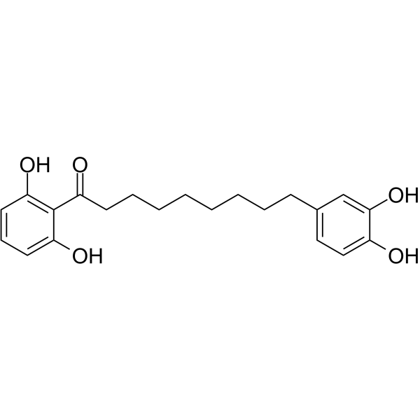
HY-N8518
Malabaricone CSphingomyelin Synthase Inhibitor -

HY-N8411R
Aromadendrene (Standard)Reference Standard -

HY-W743654
Capsorubin -
HY-106784A
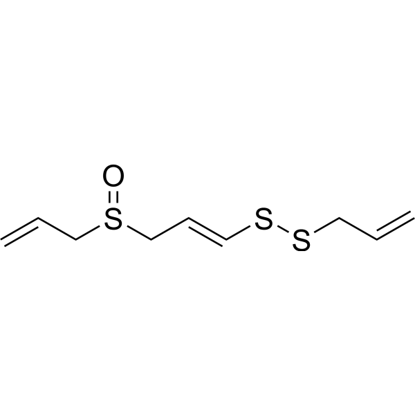
(E)-AjoeneNeuroprotective Agent -
HY-14892A
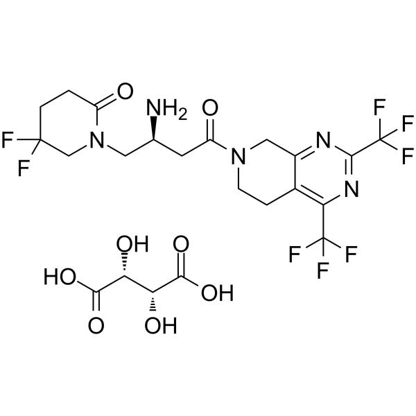
Gemigliptin tartrateDPP-4 Inhibitor -

HY-127020
DeoxyenterocinBacterial Metabolite -
HY-125568
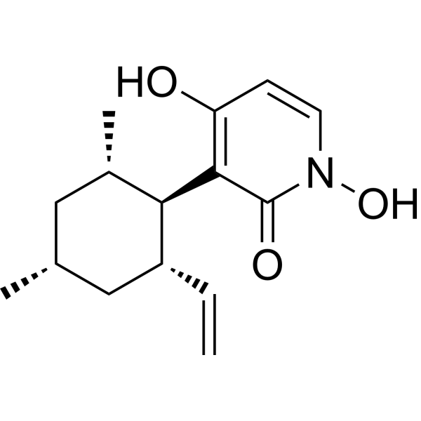
PyridoxatinFree Radical Scavenger -
HY-N8517
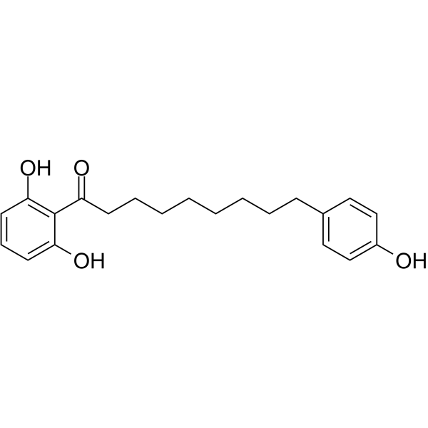
Malabaricone Bα-Glucosidase Inhibitor -

HY-N8518R
Malabaricone C (Standard)Sphingomyelin Synthase Inhibitor -

HY-W743654R
Capsorubin (Standard)


Biologische Aktivität
Beschreibung
In Vitro
Diallyl tetrasulfide (25 μg/mL; 3 h) inhibits lipid peroxidation when co-incubated with rat liver microsomes treated with ascorbic acid/Fe2+[2].
Diallyl tetrasulfide (5-50 μg/mL; 18 h) dose-dependently protects Vero cells against cadmium-induced loss of cell viability[4].
Diallyl tetrasulfide (40 μg/mL; 18 h) reduces the cadmium-induced apoptosis rate of Vero cells by 75%, decreases the intracellular accumulation of superoxide anions and hydrogen peroxide, and protects cells from cadmium-induced loss of mitochondrial membrane potential[4].
Diallyl tetrasulfide (10-40 μg/mL; 30 min) significantly reduces cadmium-induced intracellular ROS production in Vero cells[4].
MedChemExpress (MCE) has not independently confirmed the accuracy of these methods. They are for reference only. Further protocols information, click here.
-
Cell Line:Vero cells
-
Concentration:5, 10, 20, 30, 40, 50 μg/mL
-
Incubation Time:18 h
-
Result:Attenuated cadmium-induced suppression of cell viability in a dose-dependent manner.
Reached 87% cell survival rate at 50 μg/mL.
Showed a highly significant increase to 86% viability at 40 μg/mL.
Did not affect cell viability when used alone at 50 μg/mL.
-
Cell Line:Vero cells
-
Concentration:40 μg/mL
-
Incubation Time:18 h
-
Result:Prevented cadmium-induced apoptotic nuclear changes (condensed or fragmented nuclei).
Reduced cadmium-induced apoptosis by 75% relative to cadmium-only treated cells.
Showed intact, healthy nuclei with no significant increase in apoptosis compared to control when used alone at 40 μg/mL.
In Vivo
MedChemExpress (MCE) has not independently confirmed the accuracy of these methods. They are for reference only.
-
Animal Model:Wistar rats (male, initial body weight 170-200 g, cadmium-induced neurotoxicity/Liver oxidative damage model)[1]
-
Dosage:10 mg/kg; 20 mg/kg; 40 mg/kg
-
Administration:p.o.; daily; 3 weeks
-
Result:Reversed brain acetylcholinesterase activity to near normal levels.
Reduced brain lipid peroxidation markers to 14.10 μmol/g tissue (TBARS) and 1.20 mmol/g tissue (LOOH).
Reduced brain protein carbonyl levels to 4.25 nmol/mg protein.
Increased brain reduced glutathione levels to 2.65 μg/mg protein and total sulphydryl groups to 7.06 μg/mg protein.
Restored brain superoxide dismutase activity to 8.24 units, catalase activity to 3.79 μmol H2O2 consumed/min, glutathione peroxidase activity to 3.05 μg glutathione consumed/min/mg protein, and glutathione-S-transferase activity to 5.78 μmol CDNB-GSH conjugate formed/min/mg protein.
Restored brain total ATPases activity to 1.35 μg Pi liberated/min/mg protein, Na+K+-ATPase activity to 0.36 μg Pi liberated/min/mg protein, Ca2+-ATPase activity to 0.384 μg Pi liberated/min/mg protein, and Mg2+-ATPase activity to 0.28 μg Pi liberated/min/mg protein.
Normalized serum activities of hepatic marker enzymes (aspartate transaminase, alanine transaminase, alkaline phosphatase, lactate dehydrogenase) in a dose-dependent manner, with greater efficacy at 40 mg/kg than 10 mg/kg and 20 mg/kg.
Reduced liver cadmium accumulation from 500 μg/g wet tissue to 200 μg/g wet tissue.
Decreased liver thiobarbituric acid reactive substances to 0.125 nmol/mg protein, hydroperoxides to 0.96 mmol/g tissue, and protein carbonyl levels to 3.19 nmol/mg protein.
Restored liver non-enzymic antioxidant levels: reduced glutathione to 3.03 μg/mg protein, total thiols to 13.65 μg/mg protein, vitamin C to 1.20 μmol/mg tissue, and vitamin E to 0.61 μmol/mg tissue.
Restored liver enzymic antioxidant and glutathione metabolising enzyme activities: superoxide dismutase to 6.30 U/mg protein, catalase to 72.86 U/mg protein, glutathione peroxidase to 5.43 U/mg protein, glutathione-S-transferase to 6.07 U/mg protein, glutathione reductase to 0.43 U/mg protein, and glucose-6-phosphate dehydrogenase to 1.88 U/mg protein.
Improved liver histopathology, resulting in normal hepatocytes with mild portal inflammation.


Chemical Information
-
CAS. Nr. 2444-49-7
-
Molecular Weight 210.40
-
Formel C6H10S4
-
SMILES
C=CCSSSSCC=C
-
Structure Classification
-
Initial Source
-
Versand
Room temperature in continental US; may vary elsewhere.
-
Speicherung
Please store the product under the recommended conditions in the Certificate of Analysis.

Protokoll
-
Neurotoxicity Study
This protocol assesses in vitro neurotoxicity by combining neuronal viability, mitochondrial/metabolic activity, neurite outgrowth, and optional neuronal network function readouts. Calcein-AM or resazurin/PrestoBlue readouts estimate viable or metabolically active cells; βIII-tubulin immunofluorescence detects neuronal morphology and neurite networks; TMRE detects mitochondrial membrane potential; and MEA recordings detect functional changes in neuronal network activity.
-
Apoptosis
Apoptosis, also called programmed cell death, is generally characterized by distinct morphological characteristics.
-
TUNEL staining for apoptotic DNA fragmentation
TUNEL staining detects DNA strand breaks by using terminal deoxynucleotidyl transferase to add labeled nucleotides to exposed 3′-OH DNA termini, generating either microscopic staining in fixed cells or tissue sections, or fluorescence/cytometric signal in cell suspensions. TUNEL positivity reflects DNA fragmentation but should not be interpreted alone as definitive apoptosis, because TUNEL can also label necrotic, autolytic, mechanically damaged, or DNA-repair-associated DNA breaks.
-
Cell Cytotoxicity Assay
Cytotoxicity assays are usually based on the assessment of cell membrane damage, which can also be indirectly detected by measuring cell viability. Detection methods include MTT assay, CKK-8 assay, LDH assay and ATP assay, etc.
-
Mammalian live/dead viability and cytotoxicity staining
Live/dead viability and cytotoxicity staining assays are based on the simultaneous detection of intracellular esterase activity in metabolically active (viable) cells and membrane integrity loss in non-viable cells. In commonly used dual-staining approaches, membrane-permeant fluorogenic substrates are converted by intracellular esterases into fluorescent products in live cells, while impermeant DNA-binding dyes selectively enter cells with compromised plasma membranes and label nucleic acids in dead or dying cells, enabling discrimination between viable and non-viable populations by fluorescence microscopy or flow cytometry.
-
Annexin V plus membrane-impermeant dye apoptosis staining
Annexin V-based apoptosis assays rely on the detection of phosphatidylserine (PS) externalization from the inner leaflet of the plasma membrane to the outer leaflet, an early biochemical hallmark of apoptosis. Fluorescently labeled Annexin V binds PS in a calcium-dependent manner, enabling identification of early apoptotic cells by flow cytometry or fluorescence microscopy. When combined with a membrane-impermeant DNA-binding dye (e. g. , propidium iodide), this approach allows discrimination between viable (Annexin V−/dye−), early apoptotic (Annexin V+/dye−), and late apoptotic or necrotic (Annexin V+/dye+) cell populations by assessing membrane integrity and PS exposure.
-
ROS/oxidative-stress fluorescent staining
ROS/oxidative-stress fluorescent staining uses cell-permeant fluorogenic probes that become fluorescent after oxidation inside cells or tissues; commonly used examples include DCFH-DA/DCFDA for broad cellular oxidant detection, DHE for superoxide-related signal detection, MitoSOX for mitochondrial superoxide-related signal detection, and CellROX probes for oxidative-stress-associated fluorescence readouts. The assay detects probe oxidation rather than a single ROS species unless the probe and analysis method have been chemically validated for that species. DCFH-DA enters cells, is deacetylated by intracellular esterases to DCFH, and produces fluorescent DCF after oxidation, so the readout is used as an operational measure of total cellular oxidative stress rather than a species-specific ROS measurement. DHE and MitoSOX can report superoxide-related oxidation, but red fluorescence alone can include non-specific ethidium-like oxidation products; HPLC or optimized spectral approaches are
-
Apoptosis Solutions
Apoptosis is a regulated, generally non-lytic cell-death pathway that removes unwanted, damaged, infected, or abnormal cells through coordinated morphological changes, caspase activation, DNA fragmentation, and membrane remodeling. The intrinsic apoptosis pathway is controlled mainly by mitochondrial outer membrane permeabilization, BCL-2 family proteins, cytochrome c release, apoptosome formation, caspase-9 activation, and downstream executioner caspase-3/7 activation. The extrinsic apoptosis pathway is initiated by death receptors such as Fas, TNFR, and TRAIL receptors, which recruit adaptor proteins and activate caspase-8 before engaging executioner caspases or mitochondrial amplification through BID cleavage. Apoptosis is linked to many phenotypes, including cancer cell killing, tissue homeostasis, immune regulation, neurodegeneration, infection response, and treatment-induced cytotoxicity; unresolved questions include how apoptosis interacts with necroptosis, pyroptosis, ferroptos
-
CCK-8/WST-8 Cell Proliferation Assay
The CCK-8/WST-8 assay is based on the reduction of the water-soluble tetrazolium salt WST-8 to a water-soluble formazan product by cellular dehydrogenases in metabolically active cells, where the generated formazan amount is proportional to the number of living cells and is quantified by measuring absorbance in the visible range, providing a colorimetric readout for cell viability and proliferation assessment. This class of tetrazolium-based assays improves upon earlier MTT-based systems by producing a water-soluble formazan, eliminating the need for organic solubilization steps and enabling direct spectrophotometric measurement in culture medium.
-
Ferroptosis Solutions
Ferroptosis is an iron-dependent, non-apoptotic form of regulated cell death characterized by lethal lipid peroxidation and sensitivity to suppression by iron chelators or lipophilic radical-trapping antioxidants. The core pathway links cystine uptake through system Xc−, glutathione availability, GPX4-dependent detoxification of phospholipid hydroperoxides, iron-dependent oxidative reactions, and polyunsaturated-phospholipid metabolism into a cell-death program that is biochemically and morphologically distinct from apoptosis, necrosis, and autophagy. The ferroptosis pathway is experimentally linked to phenotype through chemical and genetic perturbation. Erastin induces ferroptosis by inhibiting cystine uptake through system Xc− and weakening antioxidant defenses, while GPX4 inhibition or depletion causes lipid peroxide accumulation and ferroptotic cancer-cell death. ACSL4 and oxidizable arachidonoyl- or adrenoyl-containing phosphatidylethanolamines shape ferroptosis sensitivity by con
-
MTT Cell Proliferation Assay
The MTT assay is a colorimetric endpoint assay for estimating viable cell number, cell growth, cytotoxicity, or cell activation in cultured mammalian cells. Living cells reduce the yellow tetrazolium salt MTT into purple/blue formazan, while dead cells do not generate the same signal; the resulting color can be quantified with a multiwell spectrophotometer. MTT reduction is commonly interpreted as a readout of metabolic activity that often correlates with viable cell number, but it should not be treated as a direct cell-counting method unless the assay is optimized for the cell type and experimental condition. Studies show that MTT reduction can involve mitochondrial and non-mitochondrial reducing systems, and formazan may accumulate in intracellular lipid droplets rather than simply marking mitochondria.
-
Cell Viability Determination by MTT Colorimetric Assay
The following protocol uses the MTT colorimetric assay as a classic literature-established method for assessing cell viability/metabolic activity in cultured mammalian cells. MTT[3-(4,5-dimethylthiazol-2-yl)-2,5-diphenyltetrazolium bromide] is reduced by metabolically active cells to a colored formazan product; the amount of formazan is quantified spectrophotometrically and provides an indirect measure of metabolically active viable cells. Importantly, MTT reduction reflects cellular oxidoreductase/metabolic activity rather than an absolute direct count of living cells, so changes in cellular metabolism can alter the signal independently of cell number.


Reinheit & Dokumentation
Verweise
[1]. Pari L, et al. Diallyl tetrasulfide improves cadmium induced alterations of acetylcholinesterase, ATPases and oxidative stress in brain of rats. Toxicology. 2007;234(1-2):44-50. [Content Brief]
[3]. Murugavel P, et al. Effects of diallyl tetrasulfide on cadmium-induced oxidative damage in the liver of rats. Hum Exp Toxicol. 2007;26(6):527-534. [Content Brief]
[4]. Murugavel P, et al. Cadmium induced mitochondrial injury and apoptosis in vero cells: protective effect of diallyl tetrasufide from garlic. Int J Biochem Cell Biol. 2007;39(1):161-170. [Content Brief]

Calculators
Konzentration (Stammlösung) × Volumen (Stammlösung) = Konzentration (Ziellösung) × Volumen (Ziellösung)


Diallyl tetrasulfide Related Classifications
HY-N9515 Related Classifications
 Powered by Bioz