



ERX-41
Based on 1 publication(s) in Google Scholar
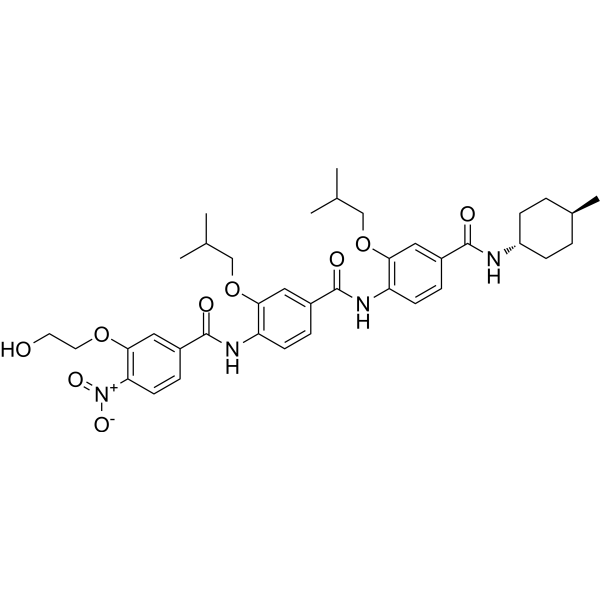
ERX-41 is an orally active and stereospecific small molecule targeting to lysosomal acid lipase A (LIPA). ERX-41 induces endoplasmic reticulum (ER) stress resulting in cell death, indicating a function independent of LIPA but dependent on its ER localization. ERX-41 involves in a targeted strategy for solid tumors.
Nur für Forschungszwecke. Wir verkaufen nicht an Patienten.
- Reinheit : 98.25%
- CAS. Nr.: 2440087-54-5
- Formel: C38H48N4O9
- Molecular Weight:704.81
-
Speicherung:
4°C, protect from light, stored under nitrogen
* In solvent : -80°C, 6 months; -20°C, 1 month (protect from light, stored under nitrogen)

To place orders, for customer services and technical support, please contact: MedChemExpress USA
Tel: 609-228-6898 E-mail: [email protected] [email protected]


-
 Biologische Aktivität
-
Chemical Information
-
Lösungsmittel & Löslichkeit
- Protokoll
- Reinheit & Dokumentation
- Verweise
-
 Help & FAQs


Publications Citing Use of MedChemExpress (MCE) ERX-41
More

Biologische Aktivität
Beschreibung
IC50 & Target
Lysosomal acid lipase A (LIPA)[1]
In Vitro
ERX-41 (1 μM; 0-30 h) induces cell death in MDA-MB-231 without significant effect against normal human mammary epithelial cells (HMECs)[1].
ERX-41 (1 μM; 2 h and 4 h) induces dramatic ER dilation within 4 h, and results disorganization of the peripheral ER network within 2 h[1].
ERX-41 (1 μM; 0.5-4 h) induces downstream unfolded protein response (UPR) pathways via induction of phosphorylated protein kinase R-like ER kinase (p-PERK) and phosphorylated eukaryotic translation initiation factor 2 subunit 1 (p-eIF2-α), and by expression of CCAAT-enhancer-binding homologous protein (CHOP) and phosphorylated inositol-requiring enzyme 1-α (IRE1-α) in TNBC[1].
MedChemExpress (MCE) has not independently confirmed the accuracy of these methods. They are for reference only.
-
Cell Line:MDA-MB-231 and HMEC cells
-
Concentration:1 μM
-
Incubation Time:0, 20 h, 30 h
-
Result:Showed potent antiproliferative activity against TNBC cell within 30 h.
-
Cell Line:SUM-159 cells
-
Concentration:1 μM
-
Incubation Time:0.5 h, 1 h, 2 h, and 4 h
-
Result:Increased the protein level of p-PERK, p-eIF2-α and CHOP at 4 h.
-
Cell Line:SUM-159 and MDA-MB-436
-
Concentration:0.125, 0.25, 0.50, 1.0, 2.0, and 4.0 μM
-
Incubation Time:0h and 30 h
-
Result:Caused cell death.
In Vivo
ERX-41 (10 mg/kg; p.o.; single dose) significantly inhibits the progression without changing body weight in mouse model with MDA-MB-231 xenografts[1].
ERX-41 (10 mg/kg; p.o. or i.p.; single dose) significantly inhibits the progression in mouse model with MDA-MB-231 s.c. xenografts[1].
MedChemExpress (MCE) has not independently confirmed the accuracy of these methods. They are for reference only.
-
Animal Model:Mouse models with D2A1 xenografts and MDA-MB-231 xenografts (s.c.), respectively[1]
-
Dosage:10 mg/kg
-
Administration:Oral gavage; once daily for 25 days
-
Result:Reduced tumor growth against MDA-MB-231 xenograft without affecting body weight. And enhanced p-PERK and p-eIF2-α staining within 24 h.
Significantly reduced the growth of D2A1 xenografts in syngeneic mice without affecting body weight.


Chemical Information
-
CAS. Nr. 2440087-54-5
-
Appearance Solid
-
Molecular Weight 704.81
-
Formel C38H48N4O9
-
Color Off-white to light yellow
-
SMILES
O=C(C1=CC(OCCO)=C(C=C1)[N+]([O-])=O)NC2=C(C=C(C=C2)C(NC3=C(C=C(C=C3)C(N[C@H]4CC[C@@H](CC4)C)=O)OCC(C)C)=O)OCC(C)C
-
Versand
Room temperature in continental US; may vary elsewhere.
-
Speicherung
4°C, protect from light, stored under nitrogen
* In solvent : -80°C, 6 months; -20°C, 1 month (protect from light, stored under nitrogen)


Publications (1)
-
Journal Impact Factor
-
Most Recent
-
Cancer Genet
Downregulation of FAM134B suppresses thyroid cell carcinoma development by modulating endoplasmic reticulum stress and autophagy. [Abstract]2026 Jun:304-305:114-125. PMID: 42033846


Lösungsmittel & Löslichkeit
In Vitro:
DMSO : 25 mg/mL (35.47 mM; Need ultrasonic; Hygroscopic DMSO has a significant impact on the solubility of product, please use newly opened DMSO)
Please refer to the solubility information to select the appropriate solvent. Once prepared, please aliquot and store the solution to prevent product inactivation from repeated freeze-thaw cycles.
Storage method and period of stock solution: -80°C, 6 months; -20°C, 1 month (protect from light, stored under nitrogen). When stored at -80°C, please use it within 6 months. When stored at -20°C, please use it within 1 month.
Please refer to the solubility information to select the appropriate solvent. Once prepared, please aliquot and store the solution to prevent product inactivation from repeated freeze-thaw cycles.
Storage method and period of stock solution: -80°C, 6 months; -20°C, 1 month (protect from light, stored under nitrogen). When stored at -80°C, please use it within 6 months. When stored at -20°C, please use it within 1 month.
Konzentration (Stammlösung) × Volumen (Stammlösung) = Konzentration (Ziellösung) × Volumen (Ziellösung)
In Vivo:
Select the appropriate dissolution method based on your experimental animal and administration route.
- For the following dissolution methods, please ensure to first prepare a clear stock solution using an In Vitro approach and then sequentially add co-solvents:
- To ensure reliable experimental results, the clarified stock solution can be appropriately stored based on storage conditions. As for the working solution for In Vivo experiments, it is recommended to prepare freshly and use it on the same day.
- The percentages shown for the solvents indicate their volumetric ratio in the final prepared solution. If precipitation or phase separation occurs during preparation, heat and/or sonication can be used to aid dissolution.
In Vivo Dissolution Calculator
Please enter the basic information of animal experiments:
-
-
-
-
Recommended: Prepare an additional quantity of animals to account for potential losses during experiments.
Please enter your animal formula composition:
-
%DMSO +
Recommended: Keep the proportion of DMSO in working solution below 2% if your animal is weak.
-
%+
-
+%Tween-80 + +
-
%Saline +
The co-solvents required include: DMSO, . All of co-solvents are available by MedChemExpress (MCE). , Tween 80. All of co-solvents are available by MedChemExpress (MCE).
Working solution concentration: 0.22 mg/mL
Method for preparing stock solution: mg drug dissolved in μL DMSO. Stock solution concentration: mg/mL. * In solvent : -80°C, 6 months; -20°C, 1 month (protect from light, stored under nitrogen)
1. Take μL DMSO stock solution;
2. Add μL .
μL , mix evenly;
3. Then add μL Tween 80, mix evenly;
4. Then add μL
Please ensure that the stock solution in the first step is dissolved to a clear state, and add co-solvents in sequence. You can use ultrasonic heating (ultrasonic cleaner, recommended frequency 20-40 kHz), vortexing, etc. to assist dissolution.


Reinheit & Dokumentation
-
Data Sheet (275 KB)
-
SDS (252 KB)
- English - EN (252 KB)
- Français - FR (252 KB)
- Deutsch - DE (252 KB)
- Norwegian - NO (252 KB)
- Español - ES (252 KB)
- Swedish - SV (252 KB)
- Italian - IT (252 KB)
- Korean - KR (252 KB)
- Portuguese - PT (252 KB)
-
Handling Instructions (2659 KB)
Verweise


Complete Stock Solution Preparation Table
Please refer to the solubility information to select the appropriate solvent. Once prepared, please aliquot and store the solution to prevent product inactivation from repeated freeze-thaw cycles.
Storage method and period of stock solution: -80°C, 6 months; -20°C, 1 month (protect from light, stored under nitrogen). When stored at -80°C, please use it within 6 months. When stored at -20°C, please use it within 1 month.
| Optional Solvent | Concentration Solvent Mass | 1 mg | 5 mg | 10 mg | 25 mg |
|---|---|---|---|---|---|
| DMSO | 1 mM | 1.4188 mL | 7.0941 mL | 14.1882 mL | 35.4706 mL |
| 5 mM | 0.2838 mL | 1.4188 mL | 2.8376 mL | 7.0941 mL | |
| 10 mM | 0.1419 mL | 0.7094 mL | 1.4188 mL | 3.5471 mL | |
| 15 mM | 0.0946 mL | 0.4729 mL | 0.9459 mL | 2.3647 mL | |
| 20 mM | 0.0709 mL | 0.3547 mL | 0.7094 mL | 1.7735 mL | |
| 25 mM | 0.0568 mL | 0.2838 mL | 0.5675 mL | 1.4188 mL | |
| 30 mM | 0.0473 mL | 0.2365 mL | 0.4729 mL | 1.1824 mL |


ERX-41 Related Classifications
HY-148755 Related Classifications
 Powered by Bioz