



Estramustine phosphate
Based on 1 publication(s) in Google Scholar
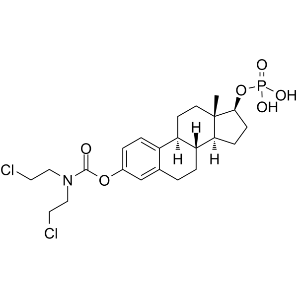
Estramustine phosphate, an estradiol analog, is an orally active antimicrotubule chemotherapy agent. Estramustine phosphate depolymerises microtubules by binding to microtubule associated proteins (MAPs) and/or to tubulin. Estramustine phosphate can interfere mitosis, trigger cell death and induce apoptosis, which can be used for the research of cancer like prostate cancer.
Nur für Forschungszwecke. Wir verkaufen nicht an Patienten.
- CAS. Nr.: 4891-15-0
- Formel: C23H32Cl2NO6P
- Molecular Weight:520.38
-
Speicherung:
Please store the product under the recommended conditions in the Certificate of Analysis.

To place orders, for customer services and technical support, please contact: MedChemExpress USA
Tel: 609-228-6898 E-mail: [email protected] [email protected]


-
 Biologische Aktivität
-
Chemical Information
- Protokoll
- Reinheit & Dokumentation
- Verweise
-
 Help & FAQs


Publications Citing Use of MedChemExpress (MCE) Estramustine phosphate
More

Biologische Aktivität
Beschreibung
In Vitro
Estramustine phosphate (1 µg/mL, 48 h) suppresses PC3 cell growth[1]. Estramustine phosphate (2 µg/mL, 48 h) elevates phosphatidylserine eversion amount on PC3 cells and induces PC3 cell apoptosis through reducing miR-31[1]. Estramustine phosphate (0-40 µM, 24-72 h) inhibits cell proliferation and tubulin cytoskeleton in RAW 264.7 cells[2]. Estramustine phosphate (10 µM, 24 h) inhibits TGF-β-induced RAW 264.7 cell migration, as well as TGF-β-induced uPA production by inhibiting Smad3 activation[2].
MedChemExpress (MCE) has not independently confirmed the accuracy of these methods. They are for reference only.
-
Cell Line:PC3 cells
-
Concentration:1 µg/ mL
-
Incubation Time:48 h
-
Result:Suppressed PC3 cell growth.
-
Cell Line:RAW 264.7 cells
-
Concentration:10 µM
-
Incubation Time:24 h
-
Result:Disrupted the interphase microtubules.
-
Cell Line:18 h of TGF-β treated RAW 264.7 cells
-
Concentration:10 µM
-
Incubation Time:24 h
-
Result:Inhibited TGF-β-Induced Cell Migration.
In Vivo
MedChemExpress (MCE) has not independently confirmed the accuracy of these methods. They are for reference only.
-
Animal Model:Human prostate cancer xenograft PAC120[3]
-
Dosage:4 or 12 mg/kg, a daily dose for 2 weeks.
-
Administration:Intraperitoneal injection
-
Result:Inhibited PAC120 tumor growth 53% by day 35.
Clinical Trial
| NCT Number | Sponsor | Condition | Start Date |
Phase

|
|---|---|---|---|---|
| NCT01329991 | 2011-05 | PHASE1 |


Chemical Information
-
CAS. Nr. 4891-15-0
-
Molecular Weight 520.38
-
Formel C23H32Cl2NO6P
-
SMILES
O=C(OC1=CC=C2[C@@]3([H])CC[C@]4(C)[C@@H](OP(O)(O)=O)CC[C@@]4([H])[C@]3([H])CCC2=C1)N(CCCl)CCCl
-
Versand
Room temperature in continental US; may vary elsewhere.
-
Speicherung
Please store the product under the recommended conditions in the Certificate of Analysis.


Publications (1)
-
Journal Impact Factor
-
Most Recent
-
Cell Rep Med
CAN-Scan: A multi-omic phenotype-driven precision oncology platform identifies prognostic biomarkers of therapy response for colorectal cancer. [Abstract]2025 Apr 15;6(4):102053. PMID: 40187357


Reinheit & Dokumentation
Verweise
[1]. C Wei, et al. Estramustine phosphate induces prostate cancer cell line PC3 apoptosis by down-regulating miR-31 levels. Eur Rev Med Pharmacol Sci. 2018 Jan;22(1):40-45. [Content Brief]
[2]. Sonja S Mojsilovic, et al. Estramustine Phosphate Inhibits TGF- β-Induced Mouse Macrophage Migration and Urokinase-Type Plasminogen Activator Production. Anal Cell Pathol (Amst). 2018 Sep 2;2018:3134102. [Content Brief]
[3]. Stephane Oudard, et al. Activity of docetaxel with or without estramustine phosphate versus mitoxantrone in androgen dependent and independent human prostate cancer xenografts. J Urol. 2003 May;169(5):1729-34. [Content Brief]

Calculators
Konzentration (Stammlösung) × Volumen (Stammlösung) = Konzentration (Ziellösung) × Volumen (Ziellösung)


Estramustine phosphate Related Classifications
HY-13627A Related Classifications
 Powered by Bioz