



ABT-510
Based on 1 publication(s) in Google Scholar
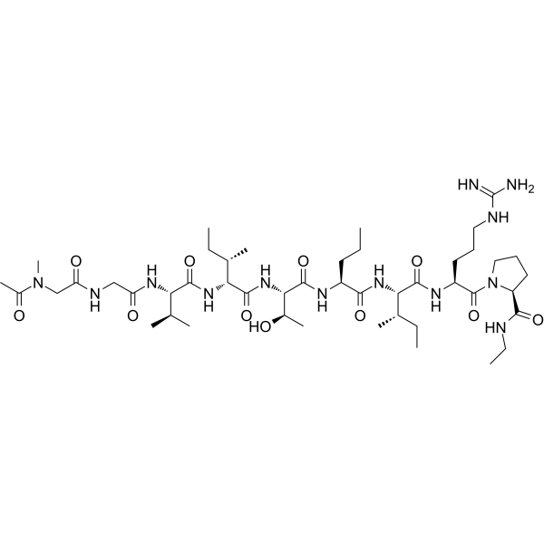
ABT-510 is an anti-angiogenic TSP peptide (Thrombospondin-1 analogue) that induces apoptosis and inhibits ovarian tumour growth in an orthotopic, syngeneic model of epithelial ovarian cancer. ABT-510 also reduces angiogenesis and inflammatory responses in a murine model of inflammatory bowel disease. ABT-510 can be used in studies of cancer (particularly epithelial ovarian cancer) and inflammatory bowel disease (IBD).
For research use only. We do not sell to patients.
- Purity : 99.87%
- CAS No.: 251579-55-2
- Formula: C46H83N13O11
- Molecular Weight:994.23
-
Storage:
Sealed storage, away from moisture.
Powder -80°C, 2 years , -20°C, 1 year* In solvent : -80°C, 6 months; -20°C, 1 month (sealed storage, away from moisture)

To place orders, for customer services and technical support, please contact: MedChemExpress USA
Tel: 609-228-6898 E-mail: [email protected] [email protected]


-
 Biological Activity
-
Chemical Information
-
Solvent & Solubility
- Protocols
- Purity & Documentation
- References
-
 Help & FAQs


Publications Citing Use of MedChemExpress (MCE) ABT-510
More

Biological Activity
Description
In Vitro
ABT-510 (1, 5, 10, 20, 50 nM; 24 h) induces apoptosis in ID8 cells and (50 nM; 24 h) increases the incidence of apoptosis in the human epithelial cancer cell lines SKOV3, OVCAR3, and CAOV3[1].
ABT-510 (0-10 μM; 7 days) inhibits NO-stimulated vascular cell outgrowth into and invasion through extracellular matrix. ABT-510 blocks tumor-driven vascular cell outgrowth, NO-driven cGMP flux, and CD36-mediated fatty acid uptake. [3].
MedChemExpress (MCE) has not independently confirmed the accuracy of these methods. They are for reference only. Further protocols information, click here.
-
Cell Line:ID8, SKOV3, OVCAR3, and CAOV3 cells
-
Concentration:1, 5, 10, 20, 50 nM
-
Incubation Time:24 h
-
Result:Induced ID8 cells apoptosis and increased in apoptosis in the human EOC cell lines SKOV3, OVCAR3, and CAOV3.
-
Cell Line:Tissue biopsies of B16F10 melanoma tumors grown in C57BL/6 mice
-
Concentration:0-10 μM
-
Incubation Time:7 days
-
Result:Inhibited NO-stimulated vascular cell outgrowth into and invasion through extracellular matrix.
In Vivo
ABT-510 (60 mg/kg; osmotic minipumps for s.c.; single daily for 7 days) decreases angiogenesis and inflammation in a murine model of inflammatory bowel disease[2].
MedChemExpress (MCE) has not independently confirmed the accuracy of these methods. They are for reference only.
-
Animal Model:TSP-1-Null mice (C57BL/6 background; orthotopic, syngeneic model of epithelial ovarian cancer)[1].
-
Dosage:100 mg/kg
-
Administration:Intraperitoneal injection; single daily for 90 days
-
Result:Reduced ovarian tumor growth in wild-type and TSP-1-Null Mice.
Significantly reduced the volume of ascites and completely abolished the formation of peritoneal lesions.
Reversed ovarian tumor hypervascularization and increased the proportion of mature blood vessels.
-
Animal Model:TSP-1-Null mice (C57BL/6 background; 6-week-old; DSS-induced inflammatory bowel disease murine model)[2].
-
Dosage:60 mg/kg
-
Administration:Subcutaneously implanted osmotic minipumps (0.5µL/h); single daily for 7 days
-
Result:Significantly delayed DSS-induced bleeding and improved the overall severity of disease.
Significantly diminished inflammation grading and angiogenesis


Chemical Information
-
CAS No. 251579-55-2
-
Appearance Solid
-
Molecular Weight 994.23
-
Formula C46H83N13O11
-
Color White to off-white
-
Sequence
Ac-{Sar}-Gly-Val-{D-Allo-Ile}-Thr-{Nva}-Ile-Arg-{Pro-NHEt}
-
Sequence Shortening
Ac-{Sar}-GV-{D-Allo-Ile-T-{Nva}-IR-{Pro-NHEt}
-
Shipping
Room temperature in continental US; may vary elsewhere.
-
Storage
Sealed storage, away from moisture
Powder -80°C 2 years -20°C 1 year * In solvent : -80°C, 6 months; -20°C, 1 month (sealed storage, away from moisture)


Publications (1)
-
Journal Impact Factor
-
Most Recent
-
Int J Biol Macromol
Truncated thrombospondin-1 polypeptide alleviates non-alcoholic fatty liver disease induced by palmitic acid and high-fat diets. [Abstract]2025 Oct 17;331(Pt 1):148308. PMID: 41109376


Solvent & Solubility
In Vitro:
DMSO : 25 mg/mL (25.15 mM; Need ultrasonic; Hygroscopic DMSO has a significant impact on the solubility of product, please use newly opened DMSO)
Please refer to the solubility information to select the appropriate solvent. Once prepared, please aliquot and store the solution to prevent product inactivation from repeated freeze-thaw cycles.
Storage method and period of stock solution: -80°C, 6 months; -20°C, 1 month (sealed storage, away from moisture). When stored at -80°C, please use it within 6 months. When stored at -20°C, please use it within 1 month.
Please refer to the solubility information to select the appropriate solvent. Once prepared, please aliquot and store the solution to prevent product inactivation from repeated freeze-thaw cycles.
Storage method and period of stock solution: -80°C, 6 months; -20°C, 1 month (sealed storage, away from moisture). When stored at -80°C, please use it within 6 months. When stored at -20°C, please use it within 1 month.
Concentration (start) × Volume (start) = Concentration (final) × Volume (final)

Protocols
-
Apoptosis
Apoptosis, also called programmed cell death, is generally characterized by distinct morphological characteristics.
-
TUNEL staining for apoptotic DNA fragmentation
TUNEL staining detects DNA strand breaks by using terminal deoxynucleotidyl transferase to add labeled nucleotides to exposed 3′-OH DNA termini, generating either microscopic staining in fixed cells or tissue sections, or fluorescence/cytometric signal in cell suspensions. TUNEL positivity reflects DNA fragmentation but should not be interpreted alone as definitive apoptosis, because TUNEL can also label necrotic, autolytic, mechanically damaged, or DNA-repair-associated DNA breaks.
-
Human pluripotent stem cell endothelial-cell differentiation
Human pluripotent stem cell endothelial differentiation is based on stepwise developmental patterning: early activation of WNT/GSK3β inhibition promotes mesodermal or vascular progenitor entry, followed by endothelial specification using VEGF-related signaling, BMP4, FGF2, Notch modulation, or cAMP depending on the published protocol. Endothelial differentiation is read out by acquisition of CD31, CD34, VE-cadherin/CD144, KDR/VEGFR2, vWF, Tie2, NOS3, acetylated LDL uptake, tube/network formation, barrier function, and in vivo vessel-forming capacity where tested.
-
Vascular/Branching Fractal Analysis
Vascular/branching fractal analysis quantifies the geometric complexity of vessel trees or vascular networks from segmented 2D images, commonly by converting vessels into binary and/or skeletonized maps and estimating fractal dimension using box-counting or related approaches. Fractal dimension is interpreted as an image-derived readout of vascular branching complexity, space filling, or density, and has been applied to retinal photographs, fluorescein angiography, OCT angiography, capillary perfusion maps, and in vitro Matrigel angiogenesis networks. The assay readout is generated from vessel-positive pixels after image preprocessing, vessel segmentation, binarization, and optional skeletonization; reported outputs include fractal dimension, vessel density, branchpoint density, endpoint density, vessel length density, tortuosity, and generation-based branching metrics when VESGEN-style analysis is used. The biological interpretation is limited to quantitative vascular patterning and s
-
Annexin V plus membrane-impermeant dye apoptosis staining
Annexin V-based apoptosis assays rely on the detection of phosphatidylserine (PS) externalization from the inner leaflet of the plasma membrane to the outer leaflet, an early biochemical hallmark of apoptosis. Fluorescently labeled Annexin V binds PS in a calcium-dependent manner, enabling identification of early apoptotic cells by flow cytometry or fluorescence microscopy. When combined with a membrane-impermeant DNA-binding dye (e. g. , propidium iodide), this approach allows discrimination between viable (Annexin V−/dye−), early apoptotic (Annexin V+/dye−), and late apoptotic or necrotic (Annexin V+/dye+) cell populations by assessing membrane integrity and PS exposure.
-
DSS-Induced Colitis
Dextran sulfate sodium (DSS)-induced colitis is generated by administering DSS in mouse drinking water, producing epithelial injury, barrier disruption, weight loss, diarrhea, fecal blood, colon shortening, histologic mucosal damage, and inflammatory mediator changes; the model is mainly used to study acute or chronic intestinal inflammation resembling selected features of ulcerative colitis. DSS injury is interpreted through clinical and tissue readouts rather than a single molecular endpoint: daily body weight, stool consistency, and bleeding are combined into a disease activity index, while colon length, histology, cytokines, myeloperoxidase activity, intestinal permeability, and tight-junction markers provide complementary measures of inflammation and barrier damage.
-
TNBS-Induced Colitis
TNBS-induced colitis is produced by intrarectal delivery of 2,4,6-trinitrobenzene sulfonic acid in ethanol, where ethanol disrupts the mucosal barrier and TNBS haptenates colonic proteins, generating immune-mediated colonic inflammation with weight loss, diarrhea, ulceration, transmural injury, inflammatory-cell infiltration, and cytokine responses. The model is used as an experimental intestinal inflammation model with Crohn’s disease–like features, especially when Th1-type responses, IL-12–dependent inflammation, chronic relapsing inflammation, or fibrosis-related endpoints are studied.
-
Endothelial Tube Formation Assay
Endothelial tube formation assay evaluates the ability of endothelial cells to attach, migrate, align, and organize into capillary-like networks when cultured on gelled basement membrane extract or Matrigel; the readout is the morphology and quantity of tube-like networks, which reflects an in vitro endothelial morphogenesis step related to angiogenesis. Basement membrane extract/Matrigel provides laminin-rich extracellular matrix cues that support endothelial differentiation into capillary-like structures, but it can contain biologically active growth factors, so growth-factor-reduced matrix is preferred when testing defined angiogenic stimulators or inhibitors.
-
Apoptosis Solutions
Apoptosis is a regulated, generally non-lytic cell-death pathway that removes unwanted, damaged, infected, or abnormal cells through coordinated morphological changes, caspase activation, DNA fragmentation, and membrane remodeling. The intrinsic apoptosis pathway is controlled mainly by mitochondrial outer membrane permeabilization, BCL-2 family proteins, cytochrome c release, apoptosome formation, caspase-9 activation, and downstream executioner caspase-3/7 activation. The extrinsic apoptosis pathway is initiated by death receptors such as Fas, TNFR, and TRAIL receptors, which recruit adaptor proteins and activate caspase-8 before engaging executioner caspases or mitochondrial amplification through BID cleavage. Apoptosis is linked to many phenotypes, including cancer cell killing, tissue homeostasis, immune regulation, neurodegeneration, infection response, and treatment-induced cytotoxicity; unresolved questions include how apoptosis interacts with necroptosis, pyroptosis, ferroptos
-
Endothelial Cell Migration/Angiogenic Sprouting Assay
Endothelial cell migration and angiogenic sprouting assays are in vitro (and partially ex vivo-adapted) functional models that quantify the ability of endothelial cells to undergo coordinated migration, extracellular matrix invasion, and multicellular organization into capillary-like sprouts in response to pro-angiogenic stimuli such as VEGF, bFGF, or conditioned microenvironments. These assays are used to model early angiogenic events including tip-cell formation, directional migration, and lumen-like sprout extension, which collectively reflect angiogenic activation and vascular morphogenesis processes observed in vivo.


Purity & Documentation
-
Data Sheet (278 KB)
-
SDS (394 KB)
- English - EN (394 KB)
- Français - FR (394 KB)
- Deutsch - DE (394 KB)
- Norwegian - NO (394 KB)
- Español - ES (394 KB)
- Swedish - SV (394 KB)
- Italian - IT (394 KB)
- Korean - KR (394 KB)
- Portuguese - PT (394 KB)
-
Handling Instructions (2659 KB)
References
[1]. Greenaway J, et al. ABT-510 induces tumor cell apoptosis and inhibits ovarian tumor growth in an orthotopic, syngeneic model of epithelial ovarian cancer. Mol Cancer Ther. 2009 Jan;8(1):64-74. [Content Brief]
[2]. Punekar S, et al. Thrombospondin 1 and its mimetic peptide ABT-510 decrease angiogenesis and inflammation in a murine model of inflammatory bowel disease. Pathobiology. 2008;75(1):9-21. [Content Brief]
[3]. Isenberg JS, et.al. Differential effects of ABT-510 and a CD36-binding peptide derived from the type 1 repeats of thrombospondin-1 on fatty acid uptake, nitric oxide signaling, and caspase activation in vascular cells. Biochem Pharmacol. 2008 Feb 15;75(4):875-82. [Content Brief]


Complete Stock Solution Preparation Table
Please refer to the solubility information to select the appropriate solvent. Once prepared, please aliquot and store the solution to prevent product inactivation from repeated freeze-thaw cycles.
Storage method and period of stock solution: -80°C, 6 months; -20°C, 1 month (sealed storage, away from moisture). When stored at -80°C, please use it within 6 months. When stored at -20°C, please use it within 1 month.
| Optional Solvent | Concentration Solvent Mass | 1 mg | 5 mg | 10 mg | 25 mg |
|---|---|---|---|---|---|
| DMSO | 1 mM | 1.0058 mL | 5.0290 mL | 10.0580 mL | 25.1451 mL |
| 5 mM | 0.2012 mL | 1.0058 mL | 2.0116 mL | 5.0290 mL | |
| 10 mM | 0.1006 mL | 0.5029 mL | 1.0058 mL | 2.5145 mL | |
| 15 mM | 0.0671 mL | 0.3353 mL | 0.6705 mL | 1.6763 mL | |
| 20 mM | 0.0503 mL | 0.2515 mL | 0.5029 mL | 1.2573 mL | |
| 25 mM | 0.0402 mL | 0.2012 mL | 0.4023 mL | 1.0058 mL |


ABT-510 Related Classifications
HY-13545 Related Classifications
 Powered by Bioz