



Codrituzumab
Based on 1 publication(s) in Google Scholar
Codrituzumab (GC33) is a humanized monoclonal antibody targeting human GPC3 (glypican-3), with high affinity (Kd of 0.673 nM). GPC3 is an oncofetal protein expressed on the cell surface of hepatocellular carcinoma (HCC). Codrituzumab induces antibody-dependent cellular cytotoxicity (ADCC) and inhibits tumor growth.
For research use only. We do not sell to patients.
- Purity : 99.07%
- CAS No.: 1365267-33-9
- Molecular Weight:145.58 kDa
-
Storage:
Please store the product under the recommended conditions in the Certificate of Analysis.


To place orders, for customer services and technical support, please contact: MedChemExpress USA
Tel: 609-228-6898 E-mail: [email protected] [email protected]


-
 Biological Activity
-
Chemical Information
- Protocols
- Purity & Documentation
- References
-
 Help & FAQs


-

HY-138298
Trastuzumab deruxtecan (solution)HER2 ADC -

HY-P99001
Human IgG1 kappa, Isotype ControlIgG1κ Isotype Control -

HY-P9902
PembrolizumabPD-1 Inhibitor -

HY-P99058
ZolbetuximabClaudin-18.2 Inhibitor -

HY-P99041
PanitumumabAnti-EGFR Monoclonal Antibody -

HY-P9904
AtezolizumabAnti-PD-L1 Antibody -

HY-P9966
EnoblituzumabB7-H3 mAb -
HY-P990840
Anti-GPC3 Antibody (YP7)Anti-GPC3 Antibody
Publications Citing Use of MedChemExpress (MCE) Codrituzumab
More

Biological Activity
Description
Isotype
Human IgG1 kappa
Recommend Isotype Controls
Species Reactivity
Human
IC50 & Target
GPC3
In Vitro
Codrituzumab (once-weekly for 21 days) combined with PD-L1 mAb demonstrates a marked antitumor effect, enhancing tumor infiltration of PD-L1-positive immune cells, such as macrophages and multinucleated giant cells in the Hepa1-6/hGPC3 mice model[1].
MedChemExpress (MCE) has not independently confirmed the accuracy of these methods. They are for reference only.
- Cell Cytotoxicity Assay
- Mammalian live/dead viability and cytotoxicity staining
- Patient-Derived Xenograft (PDX)
- Patient-Derived Orthotopic Xenograft (PDOX)
- Liver Cancer Modeling
- Subcutaneous Cell-Line-Derived Xenograft
- Orthotopic Cell-Line Xenograft
- Cell Viability Determination by MTT Colorimetric Assay
Clinical Trial
| NCT Number | Sponsor | Condition | Start Date |
Phase

|
|---|---|---|---|---|
| NCT01329991 | 2011-05 | PHASE1 |
Gene ID
Accession
Conjugated
Unconjugated
Reconsititution
The product can be reconstituted/diluted with sterile PBS or saline.
Format
-
Human IgG1 kappa
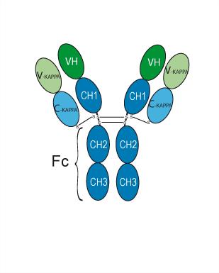
Application
ELISA, FACS, Functional assay
Verified Bioactivity
-
 Immobilized Glypican-3/GPC3 Protein, Human (HEK293, His, HY-P70487) can bind Codrituzumab. The EC50 for this effect is 13.3 ng/mL.
 -
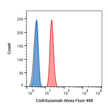 Flow cytometric analysis of 1X10⁶ Jurkat cells with Codrituzumab (HY-P99013, red). Cells were fixed with 4% paraformaldehyde and permeabilised with 90% methanol. Then stained with the primary antibody at 1/200 dilution for an hour at 4℃. Alexa Fluor 488-conjugated AffiniPure Goat Anti-Human IgG H&L (HY-P83776) was used as the secondary antibody at 1/1,000 dilution for 30 minutes at 4℃. Human IgG1 kappa Isotype Control (HY-P99001, blue) was used as the isotype control, cells without incubation with primary antibody were used as the unlabeled control (black).
 -
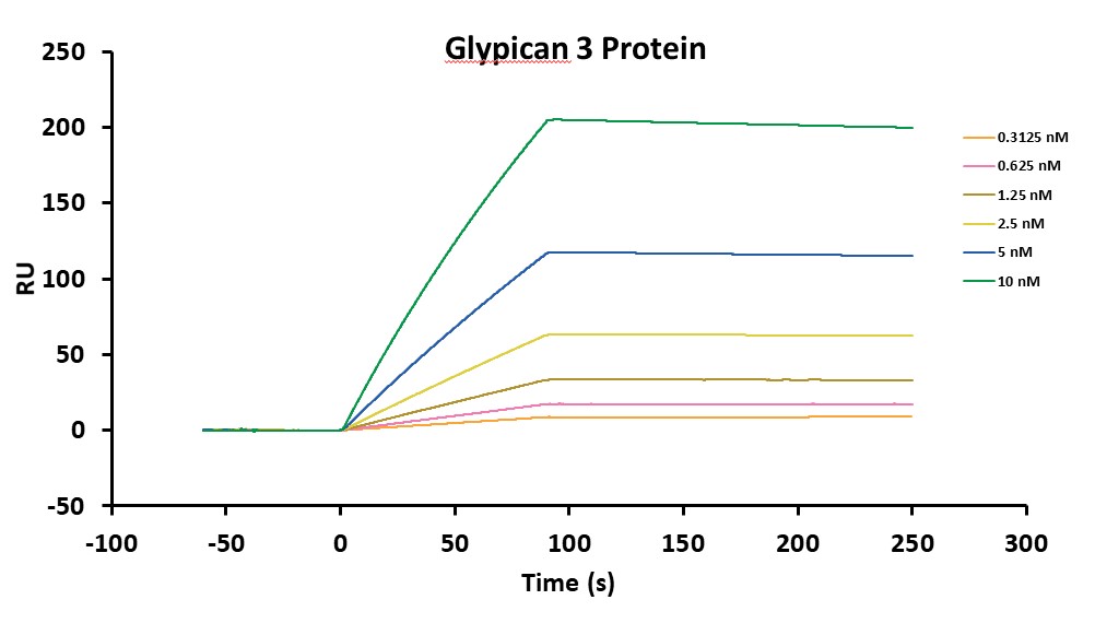 Loaded Codrituzumab on Sensor Chip Protein A, can bind Glypican-3/GPC3 Protein, Human (HEK293, HY-P70487) with an affinity constant of 2.15E-10 M as determined in SPR assay.


Chemical Information
-
CAS No. 1365267-33-9
-
Appearance Liquid
-
Molecular Weight 145.58 kDa
-
Color Colorless to light yellow
-
SMILES
[Codrituzumab]
-
Synonyms
GC33; RO5137382
-
Shipping
Shipping with dry ice.
-
Formulation
Please refer to the lot-specific COA for specific buffer information.
-
Storage
Please store the product under the recommended conditions in the Certificate of Analysis.


Publications (1)
-
Journal Impact Factor
-
Most Recent
-
Small Sci
Therapeutic Glypican-3 CRISPR Genome-Editing Using UltraLarge Porous Silica Nano-Depot for the Treatment of Hepatocellular Carcinoma. [Abstract]2024 Nov 21;5(4):2400447. PMID: 40657181


Purity & Documentation
-
Data Sheet (274 KB)
-
SDS (251 KB)
- English - EN (251 KB)
- Français - FR (251 KB)
- Deutsch - DE (251 KB)
- Norwegian - NO (251 KB)
- Español - ES (251 KB)
- Swedish - SV (251 KB)
- Italian - IT (251 KB)
- Korean - KR (251 KB)
- Portuguese - PT (251 KB)
-
Inhibitory Antibodies User Guide (603 KB)
References
[2]. Chen G, et al. Combining expression of GPC3 in tumors and CD16 on NK cells from peripheral blood to identify patients responding to codrituzumab. Oncotarget. 2018 Jan 2;9(12):10436-10444. [Content Brief]
[3]. Carrasquillo JA, et al. I-124 codrituzumab imaging and biodistribution in patients with hepatocellular carcinoma. EJNMMI Res. 2018 Mar 5;8(1):20. [Content Brief]
[4]. Abou-Alfa GK, et al. Randomized phase II placebo controlled study of codrituzumab in previously treated patients with advanced hepatocellular carcinoma. J Hepatol. 2016 Aug;65(2):289-95. [Content Brief]

Calculators
Concentration (start) × Volume (start) = Concentration (final) × Volume (final)


Codrituzumab Related Classifications
HY-P99013 Related Classifications
 Powered by Bioz