



Ganoderic acid D
Based on 2 publication(s) in Google Scholar
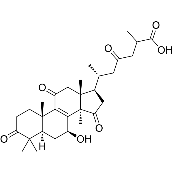
Ganoderic acid D, a highly oxygenated tetracyclic triterpenoid, is the major active component of Ganoderma lucidum. Ganoderic acid D upregulates the protein expression of SIRT3 and induces the deacetylated cyclophilin D (CypD) by SIRT3. Ganoderic acid D inhibits the energy reprogramming of colon cancer cells including glucose uptake, lactate production, pyruvate and acetyl-coenzyme production in colon cancer cells. Ganoderic acid D induces HeLa human cervical carcinoma apoptosis.
For research use only. We do not sell to patients.
- Purity : 99.18%
- CAS No.: 108340-60-9
- Formula: C30H42O7
- Molecular Weight:514.65
-
Storage:Powder -20°C, 3 years , 4°C, 2 years ; In solvent -80°C, 6 months , -20°C, 1 month

To place orders, for customer services and technical support, please contact: MedChemExpress USA
Tel: 609-228-6898 E-mail: [email protected] [email protected]


-
 Biological Activity
-
Chemical Information
-
Solvent & Solubility
- Protocols
- Purity & Documentation
- References
-
 Help & FAQs


-
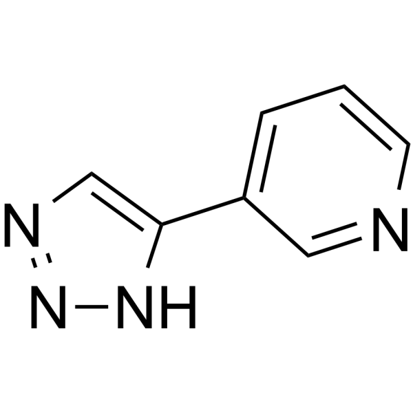
HY-108331
3-TYPSIRT3 Inhibitor -
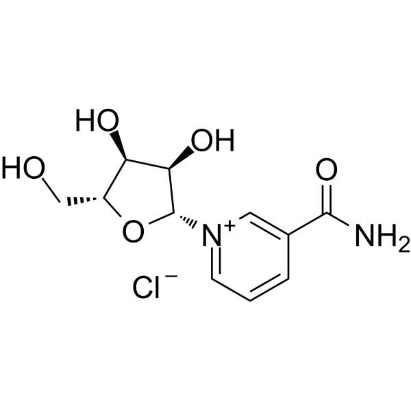
HY-123033A
Nicotinamide riboside chlorideEndogenous Metabolite -
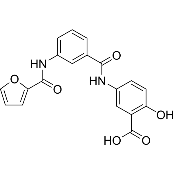
HY-107454
OSS_128167SIRT6 Inhibitor -
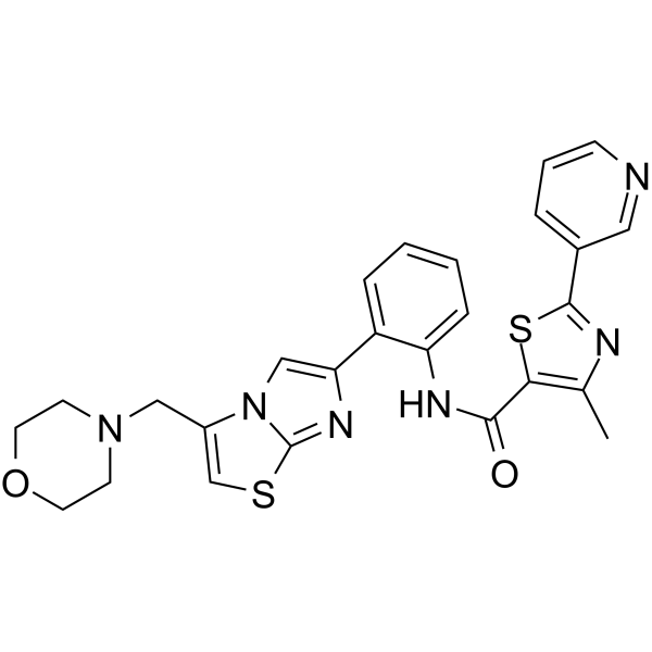
HY-15262
SRT 2104SIRT1 Activator -
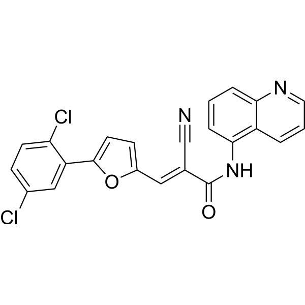
HY-100578
AGK2SIRT2 Inhibitor -
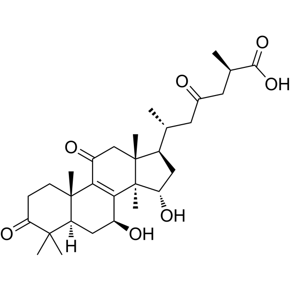
HY-N1447
Ganoderic acid AAutophagy Inducer -
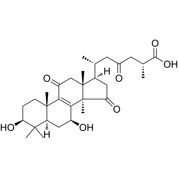
HY-N2006
Ganoderic acid BHIV Protease Inhibitor -
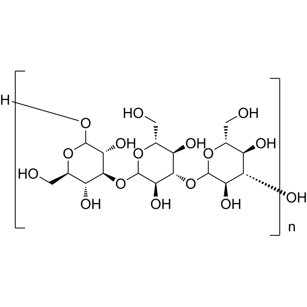
HY-119109
LaminaranImmunotherapy Agent -

HY-Y1310A
Sodium alginate (Viscosity 200±20mpa.s)Biochemical Assay Reagent -
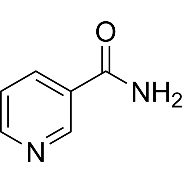
HY-B0150
NicotinamideSIRT Inhibitor -
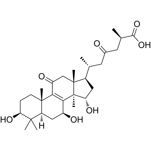
HY-N1517
Ganoderic acid C2Aldose Reductase Inhibitor -
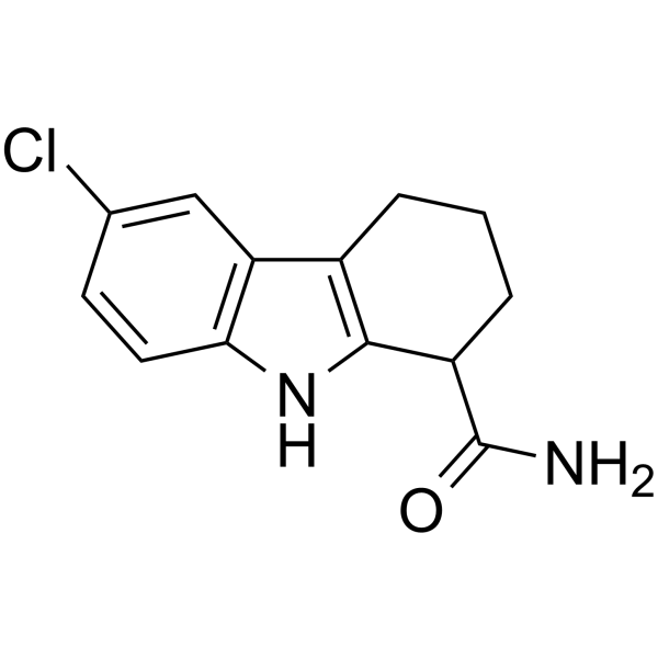
HY-15452
SelisistatSirT1/Sir2 Inhibitor
Publications Citing Use of MedChemExpress (MCE) Ganoderic acid D
More

Biological Activity
Description
IC50 & Target
[1]|
SIRT3 |
Cellular Effect
|
Cell Line

|
Type | Value | Description | References |
|---|---|---|---|---|
| Bel-7402 | IC50 |
7.25 μM
Compound: 10
|
Cytotoxicity against human Bel7402 cells
Cytotoxicity against human Bel7402 cells
|
[PMID: 23092389] |
| BV-2 | IC50 |
10.86 μM
Compound: 14
|
Antiinflammatory activity in mouse BV2 cells assessed as inhibition of LPS-induced NO production incubated for 24 hrs measured after 15 mins by Griess assay
Antiinflammatory activity in mouse BV2 cells assessed as inhibition of LPS-induced NO production incubated for 24 hrs measured after 15 mins by Griess assay
|
[PMID: 27335254] |
| HepG2 | IC50 |
22.9 μM
Compound: 12
|
Anticancer activity against human HepG2 cells assessed as cell viability after 48 hrs by MTT assay
Anticancer activity against human HepG2 cells assessed as cell viability after 48 hrs by MTT assay
|
[PMID: 24974349] |
| P388 | IC50 |
7.25 μM
Compound: 10
|
Cytotoxicity against mouse P388 cells
Cytotoxicity against mouse P388 cells
|
[PMID: 23092389] |
| SGC-7901 | IC50 |
7.25 μM
Compound: 10
|
Cytotoxicity against human SGC7901 cells
Cytotoxicity against human SGC7901 cells
|
[PMID: 23092389] |
In Vitro
Ganoderic acid D can inhibit the growth of numerous cancer cell lines and it inhibits HeLa human cervical carcinoma cells with an IC50 of 17.3 mM[2].
Ganoderic acid D (1-50 μM; 24-72 hours) reduces the cell survival rate in a dose- and time-dependent manner[2].
Ganoderic acid D (10, 50 μM; 24, 48 hours) induces G2/M phase arrest[2].
Ganoderic acid D (10, 50 μM; 24, 48 hours) induces a morphological change typical of apoptosis in HeLa cells[2].
Ganoderic acid D (10 μM; 48 hours) up-regulates 14-3-3E and PRDX3[2].
MedChemExpress (MCE) has not independently confirmed the accuracy of these methods. They are for reference only.
-
Cell Line:HeLa human cervical carcinoma cell line (CCL-2)
-
Concentration:1, 5, 10, 20, 50 μM
-
Incubation Time:24, 48, 72 hours
-
Result:Reduced the cell survival rate in a dose- and time-dependent manner and had an IC50 value of 17.3 μM for 48 hours treatment.
-
Cell Line:HeLa human cervical carcinoma cell line (CCL-2)
-
Concentration:10, 50 μM
-
Incubation Time:24, 48 hours
-
Result:Induced G2/M phase arrest.
Displayed a cell cycle profile with an elevated G2/M cell population after 24-h treatment with 10 μM.
-
Cell Line:HeLa human cervical carcinoma cell line (CCL-2)
-
Concentration:10, 50 μM
-
Incubation Time:48 hours
-
Result:Induced a morphological change typical of apoptosis in HeLa cells.
-
Cell Line:HeLa human cervical carcinoma cell line (CCL-2)
-
Concentration:10 μM
-
Incubation Time:48 hours
-
Result:Up-regulated 14-3-3E and PRDX3.


Chemical Information
-
CAS No. 108340-60-9
-
Appearance Solid
-
Molecular Weight 514.65
-
Formula C30H42O7
-
Color White to off-white
-
SMILES
CC1(C)C(CC[C@]2(C)C3=C([C@@]4(C(C[C@@H]([C@]4(CC3=O)C)[C@H](C)CC(CC(C)C(O)=O)=O)=O)C)[C@@H](O)C[C@@]12[H])=O
-
Structure Classification
-
Initial Source
-
Shipping
Room temperature in continental US; may vary elsewhere.
-
Storage
Powder -20°C 3 years 4°C 2 years In solvent -80°C 6 months -20°C 1 month


Publications (2)
-
Journal Impact Factor
-
Most Recent
-
Apoptosis
Brazilin alleviates acute lung injury via inhibition of ferroptosis through the SIRT3/GPX4 pathway. [Abstract]2025 Apr;30(3-4):768-783. PMID: 39720978 -
Front Pharmacol
Sporoderm-Broken Spores of Ganoderma lucidum Sensitizes Ovarian Cancer to Cisplatin by ROS/ERK Signaling and Attenuates Chemotherapy-Related Toxicity. [Abstract]2022 Feb 21:13:826716. PMID: 35264959


Solvent & Solubility
In Vitro:
DMSO : 100 mg/mL (194.31 mM; Need ultrasonic; Hygroscopic DMSO has a significant impact on the solubility of product, please use newly opened DMSO)
H2O : < 0.1 mg/mL (insoluble)
Please refer to the solubility information to select the appropriate solvent. Once prepared, please aliquot and store the solution to prevent product inactivation from repeated freeze-thaw cycles.
Storage method and period of stock solution: -80°C, 6 months; -20°C, 1 month. When stored at -80°C, please use it within 6 months. When stored at -20°C, please use it within 1 month.
Please refer to the solubility information to select the appropriate solvent. Once prepared, please aliquot and store the solution to prevent product inactivation from repeated freeze-thaw cycles.
Storage method and period of stock solution: -80°C, 6 months; -20°C, 1 month. When stored at -80°C, please use it within 6 months. When stored at -20°C, please use it within 1 month.
Concentration (start) × Volume (start) = Concentration (final) × Volume (final)
In Vivo:
Select the appropriate dissolution method based on your experimental animal and administration route.
- For the following dissolution methods, please ensure to first prepare a clear stock solution using an In Vitro approach and then sequentially add co-solvents:
- To ensure reliable experimental results, the clarified stock solution can be appropriately stored based on storage conditions. As for the working solution for In Vivo experiments, it is recommended to prepare freshly and use it on the same day.
- The percentages shown for the solvents indicate their volumetric ratio in the final prepared solution. If precipitation or phase separation occurs during preparation, heat and/or sonication can be used to aid dissolution.
Add each solvent one by one: 10% DMSO 40% PEG300 5% Tween-80 45% Saline
Solubility: ≥ 2.5 mg/mL (4.86 mM); Clear solution
This protocol yields a clear solution of ≥ 2.5 mg/mL (saturation unknown).
Taking 1 mL working solution as an example, add 100 μL DMSO stock solution (25.0 mg/mL) to 400 μL PEG300, and mix evenly; then add 50 μL Tween-80 and mix evenly; then add 450 μL Saline to adjust the volume to 1 mL.
Preparation of Saline: Dissolve 0.9 g sodium chloride in ddH₂O and dilute to 100 mL to obtain a clear Saline solution.
Add each solvent one by one: 10% DMSO 90% (20% SBE-β-CD in Saline)
Solubility: ≥ 2.5 mg/mL (4.86 mM); Clear solution
This protocol yields a clear solution of ≥ 2.5 mg/mL (saturation unknown).
Taking 1 mL working solution as an example, add 100 μL DMSO stock solution (25.0 mg/mL) to 900 μL 20% SBE-β-CD in Saline, and mix evenly.
Preparation of 20% SBE-β-CD in Saline (4°C, storage for one week): 2 g SBE-β-CD powder is dissolved in 10 mL Saline, completely dissolve until clear.
In Vivo Dissolution Calculator
Please enter the basic information of animal experiments:
-
-
-
-
Recommended: Prepare an additional quantity of animals to account for potential losses during experiments.
Please enter your animal formula composition:
-
%DMSO +
Recommended: Keep the proportion of DMSO in working solution below 2% if your animal is weak.
-
%+
-
+%Tween-80 + +
-
%Saline +
The co-solvents required include: DMSO, . All of co-solvents are available by MedChemExpress (MCE). , Tween 80. All of co-solvents are available by MedChemExpress (MCE).
Working solution concentration: 0.22 mg/mL
Method for preparing stock solution: mg drug dissolved in μL DMSO. Stock solution concentration: mg/mL.
1. Take μL DMSO stock solution;
2. Add μL .
μL , mix evenly;
3. Then add μL Tween 80, mix evenly;
4. Then add μL
Please ensure that the stock solution in the first step is dissolved to a clear state, and add co-solvents in sequence. You can use ultrasonic heating (ultrasonic cleaner, recommended frequency 20-40 kHz), vortexing, etc. to assist dissolution.


Purity & Documentation
-
Data Sheet (281 KB)
-
SDS (393 KB)
- English - EN (393 KB)
- Français - FR (393 KB)
- Deutsch - DE (393 KB)
- Norwegian - NO (393 KB)
- Español - ES (393 KB)
- Swedish - SV (393 KB)
- Italian - IT (393 KB)
- Korean - KR (393 KB)
- Portuguese - PT (393 KB)
-
Handling Instructions (2659 KB)
References
[1]. Liu Z, et al. Effect of ganoderic acid D on colon cancer Warburg effect: Role of SIRT3/cyclophilin D. Eur J Pharmacol. 2018 Apr 5;824:72-77. [Content Brief]
[2]. Yue QX, et al. Proteomics characterization of the cytotoxicity mechanism of ganoderic acid D and computer-automated estimation of the possible drug target network. Mol Cell Proteomics. 2008 May;7(5):949-61. [Content Brief]


Complete Stock Solution Preparation Table
Please refer to the solubility information to select the appropriate solvent. Once prepared, please aliquot and store the solution to prevent product inactivation from repeated freeze-thaw cycles.
Storage method and period of stock solution: -80°C, 6 months; -20°C, 1 month. When stored at -80°C, please use it within 6 months. When stored at -20°C, please use it within 1 month.
| Optional Solvent | Concentration Solvent Mass | 1 mg | 5 mg | 10 mg | 25 mg |
|---|---|---|---|---|---|
| DMSO | 1 mM | 1.9431 mL | 9.7153 mL | 19.4307 mL | 48.5767 mL |
| 5 mM | 0.3886 mL | 1.9431 mL | 3.8861 mL | 9.7153 mL | |
| 10 mM | 0.1943 mL | 0.9715 mL | 1.9431 mL | 4.8577 mL | |
| 15 mM | 0.1295 mL | 0.6477 mL | 1.2954 mL | 3.2384 mL | |
| 20 mM | 0.0972 mL | 0.4858 mL | 0.9715 mL | 2.4288 mL | |
| 25 mM | 0.0777 mL | 0.3886 mL | 0.7772 mL | 1.9431 mL | |
| 30 mM | 0.0648 mL | 0.3238 mL | 0.6477 mL | 1.6192 mL | |
| 40 mM | 0.0486 mL | 0.2429 mL | 0.4858 mL | 1.2144 mL | |
| 50 mM | 0.0389 mL | 0.1943 mL | 0.3886 mL | 0.9715 mL | |
| 60 mM | 0.0324 mL | 0.1619 mL | 0.3238 mL | 0.8096 mL | |
| 80 mM | 0.0243 mL | 0.1214 mL | 0.2429 mL | 0.6072 mL | |
| 100 mM | 0.0194 mL | 0.0972 mL | 0.1943 mL | 0.4858 mL |


Ganoderic acid D Related Classifications
HY-N1511 Related Classifications
 Powered by Bioz