



IBR2
Based on 8 publication(s) in Google Scholar
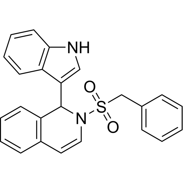
IBR2 is a potent and specific RAD51 inhibitor and inhibits RAD51-mediated DNA double-strand break repair. IBR2 disrupts RAD51 multimerization, accelerates proteasome-mediated RAD51 protein degradation, inhibits cancer cell growth and induces apoptosis.
Para uso exclusivo en investigación. No vendemos a pacientes.
- Pureza : 98.24%
- No. CAS: 313526-24-8
- Fòrmula: C24H20N2O2S
- Peso molecular:400.49
-
Almacenamiento:Powder -20°C, 3 years , 4°C, 2 years ; In solvent -80°C, 2 years , -20°C, 1 year

To place orders, for customer services and technical support, please contact: MedChemExpress USA
Tel: 609-228-6898 E-mail: [email protected] [email protected]


-
 Actividad biológica
-
Chemical Information
-
Solvente y solubilidad
- Protocolo
- Pureza y Documentación
- Referencias
-
 Help & FAQs


-
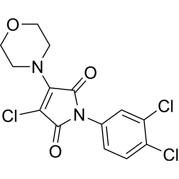
HY-15317
RI-1RAD51 Inhibitor -
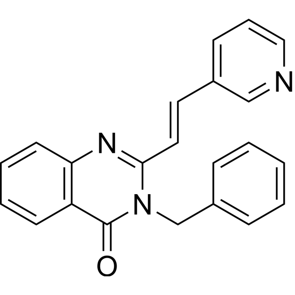
HY-101462
RAD51 Inhibitor B02RAD51 Inhibitor -
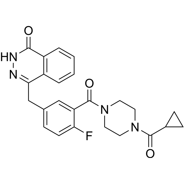
HY-10162
OlaparibPARP Inhibitor -
HY-19793
RS-1RAD51 Activator -

HY-13259
MG-132Proteasome Inhibitor -

HY-10219
RapamycinmTOR Inhibitor -

HY-10227
BortezomibProteasome Inhibitor -
HY-13629
EtoposideTopoisomerase II Inhibitor -
HY-124691
D-I03RAD52 Inhibitor -
HY-17536
SelinexorCRM1 Inhibitor -
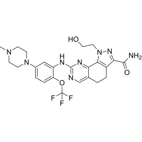
HY-15828
OnvansertibPLK1 Inhibitor -
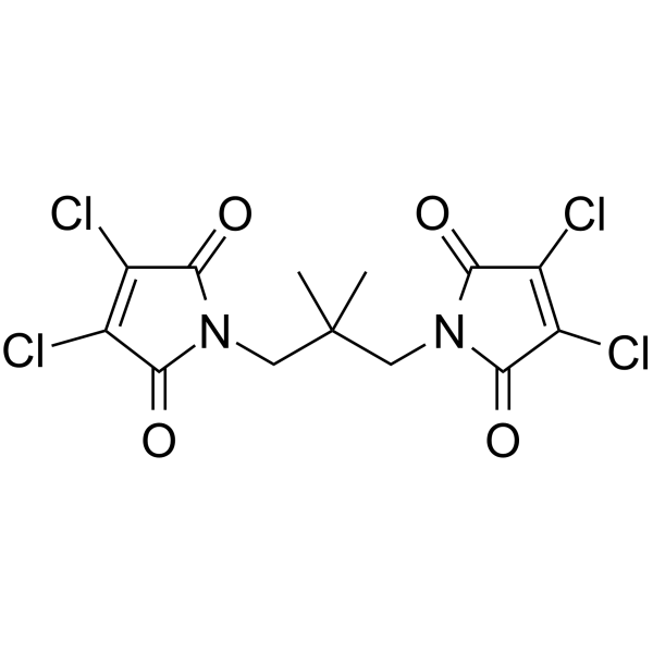
HY-110185
NSC 617145WRN Helicase Inhibitor
Publications Citing Use of MedChemExpress (MCE) IBR2
More- Sci Bull. 2025 Aug 25:S2095-9273(25)00865-5. [Abstract]
- Reprod Biol Endocrinol. 2025 Jul 3;23(1):93. [Abstract]
- J Virol. 2023 Dec 21;97(12):e0173723. [Abstract]
- Theriogenology. 2026 Jun 24:265:118052. [Abstract]
- Biomed Rep. 2023 Feb 24;18(4):28. [Abstract]
- J Surg Res. 2026 Aug 24:326:985-995. [Abstract]
- Patent. US20250049807A1.
- CUNY Hunter College. 2020 Sep.


Actividad biológica
Descripciòn
IC50 & Target
RAD51[1]
Cellular Effect
|
Cell Line

|
Type | Value | Description | References |
|---|---|---|---|---|
| MDA-MB-468 | IC50 |
14.8 μM
Compound: IBR2 (rac)
|
Cytotoxicity against human MDA-MB-468 cells assessed as growth inhibition after 4 days by XTT assay
Cytotoxicity against human MDA-MB-468 cells assessed as growth inhibition after 4 days by XTT assay
|
[PMID: 25874343] |
In Vitro
IBR2 shows interesting RAD51 inhibition activities. RAD51 is rapidly degraded in IBR2-treated cancer cells, and the homologous recombination repair is impaired, subsequently leading to cell death. The IC50 values of the original IBR2 are in the range of 12-20 μM for most tested cancer cell lines. IBR2 can inhibit the growth of triple-negative human breast cancer cell line MBA-MD-468 with an IC50 of 14.8 μM[1].
MedChemExpress (MCE) has not independently confirmed the accuracy of these methods. They are for reference only.
- Detection of Apoptosis
- TUNEL staining for apoptotic DNA fragmentation
- Annexin V plus membrane-impermeant dye apoptosis staining
- Protocol for Cell Counting and Cell Density Analysis
- Somatic Cell Culture
- Apoptosis Solutions
- CCK-8/WST-8 Cell Proliferation Assay
- Cell Counting-Based Growth Curve Assay
- MTT Cell Proliferation Assay
- Cell Viability Determination by MTT Colorimetric Assay


Chemical Information
-
No. CAS 313526-24-8
-
Appearance Solid
-
Peso molecular 400.49
-
Fòrmula C24H20N2O2S
-
Color Off-white to pink
-
SMILES
O=S(N1C(C2=CNC3=C2C=CC=C3)C4=C(C=CC=C4)C=C1)(CC5=CC=CC=C5)=O
-
Envío
Room temperature in continental US; may vary elsewhere.
-
Almacenamiento
Powder -20°C 3 years 4°C 2 years In solvent -80°C 2 years -20°C 1 year


Publications (8)
-
Journal Impact Factor
-
Most Recent
-
Sci Bull
Insufficient telomeric DNA damage response promotes chromosomal instability in aged oocytes. [Abstract]2025 Aug 25:S2095-9273(25)00865-5. PMID: 40930920 -
Reprod Biol Endocrinol
Rapamycin reduces DNA damage of in vitro matured oocytes by promoting Rad51-mediated homologous recombination. [Abstract]2025 Jul 3;23(1):93. PMID: 40611176 -
J Virol
SARS-CoV-2 exploits cellular RAD51 to promote viral propagation: implication of RAD51 inhibitor as a potential drug candidate against COVID-19. [Abstract]2023 Dec 21;97(12):e0173723. PMID: 38051260 -
Theriogenology
Farrerol improves the maturation quality of porcine oocytes derived from small follicles associated with RAD51-related DNA repair responses. [Abstract]2026 Jun 24:265:118052. PMID: 42379065 -
Biomed Rep
DNA‑PKcs phosphorylation specific inhibitor, NU7441, enhances the radiosensitivity of clinically relevant radioresistant oral squamous cell carcinoma cells. [Abstract]2023 Feb 24;18(4):28. PMID: 36926187 -
J Surg Res
RAD51 Predicts Poor Prognosis and Serves as a Therapeutic Target in Intrahepatic Cholangiocarcinoma. [Abstract]2026 Aug 24:326:985-995. PMID: 42641219 -
-


Solvente y solubilidad
In Vitro:
DMSO : ≥ 100 mg/mL (249.69 mM; Hygroscopic DMSO has a significant impact on the solubility of product, please use newly opened DMSO)
* "≥" means soluble, but saturation unknown.
Please refer to the solubility information to select the appropriate solvent. Once prepared, please aliquot and store the solution to prevent product inactivation from repeated freeze-thaw cycles.
Storage method and period of stock solution: -80°C, 2 years; -20°C, 1 year. When stored at -80°C, please use it within 2 years. When stored at -20°C, please use it within 1 year.
Please refer to the solubility information to select the appropriate solvent. Once prepared, please aliquot and store the solution to prevent product inactivation from repeated freeze-thaw cycles.
Storage method and period of stock solution: -80°C, 2 years; -20°C, 1 year. When stored at -80°C, please use it within 2 years. When stored at -20°C, please use it within 1 year.
Concentration (start) × Volume (start) = Concentration (final) × Volume (final)
In Vivo:
Select the appropriate dissolution method based on your experimental animal and administration route.
- For the following dissolution methods, please ensure to first prepare a clear stock solution using an In Vitro approach and then sequentially add co-solvents:
- To ensure reliable experimental results, the clarified stock solution can be appropriately stored based on storage conditions. As for the working solution for In Vivo experiments, it is recommended to prepare freshly and use it on the same day.
- The percentages shown for the solvents indicate their volumetric ratio in the final prepared solution. If precipitation or phase separation occurs during preparation, heat and/or sonication can be used to aid dissolution.
Add each solvent one by one: 10% DMSO 40% PEG300 5% Tween-80 45% Saline
Solubility: ≥ 2.5 mg/mL (6.24 mM); Clear solution
This protocol yields a clear solution of ≥ 2.5 mg/mL (saturation unknown).
Taking 1 mL working solution as an example, add 100 μL DMSO stock solution (25.0 mg/mL) to 400 μL PEG300, and mix evenly; then add 50 μL Tween-80 and mix evenly; then add 450 μL Saline to adjust the volume to 1 mL.
Preparation of Saline: Dissolve 0.9 g sodium chloride in ddH₂O and dilute to 100 mL to obtain a clear Saline solution.
In Vivo Dissolution Calculator
Please enter the basic information of animal experiments:
-
-
-
-
Recommended: Prepare an additional quantity of animals to account for potential losses during experiments.
Please enter your animal formula composition:
-
%DMSO +
Recommended: Keep the proportion of DMSO in working solution below 2% if your animal is weak.
-
%+
-
+%Tween-80 + +
-
%Saline +
The co-solvents required include: DMSO, . All of co-solvents are available by MedChemExpress (MCE). , Tween 80. All of co-solvents are available by MedChemExpress (MCE).
Working solution concentration: 0.22 mg/mL
Method for preparing stock solution: mg drug dissolved in μL DMSO. Stock solution concentration: mg/mL.
1. Take μL DMSO stock solution;
2. Add μL .
μL , mix evenly;
3. Then add μL Tween 80, mix evenly;
4. Then add μL
Please ensure that the stock solution in the first step is dissolved to a clear state, and add co-solvents in sequence. You can use ultrasonic heating (ultrasonic cleaner, recommended frequency 20-40 kHz), vortexing, etc. to assist dissolution.


Pureza y Documentación
-
Ficha de datos (281 KB)
-
SDS (393 KB)
- English - EN (393 KB)
- Français - FR (393 KB)
- Deutsch - DE (393 KB)
- Norwegian - NO (393 KB)
- Español - ES (393 KB)
- Swedish - SV (393 KB)
- Italian - IT (393 KB)
- Korean - KR (393 KB)
- Portuguese - PT (393 KB)
-
Instrucciones de manejo (2659 KB)
Referencias
[1]. Jiewen Zhu, et al. A novel small molecule RAD51 inactivator overcomes imatinib-resistance in chronic myeloid leukaemia. EMBO Mol Med. 2013 Mar;5(3):353-65. [Content Brief]
[2]. Zhu J, et al. Synthesis, molecular modeling, and biological evaluation of novel RAD51 inhibitors. Eur J Med Chem. 2015;96:196-208. [Content Brief]


Complete Stock Solution Preparation Table
Please refer to the solubility information to select the appropriate solvent. Once prepared, please aliquot and store the solution to prevent product inactivation from repeated freeze-thaw cycles.
Storage method and period of stock solution: -80°C, 2 years; -20°C, 1 year. When stored at -80°C, please use it within 2 years. When stored at -20°C, please use it within 1 year.
| Optional Solvent | Concentration Solvent Mass | 1 mg | 5 mg | 10 mg | 25 mg |
|---|---|---|---|---|---|
| DMSO | 1 mM | 2.4969 mL | 12.4847 mL | 24.9694 mL | 62.4235 mL |
| 5 mM | 0.4994 mL | 2.4969 mL | 4.9939 mL | 12.4847 mL | |
| 10 mM | 0.2497 mL | 1.2485 mL | 2.4969 mL | 6.2424 mL | |
| 15 mM | 0.1665 mL | 0.8323 mL | 1.6646 mL | 4.1616 mL | |
| 20 mM | 0.1248 mL | 0.6242 mL | 1.2485 mL | 3.1212 mL | |
| 25 mM | 0.0999 mL | 0.4994 mL | 0.9988 mL | 2.4969 mL | |
| 30 mM | 0.0832 mL | 0.4162 mL | 0.8323 mL | 2.0808 mL | |
| 40 mM | 0.0624 mL | 0.3121 mL | 0.6242 mL | 1.5606 mL | |
| 50 mM | 0.0499 mL | 0.2497 mL | 0.4994 mL | 1.2485 mL | |
| 60 mM | 0.0416 mL | 0.2081 mL | 0.4162 mL | 1.0404 mL | |
| 80 mM | 0.0312 mL | 0.1561 mL | 0.3121 mL | 0.7803 mL | |
| 100 mM | 0.0250 mL | 0.1248 mL | 0.2497 mL | 0.6242 mL |


IBR2 Related Classifications
HY-103710 Related Classifications
 Powered by Bioz