



Cytochalasin E

Based on 1 Customer Validation
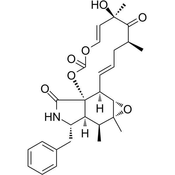
Cytochalasin E, an epoxide containing Aspergillus-derived fungal metabolite, inhibits angiogenesis and tumor growth. Cytochalasin E is a potent actin depolymerization agent, and it binds and caps the barbed end of actin filaments to prevent actin elongation.
For research use only. We do not sell to patients.
- Purity : 99.90%
- CAS No.: 36011-19-5
- Formula: C28H33NO7
- Molecular Weight:495.56
-
Storage:Powder -20°C, 3 years ; In solvent -80°C, 6 months , -20°C, 1 month

To place orders, for customer services and technical support, please contact: MedChemExpress USA
Tel: 609-228-6898 E-mail: [email protected] [email protected]


-
 Biological Activity
-
Chemical Information
-
Solvent & Solubility
- Protocols
- Purity & Documentation
- References
-
 Help & FAQs





Biological Activity
Description
Cellular Effect
|
Cell Line

|
Type | Value | Description | References |
|---|---|---|---|---|
| A549 | IC50 |
0.0062 μM
Compound: 4
|
Cytotoxicity against human A549 cells after 72 hrs by MTT method
Cytotoxicity against human A549 cells after 72 hrs by MTT method
|
[PMID: 16792402] |
| HUVEC | IC50 |
0.0114 μM
Compound: 7
|
Antiangiogenesis activity in HUVEC assessed as inhibition of VEGF-induced cell proliferation after 72 hrs by MTT assay
Antiangiogenesis activity in HUVEC assessed as inhibition of VEGF-induced cell proliferation after 72 hrs by MTT assay
|
[PMID: 21718054] |
| P388 | IC50 |
0.093 μM
Compound: 4
|
Cytotoxicity against mouse P388 cells after 72 hrs by MTT method
Cytotoxicity against mouse P388 cells after 72 hrs by MTT method
|
[PMID: 16792402] |
In Vitro
Cytochalasin E prominently inhibits the growth of A549 cells in a dose-dependent manner[3].
Cytochalasin E could induce the up-regulation of autophagy-related protein (LC3-II) and SQSTM1/p62[3].
MedChemExpress (MCE) has not independently confirmed the accuracy of these methods. They are for reference only.
- Human pluripotent stem cell endothelial-cell differentiation
- Vascular/Branching Fractal Analysis
- Endothelial Tube Formation Assay
- Patient-Derived Xenograft (PDX)
- Patient-Derived Orthotopic Xenograft (PDOX)
- Subcutaneous Cell-Line-Derived Xenograft
- Endothelial Cell Migration/Angiogenic Sprouting Assay
- Orthotopic Cell-Line Xenograft


Chemical Information
-
CAS No. 36011-19-5
-
Appearance Solid
-
Molecular Weight 495.56
-
Formula C28H33NO7
-
Color White to off-white
-
SMILES
O=C1[C@]2(O3)[C@]([C@@H]([C@](O4)(C)[C@@H]4[C@]2([H])/C=C/C[C@@H](C([C@](C)(O)/C=C/OC3=O)=O)C)C)([H])[C@H](CC5=CC=CC=C5)N1
-
Structure Classification
-
Initial Source
the fungus R. necatrix
-
Shipping
Room temperature in continental US; may vary elsewhere.
-
Storage
Powder -20°C 3 years In solvent -80°C 6 months -20°C 1 month


Solvent & Solubility
In Vitro:
DMSO : 50 mg/mL (100.90 mM; Need ultrasonic and warming; Hygroscopic DMSO has a significant impact on the solubility of product, please use newly opened DMSO)
Please refer to the solubility information to select the appropriate solvent. Once prepared, please aliquot and store the solution to prevent product inactivation from repeated freeze-thaw cycles.
Storage method and period of stock solution: -80°C, 6 months; -20°C, 1 month. When stored at -80°C, please use it within 6 months. When stored at -20°C, please use it within 1 month.
Please refer to the solubility information to select the appropriate solvent. Once prepared, please aliquot and store the solution to prevent product inactivation from repeated freeze-thaw cycles.
Storage method and period of stock solution: -80°C, 6 months; -20°C, 1 month. When stored at -80°C, please use it within 6 months. When stored at -20°C, please use it within 1 month.
Concentration (start) × Volume (start) = Concentration (final) × Volume (final)


Purity & Documentation
-
Data Sheet (276 KB)
-
SDS (477 KB)
- English - EN (477 KB)
- Français - FR (477 KB)
- Deutsch - DE (477 KB)
- Norwegian - NO (477 KB)
- Español - ES (477 KB)
- Swedish - SV (477 KB)
- Italian - IT (477 KB)
- Korean - KR (477 KB)
- Portuguese - PT (477 KB)
-
Handling Instructions (2659 KB)
References
[1]. Udagawa T, et al. Cytochalasin E, an epoxide containing Aspergillus-derived fungal metabolite, inhibits angiogenesisand tumor growth. J Pharmacol Exp Ther. 2000 Aug;294(2):421-7. [Content Brief]
[2]. Lu QY, et al. Green tea extract modulates actin remodeling via Rho activity in an in vitro multistep carcinogenicmodel. Clin Cancer Res. 2005 Feb 15;11(4):1675-83. [Content Brief]
[3]. Takanezawa Y, et al. Variation in the activity of distinct cytochalasins as autophagy inhibitiors in human lung A549 cells. Biochem Biophys Res Commun. 2017 Dec 16;494(3-4):641-647. [Content Brief]


Complete Stock Solution Preparation Table
Please refer to the solubility information to select the appropriate solvent. Once prepared, please aliquot and store the solution to prevent product inactivation from repeated freeze-thaw cycles.
Storage method and period of stock solution: -80°C, 6 months; -20°C, 1 month. When stored at -80°C, please use it within 6 months. When stored at -20°C, please use it within 1 month.
| Optional Solvent | Concentration Solvent Mass | 1 mg | 5 mg | 10 mg | 25 mg |
|---|---|---|---|---|---|
| DMSO | 1 mM | 2.0179 mL | 10.0896 mL | 20.1792 mL | 50.4480 mL |
| 5 mM | 0.4036 mL | 2.0179 mL | 4.0358 mL | 10.0896 mL | |
| 10 mM | 0.2018 mL | 1.0090 mL | 2.0179 mL | 5.0448 mL | |
| 15 mM | 0.1345 mL | 0.6726 mL | 1.3453 mL | 3.3632 mL | |
| 20 mM | 0.1009 mL | 0.5045 mL | 1.0090 mL | 2.5224 mL | |
| 25 mM | 0.0807 mL | 0.4036 mL | 0.8072 mL | 2.0179 mL | |
| 30 mM | 0.0673 mL | 0.3363 mL | 0.6726 mL | 1.6816 mL | |
| 40 mM | 0.0504 mL | 0.2522 mL | 0.5045 mL | 1.2612 mL | |
| 50 mM | 0.0404 mL | 0.2018 mL | 0.4036 mL | 1.0090 mL | |
| 60 mM | 0.0336 mL | 0.1682 mL | 0.3363 mL | 0.8408 mL | |
| 80 mM | 0.0252 mL | 0.1261 mL | 0.2522 mL | 0.6306 mL | |
| 100 mM | 0.0202 mL | 0.1009 mL | 0.2018 mL | 0.5045 mL |


Cytochalasin E Related Classifications
HY-N6772 Related Classifications
 Powered by Bioz