



Conantokin-T
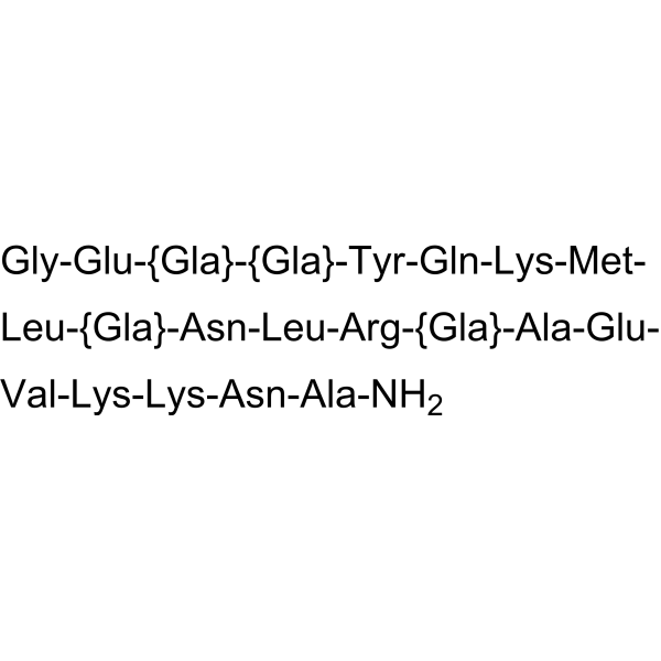
Conantokin-T is a γ-carboxyglutamate-containing, N-methyl-D-aspartate (NMDA) antagonist peptidewith an IC50 value of 2 μM. Conantokin-T inhibits NMDA receptor-mediated calcium influx in central nervous system neurons. Conantokin-T can be purified from the venom of the fish-hunting cone snail, Conus tulipa.
商品は「研究用試薬」です。人や動物の医療用・臨床診断用・食品用の製品ではありません。
研究用途以外に使用した場合、当社は一切の責任を負いかねます。
- CAS 番号: 127476-26-0
- 分子式: C110H175N31O45S
- 分子量:2683.81
-
保管条件:
Please store the product under the recommended conditions in the Certificate of Analysis.

To place orders, for customer services and technical support, please contact: MedChemExpress USA
Tel: 609-228-6898 E-mail: [email protected] [email protected]


-
 生物活性
生物活性
-
化学情報
- プロトコル
- 純度とドキュメンテーション
- 参考文献
-
 Help & FAQs


-
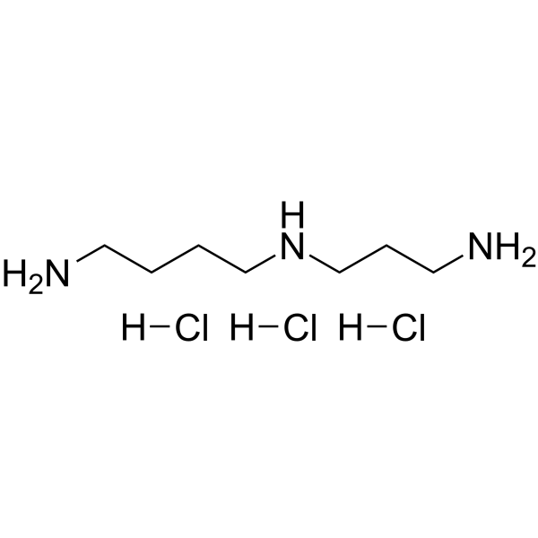
HY-B1776A
Spermidine hydrochlorideAntioxidant Agent -
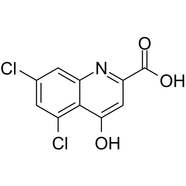
HY-100834
5,7-Dichlorokynurenic acidNMDA Receptor Antagonist -
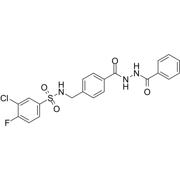
HY-13457
TCN 201NMDA Receptor Antagonist -
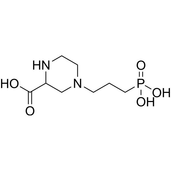
HY-100797
(RS)-CPPNMDA Antagonist -

HY-B1776AR
Spermidine hydrochloride (Standard)Antioxidant Agent -
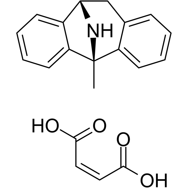
HY-15084A
(-)-Dizocilpine maleateNMDA Receptor Antagonist -
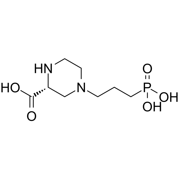
HY-100814
(R)-CPPNMDA Receptor Inhibitor -
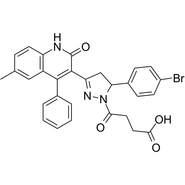
HY-107711
DQP-1105GluN1/GluN2C Antagonist -
HY-30006
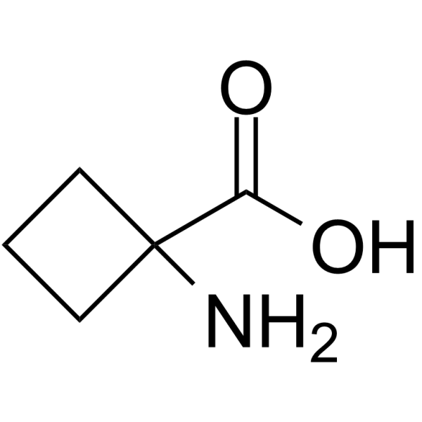
1-Aminocyclobutanecarboxylic acidCyclobutane-type Amino Acid -

HY-15086
SelfotelNMDA Antagonist -
HY-100920
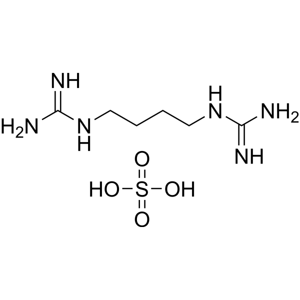
Arcaine sulfateNMDA Receptor Antagonist -
HY-101178
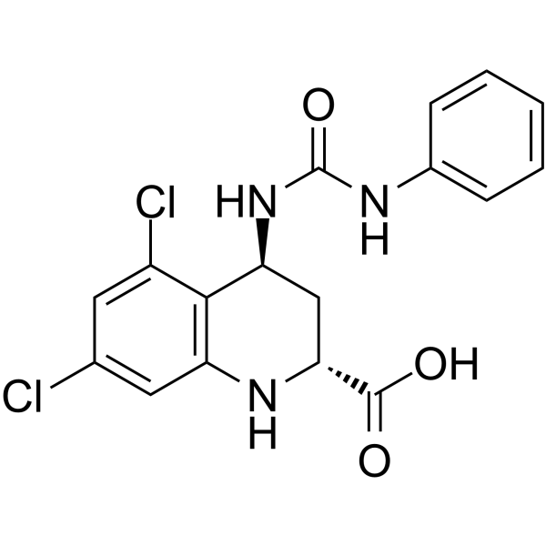
L-689560NMDA Antagonist
iGluR アイソフォーム固有の製品をすべて表示
More

生物活性
製品説明
IC50 & Target
IC50: 2 μM (NMDA)[1]
体外実験
Conantokin-T produces a partial antagonism of NMDA receptor function with an IC50 value of 2 μM[1].
MedChemExpress (MCE) has not independently confirmed the accuracy of these methods. They are for reference only. Further protocols information, click here.
体内実験
MedChemExpress (MCE) has not independently confirmed the accuracy of these methods. They are for reference only.


化学情報
-
CAS 番号 127476-26-0
-
分子量 2683.81
-
分子式 C110H175N31O45S
-
シーケンスの短縮
GE-{Gla}-{Gla}-YQKML-{Gla}-NLR-{Gla}-AEVKKNA-NH2
-
輸送条件
Room temperature in continental US; may vary elsewhere.
-
保管条件
Please store the product under the recommended conditions in the Certificate of Analysis.

プロトコル
-
Cell Cytotoxicity Assay
Cytotoxicity assays are usually based on the assessment of cell membrane damage, which can also be indirectly detected by measuring cell viability. Detection methods include MTT assay, CKK-8 assay, LDH assay and ATP assay, etc.
-
Protocol for Fluorescence In Situ Hybridization (FISH)
Fluorescence in situ hybridization detects specific DNA or RNA sequences inside fixed cells or tissue sections by hybridizing fluorescently labeled nucleic-acid probes to complementary target sequences, allowing the target’s copy number, chromosomal position, spatial distribution, or transcript abundance to be visualized microscopically. DNA-FISH detects genomic loci, chromosomal gains/losses, amplifications, deletions, and rearrangements, while RNA-FISH detects RNA molecules or transcript localization; in cancer cells, mouse tumors, neurons, organoids, macrophages, or drug-screening samples, the readout is fluorescent puncta, fusion/split signals, or localized RNA signal interpreted relative to validated controls.
-
Ca2+ Staining Technique
Ca2+ staining is an experimental technique that utilizes specific fluorescent probes (such as Fluo-4 AM, Fura-2, etc.) to qualitatively or quantitatively detect dynamic changes in intracellular Ca2+ concentrations; this is achieved by monitoring the changes in fluorescent signals generated when these probes bind to free intracellular calcium ions. The underlying principle relies primarily on the presence of chelating groups within the probe's molecular structure that possess high affinity for calcium ions.


純度とドキュメンテーション
参考文献

Calculators
濃度 (開始) × 体積 (開始) = 濃度 (終了) × 体積 (終了)


Conantokin-T 関連分類
HY-P1287 関連分類
 Powered by Bioz