



Forigerimod TFA

Based on 1 Customer Validation
Forigerimod TFA (IPP-201101 TFA) is a CD4 T-cell modulator. Forigerimod TFA is a 21-amino-acid fragment of U1 small nuclear ribonucleoprotein 70 kDa that is phosphorylated at Ser140. Forigerimod TFA can potently inhibit autophagy. Forigerimod can be used for the research of autoimmune disorders, such as systemic lupus erythematosus (SLE) .
商品は「研究用試薬」です。人や動物の医療用・臨床診断用・食品用の製品ではありません。
研究用途以外に使用した場合、当社は一切の責任を負いかねます。
- 純度 : 99.88%
- 分子式: C117H181N34O32PS.xC2HF3O2
-
保管条件:
Sealed storage, away from moisture and light, under nitrogen.
Powder -80°C, 2 years , -20°C, 1 year* In solvent : -80°C, 6 months; -20°C, 1 month (sealed storage, away from moisture and light, under nitrogen)

To place orders, for customer services and technical support, please contact: MedChemExpress USA
Tel: 609-228-6898 E-mail: [email protected] [email protected]


-
 生物活性
生物活性
-
化学情報
-
溶剤 & 溶解度
- プロトコル
- 純度とドキュメンテーション
- 参考文献
-
 Help & FAQs




生物活性
製品説明
体内実験
MedChemExpress (MCE) has not independently confirmed the accuracy of these methods. They are for reference only.
-
Animal Model:MRL/lpr mice[4]
-
Dosage:100 μg in 100 μL saline per mouse
-
Administration:Intravenous administration, single
-
Result:Corrected the cell content and distribution in MRL/lpr salivary glands.
Corrected the defective of autophagy processes in SGs of MRL/lpr mice.


化学情報
-
性状 Solid
-
分子式 C117H181N34O32PS.xC2HF3O2
-
Color White to off-white
-
別名
IPP-201101 TFA; Rigerimod TFA; Peptide P140 TFA
-
シーケンスの短縮
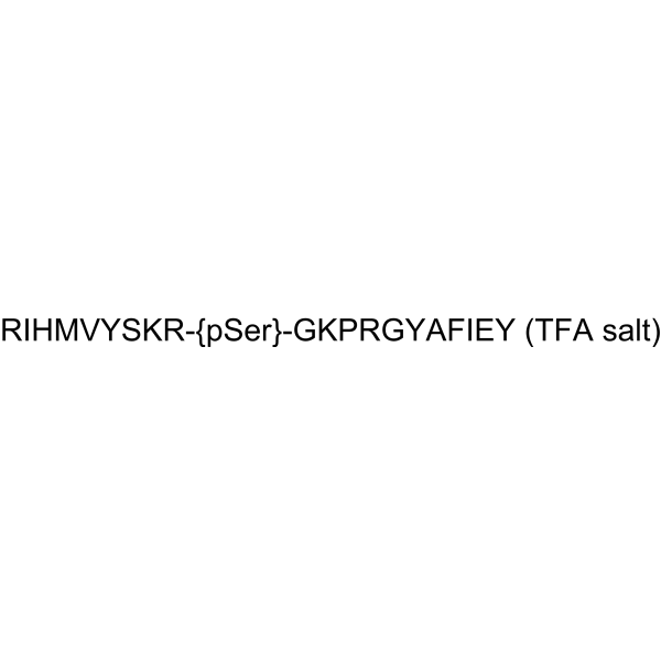
RIHMVYSKR-{pSer}-GKPRGYAFIEY (TFA)
-
輸送条件
Room temperature in continental US; may vary elsewhere.
-
保管条件
Sealed storage, away from moisture and light, under nitrogen
Powder -80°C 2 years -20°C 1 year * In solvent : -80°C, 6 months; -20°C, 1 month (sealed storage, away from moisture and light, under nitrogen)


溶剤 & 溶解度
体外:
DMSO : 100 mg/mL (Need ultrasonic; Hygroscopic DMSO has a significant impact on the solubility of product, please use newly opened DMSO)
H2O : 100 mg/mL (Need ultrasonic)


純度とドキュメンテーション
-
データシート (271 KB)
-
SDS (252 KB)
- English - EN (252 KB)
- Français - FR (252 KB)
- Deutsch - DE (252 KB)
- Norwegian - NO (252 KB)
- Español - ES (252 KB)
- Swedish - SV (252 KB)
- Italian - IT (252 KB)
- Korean - KR (252 KB)
- Portuguese - PT (252 KB)
-
取扱説明書 (2659 KB)
参考文献
[1]. Aikaterini Thanou, et al. Treatment of systemic lupus erythematosus: new therapeutic avenues and blind alleys. [Content Brief]
[2]. Sean Robinson, et al. Potential for Antigen-Specific Tolerizing Immunotherapy in Systematic Lupus Erythematosus. Front Immunol. 2021 Jul 16;12:654701. [Content Brief]
[3]. Robert Zimmer, et al. Lupuzor/P140 peptide in patients with systemic lupus erythematosus: a randomised, double-blind, placebo-controlled phase IIb clinical trial. Ann Rheum Dis. 2013 Nov;72(11):1830-5. [Content Brief]
[4]. Baihui Li, et al. Rescue of autophagy and lysosome defects in salivary glands of MRL/lpr mice by a therapeutic phosphopeptide. J Autoimmun. 2018 Jun;90:132-145. [Content Brief]

Calculators
濃度 (開始) × 体積 (開始) = 濃度 (終了) × 体積 (終了)


Forigerimod TFA 関連分類
HY-105037A 関連分類
 Powered by Bioz