



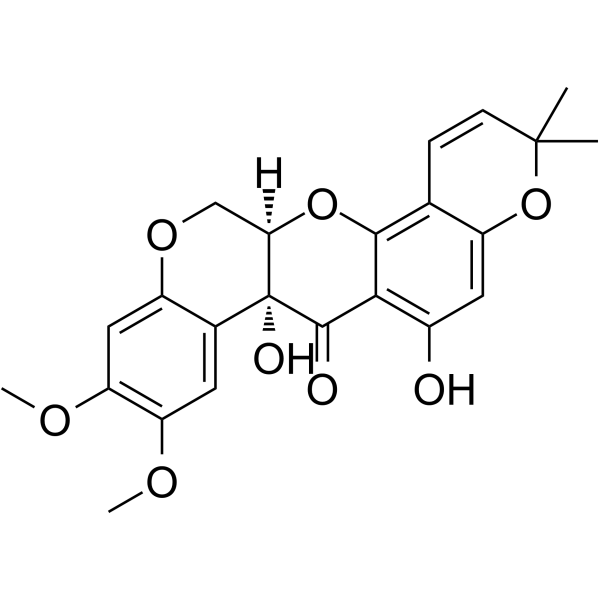
11-Hydroxytephrosin

Based on 1 Customer Validation
11-Hydroxytephrosin is a potent NF-κB inhibitor with an IC50 of 0.19 μM or 4.21 μM. 11-Hydroxytephrosin is isolated from A. fruticosa. 11-Hydroxytephrosin inhibits the activation of NF-κB. 11-Hydroxytephrosin can be used in research related to cervical cancer and inflammatory diseases.
연구목적의 판매만을 진행합니다. 환자를 대상으로 한 판매는 하지 않습니다.
- CAS No.: 72458-85-6
- 화학식: C23H22O8
- 분자량:426.42
-
보관:
-20°C, protect from light
* In solvent : -80°C, 6 months; -20°C, 1 month (protect from light)

To place orders, for customer services and technical support, please contact: MedChemExpress USA
Tel: 609-228-6898 E-mail: [email protected] [email protected]


-
 Biological Activity
-
Chemical Information
- Protocol
- 순도&문서
- References
-
 Help & FAQs




Biological Activity
제품 설명
Cellular Effect
|
Cell Line

|
Type | Value | Description | References |
|---|---|---|---|---|
| HeLa | IC50 |
0.19 μM
Compound: 2
|
Inhibition of TNF-alpha-induced NF-kappaB activation in human HeLa cells by SEAP reporter gene assay
Inhibition of TNF-alpha-induced NF-kappaB activation in human HeLa cells by SEAP reporter gene assay
|
[PMID: 18841906] |
| RAW264.7 | IC50 |
4.21 μM
Compound: 2
|
Inhibition of LPS-induced NF-kappaB activation in mouse RAW264.7 cells by SEAP reporter gene assay
Inhibition of LPS-induced NF-kappaB activation in mouse RAW264.7 cells by SEAP reporter gene assay
|
[PMID: 18841906] |
In Vitro
11-Hydroxytephrosin (24 h) potently inhibits TNF-α-induced NF-κB activation in HeLa cells with an IC50 of 0.19 μM, and shows no significant cytotoxicity at concentrations up to 30 μM[1].
11-Hydroxytephrosin (24 h) inhibits LPS-induced NF-κB activation in RAW264.7 cells with an IC50 of 4.21 μM, and shows no significant cytotoxicity at concentrations up to 30 μM[1].
MedChemExpress (MCE) has not independently confirmed the accuracy of these methods. They are for reference only. Further protocols information, click here.


Chemical Information
-
CAS No. 72458-85-6
-
Appearance Solid
-
분자량 426.42
-
화학식 C23H22O8
-
Color White to off-white
-
SMILES
O=C1[C@]2(O)[C@](COC3=CC(OC)=C(OC)C=C32)([H])OC4=C5C=CC(C)(C)OC5=CC(O)=C14
-
선적
Room temperature in continental US; may vary elsewhere.
-
보관
-20°C, protect from light
* In solvent : -80°C, 6 months; -20°C, 1 month (protect from light)

Protocol
-
Cell Cytotoxicity Assay
Cytotoxicity assays are usually based on the assessment of cell membrane damage, which can also be indirectly detected by measuring cell viability. Detection methods include MTT assay, CKK-8 assay, LDH assay and ATP assay, etc.
-
Research Protocol for Inflammation-related Diseases
The NLRP3 inflammasome is a cytosolic innate immune signaling platform that integrates priming signals and danger-signal activation to promote caspase-1 activation, maturation of IL-1β and IL-18, and gasdermin D-mediated pyroptotic cell death. The core experimental logic is to determine whether inflammatory disease phenotypes are driven by increased NLRP3 expression, ASC-containing inflammasome assembly, caspase-1 cleavage, GSDMD cleavage, and extracellular release of IL-1β/IL-18 rather than by nonspecific cell injury alone. The pathway is strongly linked to inflammation-related disease phenotypes because monosodium urate crystals activate NALP3/NLRP3 inflammasome signaling in gout-like crystal inflammation, cholesterol crystals activate NLRP3 inflammasomes in atherogenesis models, and DSS-induced intestinal inflammation has been reported to involve NLRP3 inflammasome activity. However, experimental colitis studies also show context-dependent protective effects of NLRP3 inflammasome co


순도&문서
-
Data Sheet (271 KB)
-
SDS (252 KB)
- English - EN (252 KB)
- Français - FR (252 KB)
- Deutsch - DE (252 KB)
- Norwegian - NO (252 KB)
- Español - ES (252 KB)
- Swedish - SV (252 KB)
- Italian - IT (252 KB)
- Korean - KR (252 KB)
- Portuguese - PT (252 KB)
-
Handling Instructions (2659 KB)
References

Calculators
Concentration (start) × Volume (start) = Concentration (final) × Volume (final)


11-Hydroxytephrosin Related Classifications
HY-N1022 Related Classifications
 Powered by Bioz