



Milatuzumab
Based on 7 publication(s) in Google Scholar
Milatuzumab (hLL1; MEDI-115) is a humanized anti-CD74 monoclonal antibody. CD74, a integral membrane protein, is associated with the promotion of B-cell growth and survival. Milatuzumab causes free radical oxygen generation, and loss of mitochondrial membrane potential. Milatuzumaba also decreases CD20/CD74 aggregates and cell adhesion, to lead to cell death.
연구목적의 판매만을 진행합니다. 환자를 대상으로 한 판매는 하지 않습니다.
- Purity : 99.39%
- CAS No.: 899796-83-9
- 분자량:145.66 kDa
-
보관:
Please store the product under the recommended conditions in the Certificate of Analysis.


To place orders, for customer services and technical support, please contact: MedChemExpress USA
Tel: 609-228-6898 E-mail: [email protected] [email protected]


-
 Biological Activity
-
Chemical Information
- Protocol
- 순도&문서
- References
-
 Help & FAQs


-

HY-P99001
Human IgG1 kappa, Isotype ControlIgG1κ Isotype Control -

HY-P990728
BezetabartAnti-CD74 Antibody -
HY-16141
CilengitideIntegrin Inhibitor -

HY-15141
StaurosporinePKC/PKA Inhibitor -

HY-10071
Y-27632ROCK Inhibitor -

HY-P99168
AnifrolumabIFN Receptor Antagonist -

HY-P99041
PanitumumabAnti-EGFR Monoclonal Antibody -

HY-P99262
ImalumabMIF Antibody -
HY-12885A
2’3’-c-di-AM(PS)2 (Rp,Rp) disodium saltSTING Agonist -

HY-P9965
ElotuzumabSLAMF7 Inhibitor -

HY-P99289
EpratuzumabAnti-CD22 Antibody -

HY-P990091
RiliprubartActivated C1s Inhibitor
Publications Citing Use of MedChemExpress (MCE) Milatuzumab
More- Sci Transl Med. 2026 May 27;18(851):eadv8372. [Abstract]
- Adv Sci (Weinh). 2025 Sep;12(35):e02838. [Abstract]
- J Immunother Cancer. 2026 May 14;14(5):e013255. [Abstract]
- J Immunother Cancer. 2024 Aug 6;12(8):e009024. [Abstract]
- Cell Oncol (Dordr). 2026 Apr 22;49(3):81. [Abstract]
- bioRxiv. 2026 Jan 7.
- bioRxiv. 2023 Nov 13.
 Customer Validation & Images

-
Cell Imaging/Staining
-
WB
-
IF
-
Flow Cytometry
-
Flow Cytometry


Biological Activity
제품 설명
Isotype
Human IgG1 kappa
Recommend Isotype Controls
Species Reactivity
Human
IC50 & Target
CD74
In Vitro
Milatuzumaba (5 μg/mL; 8-48 h) enhances cell death in MCL cell lines and primary patient tumor cells[1].
Milatuzumaba (5 μg/mL; 0.5-2 h) mediates the cytotoxicity partially depending on generation of ROS and loss of mitochondrial transmembrane potential in Jeko, Mino, and SP-53 cells[1].
Milatuzumaba (5 μg/mL; 4 h) inhibits NF-κB pathway and induces cell apoptosis with independent of caspase cleavage, Bcl-2 family member dysregulation, or induction of autophagy[1].
MedChemExpress (MCE) has not independently confirmed the accuracy of these methods. They are for reference only. Further protocols information, click here.
-
Cell Line:Jeko and Mino cells
-
Concentration:5 μg/mL
-
Incubation Time:4 hours
-
Result:Insignificant down-regulation of antiapoptotic proteins, such as Bax, Bcl-2, Bcl-xL, and Mcl-1.
-
Cell Line:MCL cell lines and primarypatient tumor cells
-
Concentration:5 μg/mL
-
Incubation Time:8, 24, and 48 hours
-
Result:Resulted in cell death of Jeko, Mino, SP-53, Rec-1, HBL-2, and Granta cells.
-
Cell Line:Jeko, Mino, and SP-53 cells
-
Concentration:5 μg/mL; with or without 10 mM N-acetylcysteine (HY-B0215) for 1.5 h
-
Incubation Time:0.5, 1, 1.5, and 2 hours
-
Result:Increased ROS generation as early as 0.5 hours, while peaking at 1 to 1.5 hours and reducing at 2 hours.Therefore, it resulted cell death, but reserved by nonspecific ROS scavenger.
In Vivo
MedChemExpress (MCE) has not independently confirmed the accuracy of these methods. They are for reference only.
-
Animal Model:Jeko mouse model[1]
-
Dosage:15 mg/kg/day; with or without 15 mg/kg Rituximab
-
Administration:Intraperitoneal injection; once every 3 days, starting at day 15 after engraftment
-
Result:Resulted the mean survival for the combination treated group of 44.5 days, compared with 33.5 days for Milatuzumaba treated, 28 days for control.
Clinical Trial
| NCT Number | Sponsor | Condition | Start Date |
Phase

|
|---|---|---|---|---|
| NCT01329991 | Plexxikon| | 2011-05 | PHASE1 |
Gene ID
Accession
Conjugated
Unconjugated
Reconsititution
The product can be reconstituted/diluted with sterile PBS or saline.
Format
-
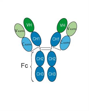
Human IgG1 kappa
신청
ELISA, FACS, Functional assay
Verified Bioactivity
-
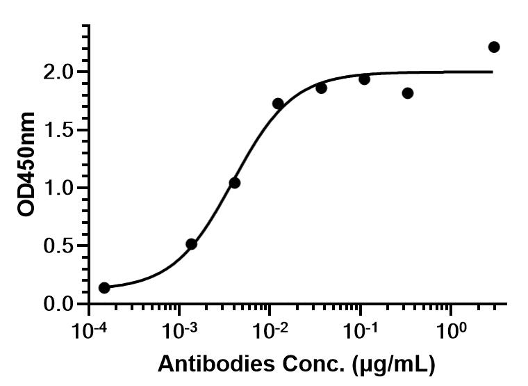 Immobilized hu-CD74-ECD-His can bind Milatuzumab. The EC50 for this effect is 3.941 ng/mL.
 -
 Flow Cytometry analysis of Raji cells labelling CD74 (red) with Milatuzumab (anti-CD74) (HY-P99731). Goat Anti-Human IgG (Alexa Fluor 488) (HY-P83776) at a dilution of 1/1000 was used as the secondary antibody. Blue-Human IgG1 kappa (HY-P99001). Black-Unlabelled control, cells without incubation with primary antibody.


Chemical Information
-
CAS No. 899796-83-9
-
Appearance Liquid
-
분자량 145.66 kDa
-
Color Colorless to light yellow
-
SMILES
[Milatuzumab]
-
Synonyms
hLL1; MEDI-115
-
선적
Shipping with dry ice.
-
Formulation
Please refer to the lot-specific COA for specific buffer information.
-
보관
Please store the product under the recommended conditions in the Certificate of Analysis.


Publications (7)
-
Journal Impact Factor
-
Most Recent
-
Sci Transl Med
CCR5 and CD74 are potential therapeutic targets for necroinflammation in preclinical cholesterol crystal embolism. [Abstract]2026 May 27;18(851):eadv8372. PMID: 42202044 -
Adv Sci (Weinh)
CD74 Blockade Disrupts Endothelial Migrasome Signaling to Prevent Inflammatory Macrophage Differentiation and Inhibit Atherosclerotic Progression. [Abstract]2025 Sep;12(35):e02838. PMID: 40548932
Milatuzumab purchased from MedChemExpress. Usage Cited in: Adv Sci (Weinh). 2025 Sep;12(35):e02838. [Abstract]
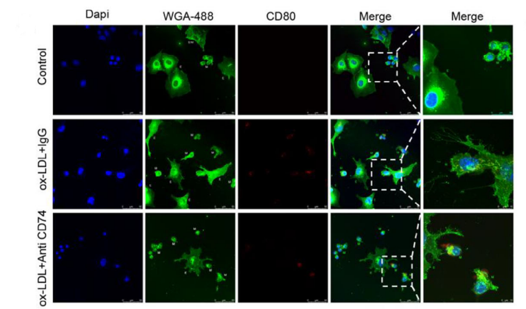
HCAEC cells were treated with ox-LDL and anti-CD74 antibody (Milatuzumab: 15 mg/kg) for 12 hours, stained with 1 µg/ml WGA-Alexa 488 and CD80, and observed under a confocal microscope.
Milatuzumab purchased from MedChemExpress. Usage Cited in: Adv Sci (Weinh). 2025 Sep;12(35):e02838. [Abstract]
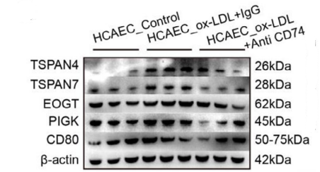
Western blot analysis of migratory bodies and M1 macrophage-related markers in HCAEC cells treated with ox-LDL and anti-CD74 antibody (Milatuzumab: 15 mg/kg) was performed.
Milatuzumab purchased from MedChemExpress. Usage Cited in: Adv Sci (Weinh). 2025 Sep;12(35):e02838. [Abstract]
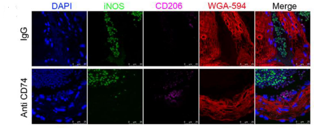
Immunofluorescence images showing M1 macrophages and migrasomes in Control, AS, and AS_Anti CD74 (Milatuzumab: 15 mg/kg) groups. Red: WGA‐594 (labeling migrasomes); Green: iNOS (labeling M1 macrophages); Magenta: CD206 (labeling M2 macrophages); Blue: DAPI (nuclei).
-
J Immunother Cancer
Targeting CD74 may mitigate immune-escape features and enhance BCMA CAR-T activity in preclinical models of relapsed/refractory multiple myeloma. [Abstract]2026 May 14;14(5):e013255. PMID: 42134899 -
J Immunother Cancer
Targeting tumor-associated macrophage-derived CD74 improves efficacy of neoadjuvant chemotherapy in combination with PD-1 blockade for cervical cancer. [Abstract]2024 Aug 6;12(8):e009024. PMID: 39107132
Milatuzumab purchased from MedChemExpress. Usage Cited in: J Immunother Cancer. 2024 Aug 6;12(8):e009024. [Abstract]
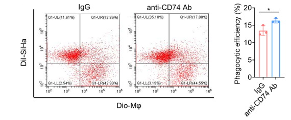
Macrophages derived from THP-1 cells treated with anti-CD74 (Milatuzumab) were co-cultured. Phagocytic efficiency was quantified as the percentage of dual-fluorescence-positive macrophages (upper right quadrant).
Milatuzumab purchased from MedChemExpress. Usage Cited in: J Immunother Cancer. 2024 Aug 6;12(8):e009024. [Abstract]
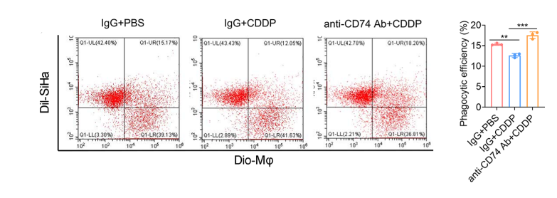
Flow cytometry was used to detect differences in phagocytic function among the control group, cisplatin group, and combination therapy group with anti-CD74 Ab (5 µg/mL). THP-1-derived macrophages were co-cultured with SiHa cells.
-
Cell Oncol (Dordr)
The MIF-CD74 axis drives colorectal cancer via glycolytic reprogramming and is targeted by a novel small-molecule inhibitor. [Abstract]2026 Apr 22;49(3):81. PMID: 42018204 -
-

Protocol
-
Mitochondrial membrane-potential fluorescent assay
Mitochondrial membrane potential fluorescent assays estimate ΔΨm in living cells using lipophilic cationic dyes such as TMRM, TMRE, rhodamine 123, and JC-1, which accumulate in mitochondria according to membrane polarization; loss of signal after FCCP or CCCP treatment is interpreted as mitochondrial depolarization. TMRM/TMRE and rhodamine 123 are commonly used for semi-quantitative live-cell microscopy or flow cytometry, while JC-1 can report a shift from red aggregate fluorescence to green monomer fluorescence during depolarization; interpretation requires controls because dye concentration, quenching mode, cell type, dye efflux, and mitochondrial mass can affect fluorescence independently of ΔΨm.
-
Mammalian live/dead viability and cytotoxicity staining
Live/dead viability and cytotoxicity staining assays are based on the simultaneous detection of intracellular esterase activity in metabolically active (viable) cells and membrane integrity loss in non-viable cells. In commonly used dual-staining approaches, membrane-permeant fluorogenic substrates are converted by intracellular esterases into fluorescent products in live cells, while impermeant DNA-binding dyes selectively enter cells with compromised plasma membranes and label nucleic acids in dead or dying cells, enabling discrimination between viable and non-viable populations by fluorescence microscopy or flow cytometry.
-
Somatic Cell Culture
A method of simulating the in vivo environment in vitro to maintain the cell growth, differentation and main functions.
-
CCK-8/WST-8 Cell Proliferation Assay
The CCK-8/WST-8 assay is based on the reduction of the water-soluble tetrazolium salt WST-8 to a water-soluble formazan product by cellular dehydrogenases in metabolically active cells, where the generated formazan amount is proportional to the number of living cells and is quantified by measuring absorbance in the visible range, providing a colorimetric readout for cell viability and proliferation assessment. This class of tetrazolium-based assays improves upon earlier MTT-based systems by producing a water-soluble formazan, eliminating the need for organic solubilization steps and enabling direct spectrophotometric measurement in culture medium.
-
Cell Counting-Based Growth Curve Assay
Cell counting-based growth curve assays quantify cell proliferation by directly measuring changes in viable cell number over time using manual or automated counting methods such as hemocytometer-based counting or instrument-assisted cell enumeration, enabling construction of growth curves that reflect population expansion dynamics in response to culture conditions. A widely used approach is trypan blue exclusion with hemocytometer counting, where membrane-compromised (non-viable) cells take up the dye, allowing discrimination between viable and non-viable cells while simultaneously enabling total cell number quantification. Repeated sampling across time points allows estimation of proliferation rate, growth phases, and comparative growth kinetics between experimental conditions.
-
Cell Viability Determination by MTT Colorimetric Assay
The following protocol uses the MTT colorimetric assay as a classic literature-established method for assessing cell viability/metabolic activity in cultured mammalian cells. MTT[3-(4,5-dimethylthiazol-2-yl)-2,5-diphenyltetrazolium bromide] is reduced by metabolically active cells to a colored formazan product; the amount of formazan is quantified spectrophotometrically and provides an indirect measure of metabolically active viable cells. Importantly, MTT reduction reflects cellular oxidoreductase/metabolic activity rather than an absolute direct count of living cells, so changes in cellular metabolism can alter the signal independently of cell number.
-
Cell Cytotoxicity Assay
Cytotoxicity assays are usually based on the assessment of cell membrane damage, which can also be indirectly detected by measuring cell viability. Detection methods include MTT assay, CKK-8 assay, LDH assay and ATP assay, etc.
-
Membrane Protein Extraction Using Detergents and Chaotropes
Membrane protein extraction with detergents and chaotropes solubilizes lipid-bilayer-associated proteins by disrupting protein-lipid and protein-protein interactions while maintaining proteins in a soluble state for downstream electrophoresis, purification, or mass spectrometry. Chaotropes such as urea and thiourea improve solubilization of difficult proteins, while nonionic and zwitterionic detergents such as CHAPS, ASB-14, SB 3-10, MEGA-10, dodecyl maltoside, and Triton X-100 differ in extraction efficiency depending on sample type and membrane protein properties.
-
Protocol for Cell Counting and Cell Density Analysis
Cell counting and cell-density analysis estimate the number of cells in a known volume or field area. Manual hemocytometer counting uses a chamber of defined geometry to convert counted cells into cells/mL, while automated counters and image-analysis workflows detect cell objects from optical, brightfield, fluorescence, impedance, or digital-image features. Trypan blue viability counting is based on dye exclusion: viable cells with intact membranes exclude dye, while non-viable cells with compromised membranes stain blue. The readout is total cell density, viable-cell density, dead-cell density, and percent viability. Cell density can also be estimated from microscopy images by counting objects per image area, from flow cytometry using calibrated volume or reference particles, or from in situ microscopy in bioreactors after calibration against reference methods such as hemocytometer or flow cytometry.
-
Fluorescent plasma-membrane potential dye assay
Fluorescent plasma-membrane potential dye assays measure changes in cell membrane potential using voltage-sensitive dyes whose fluorescence changes when cells depolarize or hyperpolarize. Anionic bis-oxonol dyes such as DiBAC4(3) enter depolarized cells more readily and show increased fluorescence after intracellular binding, while hyperpolarization reduces dye accumulation and fluorescence. FMP/FLIPR membrane-potential dyes are used for faster, homogeneous microplate assays of ion-channel or receptor-mediated membrane-potential changes.
-
Mitochondrial membrane-potential and mitochondrial mass staining
Mitochondrial membrane potential staining measures the electrochemical polarization across the mitochondrial inner membrane in live cells using lipophilic cationic fluorescent probes; early rhodamine-based work showed that selective mitochondrial dye accumulation is lost when the mitochondrial transmembrane potential is dissipated. JC-1 reports mitochondrial polarization by shifting from green monomer fluorescence to red J-aggregate fluorescence as dye concentration increases within energized mitochondria; therefore, the red/green fluorescence ratio is used as a relative readout of mitochondrial membrane potential. TMRE or TMRM staining provides a single-channel relative readout because these cationic rhodamine esters accumulate in polarized mitochondria, and lower fluorescence indicates reduced mitochondrial polarization when acquisition and dye-loading conditions are controlled. Mitochondrial mass staining is commonly performed with MitoTracker Green FM or related MitoTracker dyes as
-
Apoptosis Solutions
Apoptosis is a regulated, generally non-lytic cell-death pathway that removes unwanted, damaged, infected, or abnormal cells through coordinated morphological changes, caspase activation, DNA fragmentation, and membrane remodeling. The intrinsic apoptosis pathway is controlled mainly by mitochondrial outer membrane permeabilization, BCL-2 family proteins, cytochrome c release, apoptosome formation, caspase-9 activation, and downstream executioner caspase-3/7 activation. The extrinsic apoptosis pathway is initiated by death receptors such as Fas, TNFR, and TRAIL receptors, which recruit adaptor proteins and activate caspase-8 before engaging executioner caspases or mitochondrial amplification through BID cleavage. Apoptosis is linked to many phenotypes, including cancer cell killing, tissue homeostasis, immune regulation, neurodegeneration, infection response, and treatment-induced cytotoxicity; unresolved questions include how apoptosis interacts with necroptosis, pyroptosis, ferroptos
-
MTT Cell Proliferation Assay
The MTT assay is a colorimetric endpoint assay for estimating viable cell number, cell growth, cytotoxicity, or cell activation in cultured mammalian cells. Living cells reduce the yellow tetrazolium salt MTT into purple/blue formazan, while dead cells do not generate the same signal; the resulting color can be quantified with a multiwell spectrophotometer. MTT reduction is commonly interpreted as a readout of metabolic activity that often correlates with viable cell number, but it should not be treated as a direct cell-counting method unless the assay is optimized for the cell type and experimental condition. Studies show that MTT reduction can involve mitochondrial and non-mitochondrial reducing systems, and formazan may accumulate in intracellular lipid droplets rather than simply marking mitochondria.


순도&문서
-
Data Sheet (262 KB)
-
SDS (251 KB)
- English - EN (251 KB)
- Français - FR (251 KB)
- Deutsch - DE (251 KB)
- Norwegian - NO (251 KB)
- Español - ES (251 KB)
- Swedish - SV (251 KB)
- Italian - IT (251 KB)
- Korean - KR (251 KB)
- Portuguese - PT (251 KB)
-
Inhibitory Antibodies User Guide (603 KB)
References

Calculators
Concentration (start) × Volume (start) = Concentration (final) × Volume (final)


Milatuzumab Related Classifications
HY-P99731 Related Classifications
 Powered by Bioz