



SB24011

Based on 1 Customer Validation
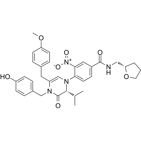
SB24011 is a STING modulator and a TRIM29-STING protein-protein interaction inhibitor. SB24011 blocks TRIM29-induced K48-linked specific ubiquitination by binding to STING, thereby upregulating intracellular STING protein levels. SB24011 enhances inflammatory cytokine expression and STING-mediated immune responses, and exhibits abscopal antitumor activity that promotes tumor regression and activates T cell infiltration. When combined with STING agonists or anti-PD1 antibodies, SB24011 synergistically enhances antitumor responses. SB24011 is suitable for research related to colon cancer and melanoma.
연구목적의 판매만을 진행합니다. 환자를 대상으로 한 판매는 하지 않습니다.
- Purity : 99.7%
- CAS No.: 1497415-41-4
- 화학식: C34H38N4O7
- 분자량:614.69
-
보관:Powder -20°C, 3 years ; In solvent -80°C, 6 months , -20°C, 1 month

To place orders, for customer services and technical support, please contact: MedChemExpress USA
Tel: 609-228-6898 E-mail: [email protected] [email protected]


-
 Biological Activity
-
Chemical Information
- Protocol
- 순도&문서
- References
-
 Help & FAQs




Biological Activity
제품 설명
In Vitro
SB24011 (5-20 μM; 24 h) shows no cytotoxicity in Raw264.7 murine macrophage-like cells[2].
SB24011 (5-20 μM; 18 h; 10 μM; 18, 24 h) upregulates cellular STING protein levels in A431 human skin squamous carcinoma cells[2].
SB24011 (20 μM; 3, 6 h) enhances cGAMP-induced activation of the STING downstream signaling pathway (phosphorylation of STING, TBK1, IRF3) in Raw264.7 murine macrophage-like cells[2].
SB24011 (20 μM; 3, 6 h) augments cGAMP-induced proinflammatory cytokine mRNA expression (ifnb, il6, il15) in Raw264.7 murine macrophage-like cells[2].
SB24011 (5-20 μM) dose-dependently augments cGAMP-induced proinflammatory cytokine mRNA expression (ifnb, il6) in Raw264.7 murine macrophage-like cells[2].
MedChemExpress (MCE) has not independently confirmed the accuracy of these methods. They are for reference only.
-
Cell Line:A431 human skin squamous carcinoma cells
-
Concentration:5-20 μM (18 h incubation); 10 μM (18, 24 h incubation)
-
Incubation Time:18 h (5,10,20 μM); 18 h, 24 h (10 μM)
-
Result:Increased cellular STING protein levels in a time- and dose-dependent manner.
Elevated the STING/actin band ratio by 2.1-, 2.9-, and 3.2-fold at 5, 10, and 20 μM, respectively, compared to vehicle.
-
Cell Line:Raw264.7 murine macrophage-like cells
-
Concentration:20 μM
-
Incubation Time:3 h, 6 h
-
Result:Enhanced cGAMP-induced phosphorylation of STING, TBK1, and IRF3 at both 3 h and 6 h post-treatment.
-
Cell Line:Raw264.7 murine macrophage-like cells
-
Concentration:20 μM
-
Incubation Time:3 h, 6 h
-
Result:Enhanced cGAMP-induced mRNA expression of ifnb (310% increase at 3 h, 30% increase at 6 h), il-6 (1100% increase at 3 h, 3500% increase at 6 h), il-15 (1800% increase at 3 h, 2700% increase at 6 h), and an additional cytokine (800% increase at 3 h, 1500% increase at 6 h) at 3 h compared to cGAMP alone.
Observed similar enhancements at 6 h.
Parmacokinetics
In Vivo
SB24011 (1-3 μg per head; intratumoral injection; first and third of four total injections; once every 2 days over 8 days) dose-dependently potentiates cGAMP-mediated anticancer immunity in wild-type C57BL/6J mice with B16F10 melanoma, reducing tumor growth and enhancing effector T cell activity[2].
SB24011 (3 μg per head; intratumoral injection; last two of three total cGAMP injections)-mediated potentiation of cGAMP anticancer efficacy is strictly dependent on functional STING in C57BL/6J mice with B16F10 melanoma[2].
SB24011 (1-3 μg per head; intratumoral injection; once every 4 days) dose-dependently potentiates anti-PD-1 antibody-mediated anticancer immunity in BALB/c mice with CT26 colorectal tumors, producing synergistic tumor growth inhibition[2].
SB24011 promotes tumor regression and T-cell infiltration in syngeneic mouse cancer models, and induces the abscopal effect when combined with anti-PD-1 treatment[3].
MedChemExpress (MCE) has not independently confirmed the accuracy of these methods. They are for reference only.
-
Animal Model:BALB/c mice (flanks implanted with CT26 murine colon carcinoma cells, tumors grown to ~100 mm3)[2]
-
Dosage:1 μg per head; 3 μg per head
-
Administration:intratumoral injection; first and third of four total injections (once every 2 days over 8 days)
-
Result:Reduced tumor volume and mass in a dose-dependent manner.
Increased the proportion of CD3+ T cells (of CD45+ cells) and CD8+ T cells (of CD45+ cells), reduced the proportion of total myeloid-derived suppressor cells (MDSCs, of CD45+ cells), and increased the proportion of CD8+ T cells expressing IFN-γ, granzyme B, and perforin (of CD45+ cells) compared to cGAMP alone.
Increased intracellular STING levels (measured as mean fluorescence intensity) in M1 macrophages and dendritic cells within the tumor microenvironment.
Induced superior distal tumor growth inhibition (abscopal effect).
- RNA extraction experimental
- Pull-down
- Immunoprecipitation
- Protocol for Bimolecular Fluorescence Complementation (BiFC) Assay
- Research Protocol for Inflammation-related Diseases
- Subcutaneous Cell-Line-Derived Xenograft
- Research Protocol for Cancer Immunology
- Co-Immunoprecipitation
- Protocol for Yeast Two-Hybrid (Y2H) Assay


Chemical Information
-
CAS No. 1497415-41-4
-
Appearance Solid
-
분자량 614.69
-
화학식 C34H38N4O7
-
Color Yellow to orange
-
SMILES
O=C(NC[C@H]1OCCC1)C2=CC=C(N3[C@H](C(C)C)C(N(CC4=CC=C(O)C=C4)C(CC5=CC=C(OC)C=C5)=C3)=O)C([N+]([O-])=O)=C2
-
선적
Room temperature in continental US; may vary elsewhere.
-
보관
Powder -20°C 3 years In solvent -80°C 6 months -20°C 1 month


순도&문서
-
Data Sheet (279 KB)
-
SDS (251 KB)
- English - EN (251 KB)
- Français - FR (251 KB)
- Deutsch - DE (251 KB)
- Norwegian - NO (251 KB)
- Español - ES (251 KB)
- Swedish - SV (251 KB)
- Italian - IT (251 KB)
- Korean - KR (251 KB)
- Portuguese - PT (251 KB)
-
Handling Instructions (2659 KB)
References

Calculators
Concentration (start) × Volume (start) = Concentration (final) × Volume (final)


SB24011 Related Classifications
HY-128481 Related Classifications
 Powered by Bioz