



Lecanemab (Mouse IgG2a)
Based on 1 publication(s) in Google Scholar
Lecanemab (Mouse IgG2a) (Mab158) is a monoclonal antibody that targets soluble β-amyloid and has the potential to reduce cognitive decline. The variable region of Lecanemab (Mouse IGG2a) is consistent with that of Lecanemab, while the constant region is of Mouse IgG2a sequence. Lecanemab (Mouse IgG2a) almost abolishes Aβ accumulation in astrocytes and rescues neurons from Aβ-induced cell death. Lecanemab (Mouse IgG2a) holds promise for research in the field of Alzheimer's disease.
For research use only. We do not sell to patients.
- Purity : 99.9%
- Molecular Weight:147.71 kDa
-
Storage:
Please store the product under the recommended conditions in the Certificate of Analysis.


To place orders, for customer services and technical support, please contact: MedChemExpress USA
Tel: 609-228-6898 E-mail: [email protected] [email protected]


-
 Biological Activity
-
Chemical Information
- Protocols
- Purity & Documentation
- References
-
 Help & FAQs


-
HY-P99978
Mouse IgG2a kappa, Isotype ControlIgG2aκ Isotype Control -

HY-P991045
Donanemab (Mouse IgG2a)monoclonal antibodies -

HY-P9967
AducanumabAnti-Aβ Monoclonal Antibody -

HY-P99859
DonanemabAnti-Aβ Antibody -

HY-P9902
PembrolizumabPD-1 Inhibitor -

HY-138298A
Trastuzumab deruxtecanHER2 ADC -

HY-P99355
Bimagrumab (WT IgG1)Anti-ACVR2B Antibody -

HY-P99058
ZolbetuximabClaudin-18.2 Inhibitor -

HY-P9999
TrontinemabAβ/TFRC Antibody -
HY-10964
VadimezanVascular Disrupting Agent -

HY-P99001
Human IgG1 kappa, Isotype ControlIgG1κ Isotype Control -

HY-P99675
IvonescimabPD-1/VEGF Bispecific Antibody
Publications Citing Use of MedChemExpress (MCE) Lecanemab (Mouse IgG2a)
More Customer Validation & Images

-
Cell Imaging/Staining
-
In Vivo Efficacy Study


Biological Activity
Description
Isotype
Mouse IgG2a kappa
Recommend Isotype Controls
Species Reactivity
Human
IC50 & Target
Amyloid Beta/Aβ
In Vitro
Lecanemab (Mouse IgG2a) (13 nM, 24 h) almost abolishes Aβ accumulation in astrocytes and rescues neurons from Aβ-induced cell death in co-cultures of astrocytes, neurons, and oligodendrocytes derived from embryonic mouse cortex[1].
Lecanemab (Mouse IgG2a) (13 nM, 24 h) still exerts Aβ-lowering effect in co-cultures pre-treated with proteasomal inhibitor MG-132 (HY-13259) or lysosomal inhibitor Bafilomycin (HY-100558)[1].
MedChemExpress (MCE) has not independently confirmed the accuracy of these methods. They are for reference only.
In Vivo
Lecanemab (Mouse IgG2a) displays very faint staining in tg-UppSwe mouse brain sections, while showing strong staining overlapping with Aβ40+Aβ42 positive areas in tg-ArcSwe and tg-Swe mouse brain sections[2].
Lecanemab (Mouse IgG2a) (10 mg/kg, i.p., once weekly, for 4/13 weeks) exhibits significant anti-Alzheimer's disease (AD) pathological effects in the tg-ArcSwe transgenic mouse model harboring the APP Arctic mutation[3].
MedChemExpress (MCE) has not independently confirmed the accuracy of these methods. They are for reference only.
-
Animal Model:tg-ArcSwe transgenic mice (11-14 months old) harboring the APP Arctic mutation[3]
-
Dosage:10 mg/kg
-
Administration:i.p., once weekly, for 4/13 weeks
-
Result:Achieved brain amyloid-β (Aβ) protofibril and cerebrospinal fluid (CSF) Aβ protofibrils/oligomers reduction.
Gene ID
Accession
Conjugated
Unconjugated
Reconsititution
The product can be reconstituted/diluted with sterile PBS or saline.
Application
ELISA, FACS, Functional assay
Verified Bioactivity
-
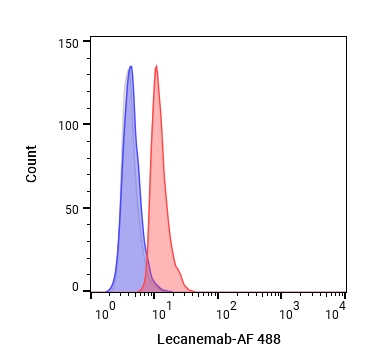 Flow cytometric analysis of 1X10⁶ HUVEC cells with Lecanemab (HY-P990110, red). Cells were fixed with 4% paraformaldehyde. Then stained with the primary antibody at 1/200 dilution for an hour at 4℃. AF 488-conjugated AffiniPure Goat Anti- Mouse IgG H&L (HY-P8005) was used as the secondary antibody at 1/1,000 dilution for 30 minutes at 4℃. Mouse IgG2a kappa (HY-P99978, blue) was used as the isotype control, cells without incubation with primary antibody were used as the unlabeled control (black).


Chemical Information
-
Appearance Liquid
-
Molecular Weight 147.71 kDa
-
Color Colorless to light yellow
-
SMILES
[Lecanemab (Mouse IgG2a)]
-
Synonyms
Mab158
-
Shipping
Shipping with dry ice.
-
Formulation
Please refer to the lot-specific COA for specific buffer information.
-
Storage
Please store the product under the recommended conditions in the Certificate of Analysis.


Publications (1)
-
Journal Impact Factor
-
Most Recent
-
Nat Commun
Chimeras co-targeting antigens and FcγRIIb trigger degradation of extracellular soluble proteins and pathological aggregates. [Abstract]2025 Dec 10. PMID: 41372196
Lecanemab (Mouse IgG2a) purchased from MedChemExpress. Usage Cited in: Nat Commun. 2025 Dec 10. [Abstract]
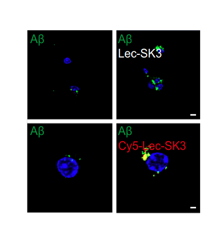
Representative confocal images showing Aβ oligomers (green) internalization in microglia treated with unlabeled Lecanemab SK3 (FcRTAC) or Cy5-labeled Lecanemab SK3. Cell nuclei were stained with DAPI (blue). Scale bar: 2 μm.
Lecanemab (Mouse IgG2a) purchased from MedChemExpress. Usage Cited in: Nat Commun. 2025 Dec 10. [Abstract]
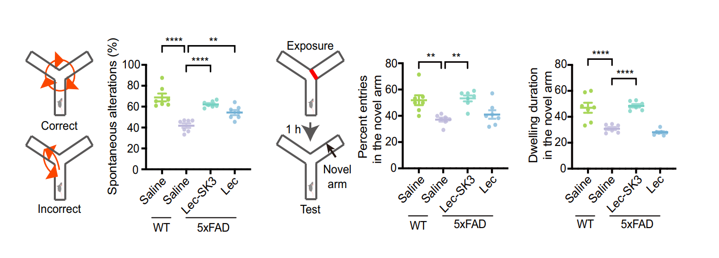
Experimental timeline of 2.5-month-old male WT or 5xFAD mice with bilateral i.c.v. injection of saline, 2 mg/kg Lecanemab SK3 or Lecanemab. Spontaneous alternation rate, exploration frequency and dwelling duration in the novel arm of the Y-maze were assessed post-injection of saline, Lecanemab SK3 or Lecanemab.


Purity & Documentation
-
Data Sheet (262 KB)
-
SDS (251 KB)
- English - EN (251 KB)
- Français - FR (251 KB)
- Deutsch - DE (251 KB)
- Norwegian - NO (251 KB)
- Español - ES (251 KB)
- Swedish - SV (251 KB)
- Italian - IT (251 KB)
- Korean - KR (251 KB)
- Portuguese - PT (251 KB)
-
Inhibitory Antibodies User Guide (603 KB)
References
[1]. Söllvander S, et al. The Aβ protofibril selective antibody mAb158 prevents accumulation of Aβ in astrocytes and rescues neurons from Aβ-induced cell death. J Neuroinflammation. 2018 Mar 28;15(1):98. [Content Brief]
[2]. Rizoska B, et al. Disease modifying effects of the amyloid-beta protofibril-selective antibody mAb158 in aged Tg2576 transgenic mice. Mol Cell Neurosci. 2024 Sep;130:103950. [Content Brief]
[3]. Tucker S, et al. The murine version of BAN2401 (mAb158) selectively reduces amyloid-β protofibrils in brain and cerebrospinal fluid of tg-ArcSwe mice. J Alzheimers Dis. 2015;43(2):575-88. [Content Brief]

Calculators
Concentration (start) × Volume (start) = Concentration (final) × Volume (final)


Lecanemab (Mouse IgG2a) Related Classifications
HY-P990110 Related Classifications
 Powered by Bioz