



M701

Based on 1 Customer Validation
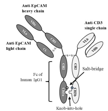
M701 is a T-cell engager bispecific humanized antibody targeting epithelial cell adhesion molecule (EpCAM) and cluster of differentiation 3 (CD3). M701 binds to EpCAM on tumor cells and CD3 on T cells, thereby linking the two cell populations to achieve targeted cytotoxicity and T cell-mediated cytotoxicity. M701 is applicable to research related to advanced epithelial solid tumors.
For research use only. We do not sell to patients.
- Purity : 99.05%
- CAS No.: 2933295-42-0
- Molecular Weight:125.8 kDa
-
Storage:
Please store the product under the recommended conditions in the Certificate of Analysis.


To place orders, for customer services and technical support, please contact: MedChemExpress USA
Tel: 609-228-6898 E-mail: [email protected] [email protected]


-
 Biological Activity
-
Chemical Information
- Protocols
- Purity & Documentation
- References
-
 Help & FAQs




Biological Activity
Description
Isotype
Bispecific T cell engager antibody
Recommend Isotype Controls
Species Reactivity
Human
IC50 & Target
CD3 & EpCAM
In Vitro
M701 (serially diluted concentrations; 1 h) binds to HCT116 colorectal carcinoma cells with a dissociation constant of 32 nM[1].
M701 (serially diluted concentrations; 1 h) binds to human T cells with a dissociation constant of 33.7 nM[1].
M701 (0-10 μg/mL; 30 min) dose-dependently bridges EpCAM-positive NCI-N87 gastric carcinoma cells and CD3-positive CIK cells, reaching up to 40% double-positive cells at 10 μg/mL[1].
M701 (serially diluted concentrations; 24 h) potently redirects CIK cell lysis to EpCAM-high HCT116, NCI-N87, and KATO-III tumor cells with EC50 values of 1.1, 1.8, and 8.5 ng/mL respectively, shows minimal activity against EpCAM-low SK-OV-3 and MDA-MB-231 cells, and has no activity against EpCAM-negative U87 cells[1].
MedChemExpress (MCE) has not independently confirmed the accuracy of these methods. They are for reference only.
-
Cell Line:EpCAM-expressing tumor cell lines (HCT116, NCI-N87, KATO-III, SK-OV-3, MDA-MB-231), U87 glioblastoma cells, cytokine-induced killer (CIK) cells
-
Concentration:Serially diluted concentrations
-
Incubation Time:24 h (37°C, 5% CO2 incubator)
-
Result:Potently redirected CIK cell lysis to EpCAM-high HCT116 cells with an EC50 of 1.1 ng/mL.
Potently redirected CIK cell lysis to EpCAM-high NCI-N87 cells with an EC50 of 1.8 ng/mL.
Potently redirected CIK cell lysis to EpCAM-high KATO-III cells with an EC50 of 8.5 ng/mL.
Showed minimal cytotoxicity against EpCAM-low SK-OV-3 cells with an EC50 of 58.7 ng/mL.
Showed minimal cytotoxicity against EpCAM-low MDA-MB-231 cells with an EC50 of 37.8 ng/mL.
Exhibited no cytotoxicity against EpCAM-negative U87 glioblastoma cells.
In Vivo
MedChemExpress (MCE) has not independently confirmed the accuracy of these methods. They are for reference only.
-
Animal Model:NOD/SCID (female, 7-8 weeks old)[1]
-
Dosage:0.1 mg/kg; 0.5 mg/kg; 2.5 mg/kg
-
Administration:i.v.; on days 0, 2, and 4
-
Result:Recorded mean tumor volume of 828.17 mm3 at 0.1 mg/kg dose on day 42.
Recorded mean tumor volume of 145.15 mm3 at 0.5 mg/kg dose on day 42.
Recorded mean tumor volume of 112.54 mm3 at 2.5 mg/kg dose on day 42.
Showed statistically significant tumor growth inhibition at 0.5 mg/kg and 2.5 mg/kg doses compared to controls.
Gene ID
Accession
Conjugated
Unconjugated
Reconsititution
The product can be reconstituted/diluted with sterile PBS or saline.
Format
-
half-IGG1/scFv-h-CH2-CH3
Application
ELISA, FACS, Functional assay
Verified Bioactivity
-
 Immobilized Human EpCAM Protein, His Tag can bind M701. The EC50 for this effect is 638.8 ng/mL.
 -
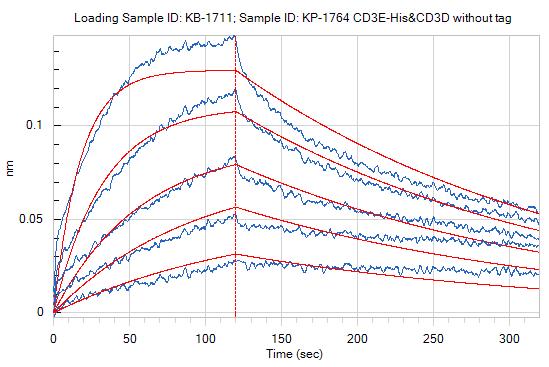 Loaded M701 on ProA biosensor, can bind CD3E-His&CD3D without tag with an affinity constant of 1.73E-08 M as determined in BLI assay.
 -
 Flow cytometric analysis of 1X10⁶ Jurkat cells labeling CD3 with M701 (HY-P991526, red). Cells were stained with the primary antibody at 1/200 dilution for an hour at 4℃. AF488-conjugated Goat Anti-Human IgG H&L (AF488) (HY-P83776) was used as the secondary antibody at 1/1,000 dilution for 30 minutes at 4℃. Human IgG1 kappa Isotype Control (HY-P99001, blue) was used as the isotype control, cells without incubation with primary antibody were used as the unlabeled control (black).


Chemical Information
-
CAS No. 2933295-42-0
-
Appearance Liquid
-
Molecular Weight 125.8 kDa
-
Color Colorless to light yellow
-
SMILES
N/A
-
Shipping
Shipping with dry ice.
-
Formulation
Please refer to the lot-specific COA for specific buffer information.
-
Storage
Please store the product under the recommended conditions in the Certificate of Analysis.


Purity & Documentation
-
Data Sheet (289 KB)
-
SDS (251 KB)
- English - EN (251 KB)
- Français - FR (251 KB)
- Deutsch - DE (251 KB)
- Norwegian - NO (251 KB)
- Español - ES (251 KB)
- Swedish - SV (251 KB)
- Italian - IT (251 KB)
- Korean - KR (251 KB)
- Portuguese - PT (251 KB)
-
Inhibitory Antibodies User Guide (603 KB)
References

Calculators
Concentration (start) × Volume (start) = Concentration (final) × Volume (final)


M701 Related Classifications
HY-P991526 Related Classifications
 Powered by Bioz