



M701

Based on 1 Customer Validation
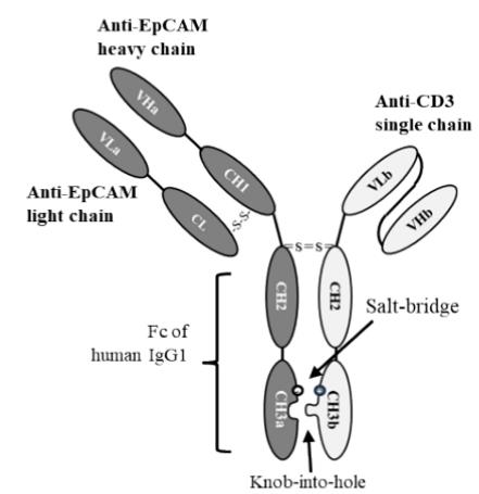
M701 is a T-cell engager bispecific humanized antibody targeting epithelial cell adhesion molecule (EpCAM) and cluster of differentiation 3 (CD3). M701 binds to EpCAM on tumor cells and CD3 on T cells, thereby linking the two cell populations to achieve targeted cytotoxicity and T cell-mediated cytotoxicity. M701 is applicable to research related to advanced epithelial solid tumors.
For research use only. We do not sell to patients.
- Purity : 99.05%
- CAS No.: 2933295-42-0
- Molecular Weight:125.8 kDa
-
Storage:
Please store the product under the recommended conditions in the Certificate of Analysis.


To place orders, for customer services and technical support, please contact: MedChemExpress USA
Tel: 609-228-6898 E-mail: [email protected] [email protected]


-
 Biological Activity
-
Chemical Information
- Protocols
- Purity & Documentation
- References
-
 Help & FAQs




Biological Activity
Description
Isotype
Bispecific T cell engager antibody
Recommend Isotype Controls
Species Reactivity
Human
IC50 & Target
CD3 & EpCAM
In Vitro
M701 (serially diluted concentrations; 1 h) binds to HCT116 colorectal carcinoma cells with a dissociation constant of 32 nM[1].
M701 (serially diluted concentrations; 1 h) binds to human T cells with a dissociation constant of 33.7 nM[1].
M701 (0-10 μg/mL; 30 min) dose-dependently bridges EpCAM-positive NCI-N87 gastric carcinoma cells and CD3-positive CIK cells, reaching up to 40% double-positive cells at 10 μg/mL[1].
M701 (serially diluted concentrations; 24 h) potently redirects CIK cell lysis to EpCAM-high HCT116, NCI-N87, and KATO-III tumor cells with EC50 values of 1.1, 1.8, and 8.5 ng/mL respectively, shows minimal activity against EpCAM-low SK-OV-3 and MDA-MB-231 cells, and has no activity against EpCAM-negative U87 cells[1].
MedChemExpress (MCE) has not independently confirmed the accuracy of these methods. They are for reference only. Further protocols information, click here.
-
Cell Line:EpCAM-expressing tumor cell lines (HCT116, NCI-N87, KATO-III, SK-OV-3, MDA-MB-231), U87 glioblastoma cells, cytokine-induced killer (CIK) cells
-
Concentration:Serially diluted concentrations
-
Incubation Time:24 h (37°C, 5% CO2 incubator)
-
Result:Potently redirected CIK cell lysis to EpCAM-high HCT116 cells with an EC50 of 1.1 ng/mL.
Potently redirected CIK cell lysis to EpCAM-high NCI-N87 cells with an EC50 of 1.8 ng/mL.
Potently redirected CIK cell lysis to EpCAM-high KATO-III cells with an EC50 of 8.5 ng/mL.
Showed minimal cytotoxicity against EpCAM-low SK-OV-3 cells with an EC50 of 58.7 ng/mL.
Showed minimal cytotoxicity against EpCAM-low MDA-MB-231 cells with an EC50 of 37.8 ng/mL.
Exhibited no cytotoxicity against EpCAM-negative U87 glioblastoma cells.
In Vivo
MedChemExpress (MCE) has not independently confirmed the accuracy of these methods. They are for reference only.
-
Animal Model:NOD/SCID (female, 7-8 weeks old)[1]
-
Dosage:0.1 mg/kg; 0.5 mg/kg; 2.5 mg/kg
-
Administration:i.v.; on days 0, 2, and 4
-
Result:Recorded mean tumor volume of 828.17 mm3 at 0.1 mg/kg dose on day 42.
Recorded mean tumor volume of 145.15 mm3 at 0.5 mg/kg dose on day 42.
Recorded mean tumor volume of 112.54 mm3 at 2.5 mg/kg dose on day 42.
Showed statistically significant tumor growth inhibition at 0.5 mg/kg and 2.5 mg/kg doses compared to controls.
Gene ID
Accession
Conjugated
Unconjugated
Reconsititution
The product can be reconstituted/diluted with sterile PBS or saline.
Format
-
half-IGG1/scFv-h-CH2-CH3
Application
ELISA, FACS, Functional assay
Verified Bioactivity
-
 Immobilized Human EpCAM Protein, His Tag can bind M701. The EC50 for this effect is 638.8 ng/mL.
 -
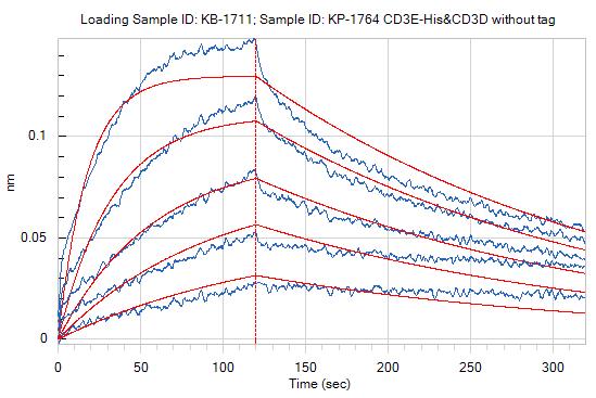 Loaded M701 on ProA biosensor, can bind CD3E-His&CD3D without tag with an affinity constant of 1.73E-08 M as determined in BLI assay.
 -
 Flow cytometric analysis of 1X10⁶ Jurkat cells labeling CD3 with M701 (HY-P991526, red). Cells were stained with the primary antibody at 1/200 dilution for an hour at 4℃. AF488-conjugated Goat Anti-Human IgG H&L (AF488) (HY-P83776) was used as the secondary antibody at 1/1,000 dilution for 30 minutes at 4℃. Human IgG1 kappa Isotype Control (HY-P99001, blue) was used as the isotype control, cells without incubation with primary antibody were used as the unlabeled control (black).


Chemical Information
-
CAS No. 2933295-42-0
-
Appearance Liquid
-
Molecular Weight 125.8 kDa
-
Color Colorless to light yellow
-
SMILES
N/A
-
Shipping
Shipping with dry ice.
-
Formulation
Please refer to the lot-specific COA for specific buffer information.
-
Storage
Please store the product under the recommended conditions in the Certificate of Analysis.

Protocols
-
Cell Cytotoxicity Assay
Cytotoxicity assays are usually based on the assessment of cell membrane damage, which can also be indirectly detected by measuring cell viability. Detection methods include MTT assay, CKK-8 assay, LDH assay and ATP assay, etc.
-
Mammalian live/dead viability and cytotoxicity staining
Live/dead viability and cytotoxicity staining assays are based on the simultaneous detection of intracellular esterase activity in metabolically active (viable) cells and membrane integrity loss in non-viable cells. In commonly used dual-staining approaches, membrane-permeant fluorogenic substrates are converted by intracellular esterases into fluorescent products in live cells, while impermeant DNA-binding dyes selectively enter cells with compromised plasma membranes and label nucleic acids in dead or dying cells, enabling discrimination between viable and non-viable populations by fluorescence microscopy or flow cytometry.
-
Research Protocol for Cancer Immunology
Cancer immunology studies how the immune system recognizes, suppresses, edits, or fails to eliminate malignant cells through tumor antigen release, antigen presentation, T-cell priming, immune trafficking, tumor-cell killing, and feedback inhibition in the tumor microenvironment. The cancer-immunity cycle links tumor antigenicity, dendritic-cell priming, CD8+ T-cell infiltration, cytotoxic function, and immune-checkpoint regulation to tumor rejection or immune escape. Immune-checkpoint pathways such as PD-1/PD-L1 and CTLA-4 suppress antitumor T-cell activity and can be therapeutically blocked, but many tumors remain resistant because of poor antigen presentation, weak T-cell infiltration, suppressive myeloid cells, regulatory T cells, and tumor-intrinsic immune-exclusion programs. Unresolved questions include which immune-cell states predict response, how tumor-intrinsic pathways exclude immune cells, how myeloid suppression limits checkpoint blockade, and which combination strategies
-
Cell Viability Determination by MTT Colorimetric Assay
The following protocol uses the MTT colorimetric assay as a classic literature-established method for assessing cell viability/metabolic activity in cultured mammalian cells. MTT[3-(4,5-dimethylthiazol-2-yl)-2,5-diphenyltetrazolium bromide] is reduced by metabolically active cells to a colored formazan product; the amount of formazan is quantified spectrophotometrically and provides an indirect measure of metabolically active viable cells. Importantly, MTT reduction reflects cellular oxidoreductase/metabolic activity rather than an absolute direct count of living cells, so changes in cellular metabolism can alter the signal independently of cell number.


Purity & Documentation
-
Data Sheet (287 KB)
-
SDS (251 KB)
- English - EN (251 KB)
- Français - FR (251 KB)
- Deutsch - DE (251 KB)
- Norwegian - NO (251 KB)
- Español - ES (251 KB)
- Swedish - SV (251 KB)
- Italian - IT (251 KB)
- Korean - KR (251 KB)
- Portuguese - PT (251 KB)
-
Inhibitory Antibodies User Guide (603 KB)
References

Calculators
Concentration (start) × Volume (start) = Concentration (final) × Volume (final)


M701 Related Classifications
HY-P991526 Related Classifications
 Powered by Bioz