



Questin

Based on 1 Customer Validation
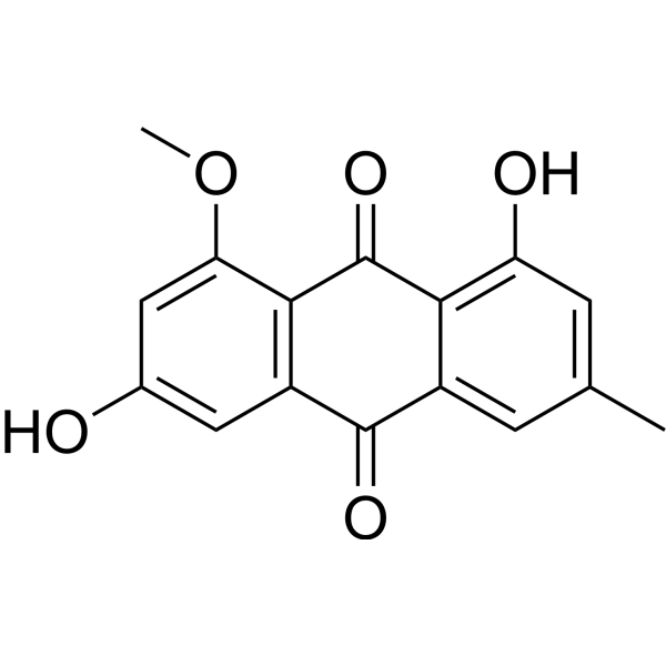
Questin is an anthraquinone compound and antibacterial agent. Questin can be isolated from marine-derived fungi and plants. Questin inhibits Cdc25B phosphatase. Questin exhibits antibacterial activity against V. harveyi, V. anguillarum, V. cholerae, and V. parahemolyticus with MIC values of 31.25 µg/mL, 62.5 µg/mL, 62.5 µg/mL, and 125 µg/mL. Questin displays antiprotozoal activity against the animal protozoan pathogen Tritrichomonas foetus, with a MIC of 12.5 µg/mL. Questin has anticancer activity against lung and colon cancer.
For research use only. We do not sell to patients.
- Purity : 97.75%
- CAS No.: 3774-64-9
- Formula: C16H12O5
- Molecular Weight:284.26
-
Storage:
4°C, protect from light
* In solvent : -80°C, 6 months; -20°C, 1 month (protect from light)

To place orders, for customer services and technical support, please contact: MedChemExpress USA
Tel: 609-228-6898 E-mail: [email protected] [email protected]


-
 Biological Activity
-
Chemical Information
- Protocols
- Purity & Documentation
- References
-
 Help & FAQs



All Parasite Isoforms
More

Biological Activity
Description
In Vitro
Questin exhibits antibacterial activity against V. harveyi, V. anguillarum, V. cholerae, and V. parahemolyticus with MIC values of 31.25 µg/mL, 62.5 µg/mL, 62.5 µg/mL, and 125 µg/mL, respectively[1].
Questin (48 h) strongly inhibits the growth of human colon cancer cells (SW620), with a GI50 value of 0.9 mg/mL[2].
Questin (50-100 μM; 24 h) shows certain toxicity to the human A549 lung cell line and reduces cell viability[3].
Questin displays a moderate level of selective antiprotozoal activity against the animal protozoan pathogen Tritrichomonas foetus, with a MIC of 12.5 µg/mL[5].
MedChemExpress (MCE) has not independently confirmed the accuracy of these methods. They are for reference only.


Chemical Information
-
CAS No. 3774-64-9
-
Appearance Solid
-
Molecular Weight 284.26
-
Formula C16H12O5
-
Color Yellow to orange
-
SMILES
O=C1C2=C(C=C(O)C=C2OC)C(C3=CC(C)=CC(O)=C13)=O
-
Structure Classification
-
Initial Source
marine-derived Aspergillus flavipes?HN4-13
-
Shipping
Room temperature in continental US; may vary elsewhere.
-
Storage
4°C, protect from light
* In solvent : -80°C, 6 months; -20°C, 1 month (protect from light)


Purity & Documentation
-
Data Sheet (271 KB)
-
SDS (252 KB)
- English - EN (252 KB)
- Français - FR (252 KB)
- Deutsch - DE (252 KB)
- Norwegian - NO (252 KB)
- Español - ES (252 KB)
- Swedish - SV (252 KB)
- Italian - IT (252 KB)
- Korean - KR (252 KB)
- Portuguese - PT (252 KB)
-
Handling Instructions (2659 KB)
References
[1]. Lei Guo, et al. Optimized production and isolation of antibacterial agent from marine Aspergillus flavipes against Vibrio harveyi. 3 Biotech. 2017 Dec;7(6):383. [Content Brief]
[2]. Choi SG, et al. Anthraquinones, Cdc25B phosphatase inhibitors, isolated from the roots of Polygonum multiflorum Thunb. Nat Prod Res. 2007 May 20;21(6):487-93. [Content Brief]
[3]. Gauthier T, et al. Trypacidin, a spore-borne toxin from Aspergillus fumigatus, is cytotoxic to lung cells. PLoS One. 2012;7(2):e29906. [Content Brief]
[4]. Liu N, et al. Structurally diverse sesquiterpenoids and polyketides from a sponge-associated fungus Aspergillus sydowii SCSIO41301. Fitoterapia. 2019 Jun;135:27-32. [Content Brief]
[5]. Chaudhary NK, et al. Banksialactones and Banksiamarins: Isochromanones and Isocoumarins from an Australian Fungus, Aspergillus banksianus. J Nat Prod. 2018 Jul 27;81(7):1517-1526. [Content Brief]

Calculators
Concentration (start) × Volume (start) = Concentration (final) × Volume (final)


Questin Related Classifications
HY-137990 Related Classifications
 Powered by Bioz