



PD-L1 Protein, Human (HEK293, His)
Based on 1 Customer Validation
PD-L1 Protein is an important immunoregulatory molecule and a member of the B7 superfamily. As the primary ligand of PD-1, PD-L1 Protein can bind to PD-1 to inhibit T-cell activation, proliferation, and cytokine secretion, and induce T-cell apoptosis. PD-L1 Protein plays a key role in tumor immune escape and immune homeostasis regulation. PD-L1 Protein, Human (HEK293, His) is a recombinant PD-L1 protein expressed by HEK293 with a C-His His tag.
- Species: Human
- Source: HEK293
-
Storage:Stored at -20°C for 2 years from date of receipt. After reconstitution, it is stable at 4°C for 1 week or -20°C for longer (with carrier protein). It is recommended to freeze aliquots at -20°C or -80°C for extended storage.


To place orders, for customer services and technical support, please contact: MedChemExpress USA
Tel: 609-228-6898 E-mail: [email protected] [email protected]


-
 Biological Activity
-
Technical Parameters
-
Product Properties
- Documentation
- References
- Help & FAQs


-

HY-P7396
PD-1 Protein, Human (HEK293, His) -
HY-13757A
Tamoxifen -
HY-P70632
PD-L1 Protein, Mouse (HEK293, His) -

HY-P9904
Atezolizumab -
HY-K0301
Cell Counting Kit-8 -
HY-P7109
EGF Protein, Human -
HY-P70254
HER2/CD340 Protein, Human (HEK293, His) -
HY-P7037B
IL-2 Protein, Human (His) -
HY-P72492
PD-L2 Protein, Human (HEK293, His) -
HY-P72259
Protein E7, HPV16 (His) -
HY-P70755
IL-7 Protein, Human (HEK293, His) -
HY-P7336
CD47 Protein, Human (HEK293, His)


Biological Activity
Description
PD-L1 Protein is an important immunoregulatory molecule and a member of the B7 superfamily. As the primary ligand of PD-1, PD-L1 Protein can bind to PD-1 to inhibit T-cell activation, proliferation, and cytokine secretion, and induce T-cell apoptosis. PD-L1 Protein plays a key role in tumor immune escape and immune homeostasis regulation. PD-L1 Protein, Human (HEK293, His) is a recombinant PD-L1 protein expressed by HEK293 with a C-His His tag[1][2][3].
Background
PD-L1 is a 33 kDa type I transmembrane glycoprotein containing 290 amino acids, with Ig- and IgC domains in its extracellular region. Under normal conditions, PD-L1 maintains immune homeostasis by binding to PD-1 on the surface of T cells to inhibit excessive immune responses. In tumors, abnormally high expression of PD-L1 binds to PD-1 to suppress T cell function, thereby helping tumors evade immune surveillance. PD-L1 may also be involved in tumor angiogenesis and microenvironmental remodeling. Currently, as a core target, PD-L1 has driven the development of tumor immune checkpoint inhibitor therapy[1][2][3].
Verified Bioactivity
1. 2 µg/mL (100 µL/well) of immoblized recombinant human PD-L1-His can bind human PD-1-Fc with a linear range of 24-390 ng/mL.
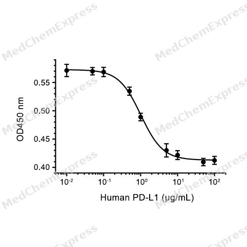
2. Measured by its ability to inhibit anti-CD3-induced proliferation of stimulated CTLL-2 mouse cytotoxic T cells. The ED50 this effect is 1.012 μg/mL, corresponding to a specific activity is 988.14 units/mg.
3. Measured by its binding ability in a functional ELISA. Immobilized Human PD-L1 at 1 μg/mL (100 μL/well) can bind Biotinylated Human PD-1 (HY-P7396). The ED50 for this effect is <0.5 μg/mL.
4. Immobilized Anti-Human PDL1 mAb at 2 μg/mL (100 μL/well) can bind Human PD-L1. The ED50 of Human PD-L1 is 1-10 ng/mL.
MCE Validation Data
-
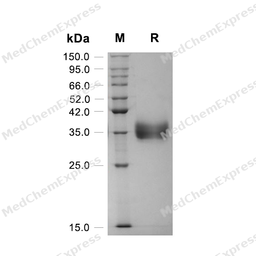
Purity - SDS-PAGE
Purity - SDS-PAGE
-
Bioactivity - Cell-Based Assay
Bioactivity - Cell-Based Assay
-
Bioactivity - SPR
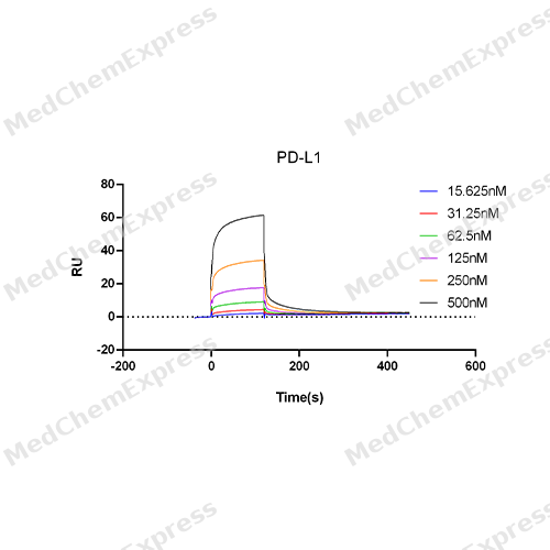
Bioactivity - SPR


Technical Parameters
-
Species Human
-
Source HEK293
-
Tag C-His
-
Accession
Q9NZQ7-1 (F19-T239)
-
Molecular Construction
-
N-term
-
PD-L1 (F19-T239)
Accession # Q9NZQ7-1 -
His
-
C-term
-
-
Protein Length
Extracellular Domain
-
Synonyms
CD274; B7 Homolog 1; Prev. PDCD1LG1; B7-H; PD-L1; PDCD1 Ligand 1; B7H1; PDCD1L1; PDL1; HPD-L1; Programmed Cell Death 1 Ligand 1; Programmed Death Ligand 1; CD274 Antigen; ADMIO5; CD274 Molecule; B7-H1
-
AA Sequence
FTVTVPKDLYVVEYGSNMTIECKFPVEKQLDLAALIVYWEMEDKNIIQFVHGEEDLKVQHSSYRQRARLLKDQLSLGNAALQITDVKLQDAGVYRCMISYGGADYKRITVKVNAPYNKINQRILVVDPVTSEHELTCQAEGYPKAEVIWTSSDHQVLSGKTTTTNSKREEKLFNVTSTLRINTTTNEIFYCTFRRLDPEENHTAELVIPELPLAHPPNERT
-
Molecular Weight
Approximately 31-42 kDa, based on SDS-PAGE under reducing conditions, due to the glycosylation.
-
Glycosylation
Yes
-
Purity
≥ 95%, as determined by reducing SDS-PAGE.


Product Properties
Lyophilized powder
1.Lyophilized from a 0.2 μm filtered solution of 20 mM PB, 150 mM NaCl, pH 7.4.
2.Lyophilized from a 0.2 μm filtered solution of 20 mM PB, 4% mannitol, 0.02% Tween 80, pH 7.4.
Please refer to the lot-specific COA for specific buffer information.
<1 EU/μg, determined by LAL method.
It is not recommended to reconstitute to a concentration less than 100 μg/mL in ddH2O. For long term storage it is recommended to add a carrier protein (0.1% BSA, 5% HSA, 10% FBS or 5% Trehalose).
Stored at -20°C for 2 years from date of receipt. After reconstitution, it is stable at 4°C for 1 week or -20°C for longer (with carrier protein). It is recommended to freeze aliquots at -20°C or -80°C for extended storage.
Room temperature in continental US; may vary elsewhere.


Documentation
-
Data Sheet (264 KB)
-
SDS (252 KB)
- English - EN (252 KB)
- Français - FR (252 KB)
- Deutsch - DE (252 KB)
- Norwegian - NO (252 KB)
- Español - ES (252 KB)
- Swedish - SV (252 KB)
- Italian - IT (252 KB)
- Korean - KR (252 KB)
- Portuguese - PT (252 KB)
-
Handling Instructions (2659 KB)


References
[1]. Chemnitz JM, et al. SHP-1 and SHP-2 associate with immunoreceptor tyrosine-based switch motif of programmed death 1 upon primary human T cell stimulation, but only receptor ligation prevents T cell activation. J Immunol. 2004 Jul 15;173(2):945-54. [Content Brief]
[2]. Han Y, et al. PD-1/PD-L1 pathway: current researches in cancer. Am J Cancer Res. 2020 Mar 1;10(3):727-742. [Content Brief]
[3]. Kythreotou A, et al. PD-L1. Journal of clinical pathology, 2018, 71(3): 189-194.

Calculators
Concentration (start) × Volume (start) = Concentration (final) × Volume (final)


 Powered by Bioz

