



SDF-1 beta/CXCL12 Protein, Human (72a.a)
Based on 2 publication(s) in Google Scholar
SDF-1 beta (Stromal-derived factor-1β, SDF-1β) is a stromal derived CXC chemokine that signal through the CXCR4 receptor. SDF-1β has chemotactic activity on B and T cells. SDF-1 beta/CXCL12 Protein, Human (72a.a) is produced in E. coli, and consists of 72 amino acids (K22-M93).
- Species: Human
- Source: E. coli
-
Storage:Stored at -20°C for 2 years from date of receipt. After reconstitution, it is stable at 4°C for 1 week or -20°C for longer (with carrier protein). It is recommended to freeze aliquots at -20°C or -80°C for extended storage.


To place orders, for customer services and technical support, please contact: MedChemExpress USA
Tel: 609-228-6898 E-mail: [email protected] [email protected]


-
 Biological Activity
-
Technical Parameters
-
Product Properties
- Documentation
- References
- Help & FAQs




Biological Activity
Description
SDF-1 beta (Stromal-derived factor-1β, SDF-1β) is a stromal derived CXC chemokine that signal through the CXCR4 receptor. SDF-1β has chemotactic activity on B and T cells[1][2]. SDF-1 beta/CXCL12 Protein, Human (72a.a) is produced in E. coli, and consists of 72 amino acids (K22-M93).
Background
The chemokine stromal-derived factor-1 (SDF-1), which is constitutively expressed in most tissues as SDF-1α and SDF-1β resulting from alternative gene splicing, regulates hematopoiesis, lymphocyte homing, B-lineage cell growth, and angiogenesis[1][2]. SDF-1β is assigned to the intercrine cytokine family (chemokines) which is characterized by four conserved cysteines that form two disulfide bonds. Furthermore its expression is found in all organs except in blood cells[3].
SDF-1β which is virtually the same as SDF-1α, except in that the fourth exon consists of only four residues attached to a C-terminus, shows very similar activity in vitro and in tissues, but is twice as potent in the blood. SDF-1α comprises 3 exons and encodes a protein of 89 amino acids whereas SDF-1β consists of 4 exons and encodes a protein of 93 amino acids. Both isoforms are highly similar regarding their sequences with the only difference of 4 additional amino acids at the C-terminus of SDF1β[1][2]. In addition, SDF-1β is shown to be a sufficient factor capable of supporting rodent B-cell lymphopoiesis. SDF-1β is expressed less abundantly and seems to be related to the vascular system. Its greater resistance to proteolysis within the blood predispose it to this role[3].
Endothelial cells of cerebral microvessels in mice express SDF-1β selectively. Its upregulation is found following focal cerebral ischemia and is associated with the infiltration of CXCR4-expressing peripheral blood cells, such as macrophages. SDF-1β also has a greater effect on angiogenesis in human microvascular endothelial cells (HMEC)[3]. Compared with SDF-1α, SDF-1β is more resistant to blood-dependent degradation, stimulates angiogenesis and is present in highly vascularized organs such as: the liver, spleen and kidneys[4].
In Vitro
Recombinant human SDF-1β (10, 25, 50 and 100 ng/mL) induces apoptosis of A549 cells[4].
Verified Bioactivity
Full biological activity determined by a chemotaxis bioassay using PHA and rHuIL-2 activated human peripheral blood T-lymphocytes is in a concentration range of 20-80 ng/ml.
MCE Validation Data
-
Purity - SDS-PAGE
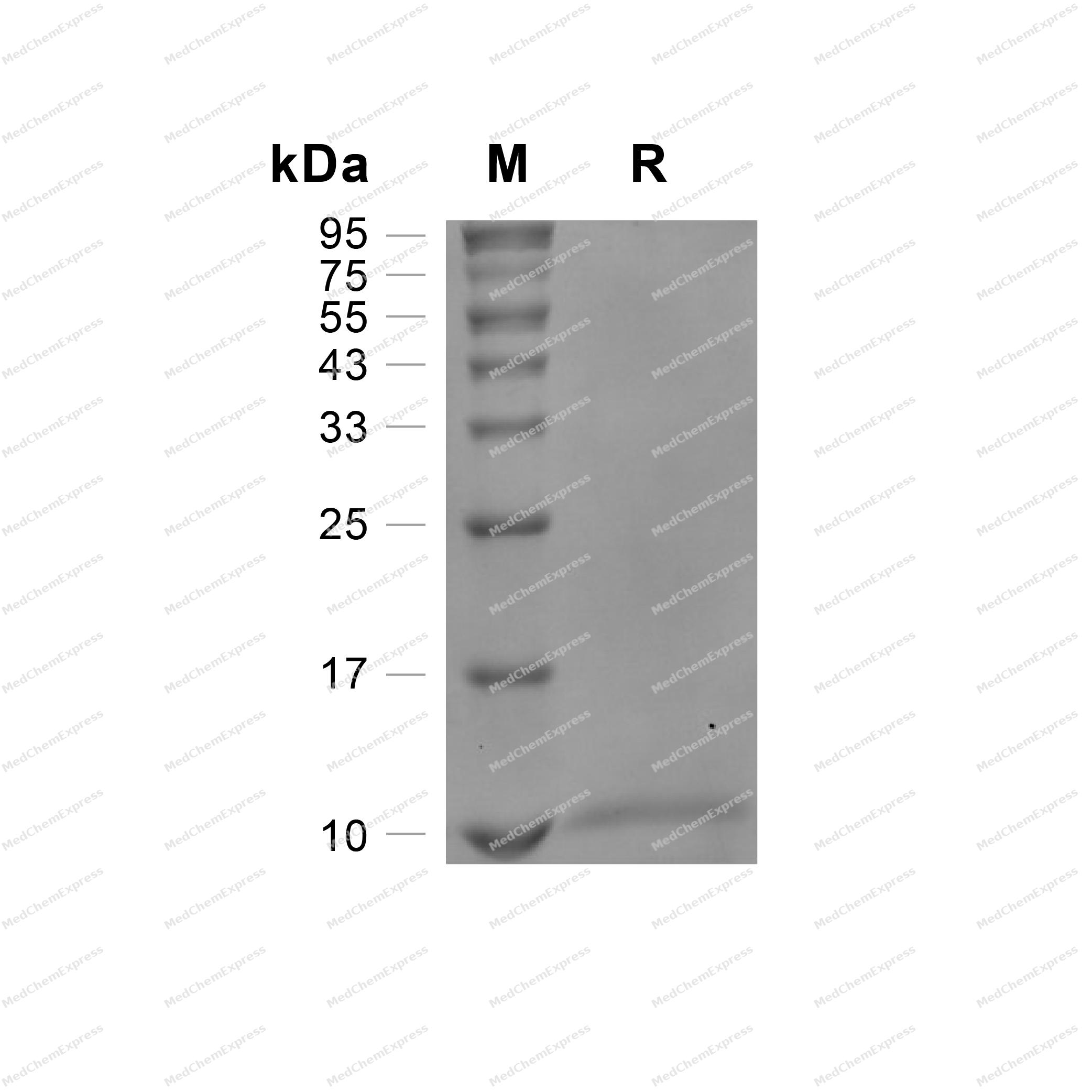
Purity - SDS-PAGE


Publications (2)
-
Journal Impact Factor
-
Most Recent


Technical Parameters
-
Species Human
-
Source E. coli
-
Tag Tag Free
-
Accession
P48061-1 (K22-M93)
-
Molecular Construction
-
N-term
-
CXCL12 (K22-M93)
Accession # P48061-1 -
C-term
-
-
Protein Length
Full Length of Isoform-1
-
Synonyms
CXCL12; Intercrine Reduced In Hepatomas; Prev. SDF1; SDF-1b; Prev. SDF1A; TLSF-A; Prev. SDF1B; TLSF-B; PBSF; IRH; Stromal Cell-Derived Factor 1; C-X-C Motif Chemokine 12; SCYB12; HSDF-1; TPAR1; SDF-1; Pre-B Cell Growth-Stimulating Factor; TLSF; Chemokine
-
AA Sequence
KPVSLSYRCPCRFFESHVARANVKHLKILNTPNCALQIVARLKNNNRQVCIDPKLKWIQEYLEKALNKRFKM
-
Predicted Molecular Mass
8.5 kDa
-
Molecular Weight
Approximately 8-12 kDa, based on SDS-PAGE under reducing conditions, due to the glycosylation.
-
Purity
≥ 95%, as determined by reducing SDS-PAGE.


Product Properties
Lyophilized powder
1.Lyophilized from a 0.22 μm filtered solution of PBS, pH 7.4.
2.Lyophilized from a 0.22 μm filtered solution of 50 mM Tris-HCl, 200 mM NaCl, 500 mM arginine, pH 8.0.
Please refer to the lot-specific COA for specific buffer information.
Note: For SPR assay, please replace the buffer. Primary amine components (e.g., Tris, imidazole) can affect protein-coupled chips.
<1 EU/μg, determined by LAL method.
It is not recommended to reconstitute to a concentration less than 100 μg/mL in ddH2O. For long term storage it is recommended to add a carrier protein (0.1% BSA, 5% HSA, 10% FBS or 5% Trehalose).
Stored at -20°C for 2 years from date of receipt. After reconstitution, it is stable at 4°C for 1 week or -20°C for longer (with carrier protein). It is recommended to freeze aliquots at -20°C or -80°C for extended storage.
Room temperature in continental US; may vary elsewhere.


Documentation
-
Data Sheet (264 KB)
-
SDS (252 KB)
- English - EN (252 KB)
- Français - FR (252 KB)
- Deutsch - DE (252 KB)
- Norwegian - NO (252 KB)
- Español - ES (252 KB)
- Swedish - SV (252 KB)
- Italian - IT (252 KB)
- Korean - KR (252 KB)
- Portuguese - PT (252 KB)
-
Handling Instructions (2659 KB)


References
[1]. Maria De La Luz Sierra, et al. Differential processing of stromal-derived factor-1alpha and stromal-derived factor-1beta explains functional diversity. Blood. 2004 Apr 1;103(7):2452-9. [Content Brief]
[2]. Kryczek I, et al. Stroma-derived factor (SDF-1/CXCL12) and human tumor pathogenesis. Am J Physiol Cell Physiol. 2007 Mar;292(3):C987-95. [Content Brief]
[3]. Zheng Jiang, et al. Contribution of SDF-1α/CXCR4 signaling to brain development and glioma progression. Neurosignals. 2013;21(3-4):240-58. [Content Brief]
[4]. Miroslaw Janowski. Functional diversity of SDF-1 splicing variants. Cell Adh Migr. 2009 Jul-Sep;3(3):243-9. [Content Brief]
[5]. Kleanthis Fytianos, et al. Anti-Fibrotic Effect of SDF-1β Overexpression in Bleomycin-Injured Rat Lung. Pharmaceutics. 2022 Aug 27;14(9):1803. [Content Brief]

Calculators
Concentration (start) × Volume (start) = Concentration (final) × Volume (final)


 Powered by Bioz

