



SIRT5 Protein, Human (Flag)
Based on 2 publication(s) in Google Scholar
SIRT5 Proteins, belonging to the sirtuin family, possess a sirtuin core domain and share homology with yeast Sir2 protein. They are classified in class III of the sirtuin family and undergo alternative splicing, resulting in multiple transcript variants. Their ubiquitous expression in tissues like the heart, liver, and 25 others suggests their involvement in diverse cellular processes. SIRT5 Protein, Human (Flag) is the recombinant human-derived SIRT5 protein, expressed by E. coli , with N-Flag labeled tag.
- Species: Human
- Source: E. coli
-
Storage:Stored at -20°C for 2 years from date of receipt. After reconstitution, it is stable at 4°C for 1 week or -20°C for longer (with carrier protein). It is recommended to freeze aliquots at -20°C or -80°C for extended storage.


To place orders, for customer services and technical support, please contact: MedChemExpress USA
Tel: 609-228-6898 E-mail: [email protected] [email protected]


-
 Biological Activity
-
Technical Parameters
-
Product Properties
- Documentation
- Help & FAQs




Biological Activity
Description
SIRT5 Proteins, belonging to the sirtuin family, possess a sirtuin core domain and share homology with yeast Sir2 protein. They are classified in class III of the sirtuin family and undergo alternative splicing, resulting in multiple transcript variants. Their ubiquitous expression in tissues like the heart, liver, and 25 others suggests their involvement in diverse cellular processes. SIRT5 Protein, Human (Flag) is the recombinant human-derived SIRT5 protein, expressed by E. coli , with N-Flag labeled tag.
Background
SIRT5, a member of the sirtuin family, shares homology with the yeast Sir2 protein and is characterized by a sirtuin core domain. The sirtuin family is divided into four classes, and while the specific functions of human sirtuins are yet to be fully elucidated, yeast sirtuin proteins are known to regulate epigenetic gene silencing and suppress recombination of rDNA. Studies suggest that human sirtuins may serve as intracellular regulatory proteins with mono-ADP-ribosyltransferase activity. SIRT5, classified within class III of the sirtuin family, undergoes alternative splicing, resulting in multiple transcript variants. The expression of SIRT5 is ubiquitous, observed in various tissues, including the heart, liver, and 25 other tissues, highlighting its likely involvement in diverse cellular processes.
Verified Bioactivity
Measured by its ability to remove the succinyl group from 25 μM fluorogenic peptide substrate incubate at room temperature for 30 minutes. The specific activity is 181.22 pmol/min/μg.
MCE Validation Data
-
Purity - SDS-PAGE
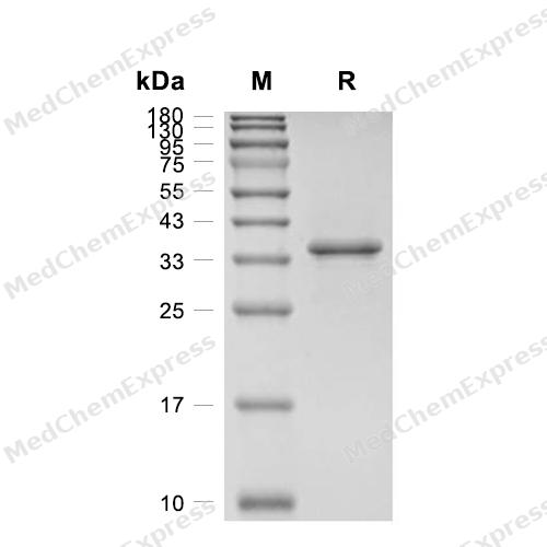
Purity - SDS-PAGE
-
Characterization - Western Blot
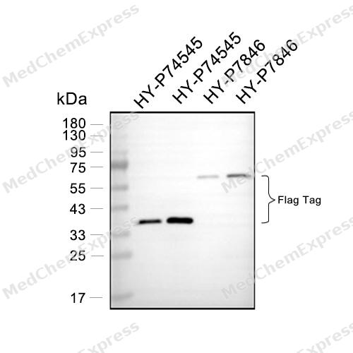
Characterization - Western Blot

Assay Procedure
Materials
Assay buffer: 25 mM Tris, 150 mM NaCl, 1 mM DTT, pH 8.0
TCNB buffer:50 mM Tris, 0.15 M NaCl, 10 mM CaCl2, 0.05% Brij-35 (w/v), pH 7.5 (TCNB)
PRSS3/Trypsin-3 Protein, Human (HEK293, His) (HY-P75985)
Activator Protein: Enterokinase Protein, Bovine (His)
Stop Solution: 8 ng/μL Trypsin, 4 mM Nicotinamide, 50 mM Tris, 100 mM NaCl, 30% (v/v) Isopropanol, pH 8.0
Beta-NAD: Dilute in ultrapure water to 100 mM
Substrate: FLUOR DE LYS?-Succinyl, Desuccinylase Substrate (5 mM)
Standard: 7-Amino-4-methylcoumarin (HY-D0027)
Procedure
1. Prepare a standard curve using AMC standards with concentrations of 10000, 5000, 2500, 1250, 625, 312.5, 156.25, 78.125, 19.53, 9.76, 0 pmol. Read the fluorescence at excitation and emission wavelengths of 380 nm and 460 nm, respectively.
2. Activation of Human Trypsin-3:
a. Dilute Bovine Enterokinase to 2 μg/mL in TCNB buffer;
b. Dilute Human Trypsin-3 to 200 μg/mL in TCNB buffer;
c. Mix equal volumes of the diluted proteins;
d. Incubate overnight at room temperature.
3. Dilute the Human SIRT5 to 10 μg/mL in assay buffer.
4. Dilute the substrate to 100 μM in assay buffer containing 2 mM beta-NAD.
5. Add 25 μL of 10 μg/mL Human SIRT5 and 25 μL of diluted substrate to the wells. For the control group, add only 25 μL of 10 μg/mL Human SIRT5.
6. Seal the plate and incubate at 37°C for 30 minutes.
7. Add 50 μL of stop solution to all wells and mix. For the control wells, add the stop solution first and then add 25 μL of substrate.
8. Seal the plate and incubate at room temperature for 15 minutes.
9. Read the fluorescence in endpoint mode at excitation and emission wavelengths of 380 nm and 460 nm, respectively.
10. Calculate specific activity:
Specific Activity (pmol/min/μg) = | Adjusted Fluorescence* (RFU) x Conversion Factor ** (pmol/RFU) |
| Incubation time (min) x amount of enzyme (μg) |
*Adjusted for Substrate Blank
**Derived using calibration standard
Per Well:
Human SIRT5: 0.025 μg
Substrate: 25 μM
beta-NAD: 0.5 mM


Publications (2)
-
Journal Impact Factor
-
Most Recent
-
Cell Death Dis
SIRT5-mediated BCAT1 desuccinylation and stabilization leads to ferroptosis insensitivity and promotes cell proliferation in glioma. [Abstract]2025 Apr 7;16(1):261. PMID: 40195331 -
Cell Rep
2026 May 28;45(6):117394. PMID: 42207644


Technical Parameters
-
Species Human
-
Source E. coli
-
Tag N-Flag
-
Accession
Q9NXA8-1/NP_036373.1 (R2-S310)
-
Molecular Construction
-
N-term
-
Flag
-
SIRT5 (R2-S310)
Accession # Q9NXA8-1/NP_036373.1 -
C-term
-
-
Protein Length
Full Length of Isoform-1
-
Synonyms
SIRT5; Sirtuin (Silent Mating Type Information Regulation 2, S.Cerevisiae, Homolog) 5; Sirtuin 5; NAD-Dependent Lysine Demalonylase And Desuccinylase Sirtuin-5, Mitochondrial; NAD-Dependent Protein Deacylase Sirtuin-5, Mitochondrial; Silent Mating Type In
-
AA Sequence
RPLQIVPSRLISQLYCGLKPPASTRNQICLKMARPSSSMADFRKFFAKAKHIVIISGAGVSAESGVPTFRGAGGYWRKWQAQDLATPLAFAHNPSRVWEFYHYRREVMGSKEPNAGHRAIAECETRLGKQGRRVVVITQNIDELHRKAGTKNLLEIHGSLFKTRCTSCGVVAENYKSPICPALSGKGAPEPGTQDASIPVEKLPRCEEAGCGGLLRPHVVWFGENLDPAILEEVDRELAHCDLCLVVGTSSVVYPAAMFAPQVAARGVPVAEFNTETTPATNRFRFHFQGPCGTTLPEALACHENETVS
-
Predicted Molecular Mass
34.7 kDa
-
Molecular Weight
Approximately 35 kDa, based on SDS-PAGE under reducing conditions.
-
Purity
≥ 95%, as determined by reducing SDS-PAGE.


Product Properties
Lyophilized powder.
1.Lyophilized from a 0.2 μm filtered solution of PBS, 1 mM DTT, pH 8.2.
2.Lyophilized from a 0.2 μm filtered solution of 20 mM PB, 150 mM NaCl, 1 mM DTT, pH 7.8 or PBS, pH 7.8, 1mM DTT, 5% trehalose, 5% mannitol and 0.01% Tween80.
3.Lyophilized from a 0.2 μm filtered solution of PBS, pH 7.8, 8% trehalose.
Please refer to the lot-specific COA for specific buffer information.
<1 EU/μg, determined by LAL method.
It is not recommended to reconstitute to a concentration less than 100 μg/mL in ddH2O.
Stored at -20°C for 2 years from date of receipt. After reconstitution, it is stable at 4°C for 1 week or -20°C for longer (with carrier protein). It is recommended to freeze aliquots at -20°C or -80°C for extended storage.
Room temperature in continental US; may vary elsewhere.


Documentation
-
Data Sheet (237 KB)
-
SDS (252 KB)
- English - EN (252 KB)
- Français - FR (252 KB)
- Deutsch - DE (252 KB)
- Norwegian - NO (252 KB)
- Español - ES (252 KB)
- Swedish - SV (252 KB)
- Italian - IT (252 KB)
- Korean - KR (252 KB)
- Portuguese - PT (252 KB)
-
Handling Instructions (2659 KB)

Calculators
Concentration (start) × Volume (start) = Concentration (final) × Volume (final)


 Powered by Bioz

