
17 Results for "

photoinduced electron transfer

" in MedChemExpress (MCE) Product Catalog:
Products (17)

17 Results for "photoinduced electron transfer" in MCE Product Catalog:

Cat. No.: HY-W007359
CAS No.: 123-90-0
Thiomorpholine is an analogue of morpholine, can be used in various applications including synthesis .
loading...
    loading...
Cat. No.: HY-D1470
CAS No.: 210832-86-3
Target:  

Fluorescent Dye

Research Areas:  

Others

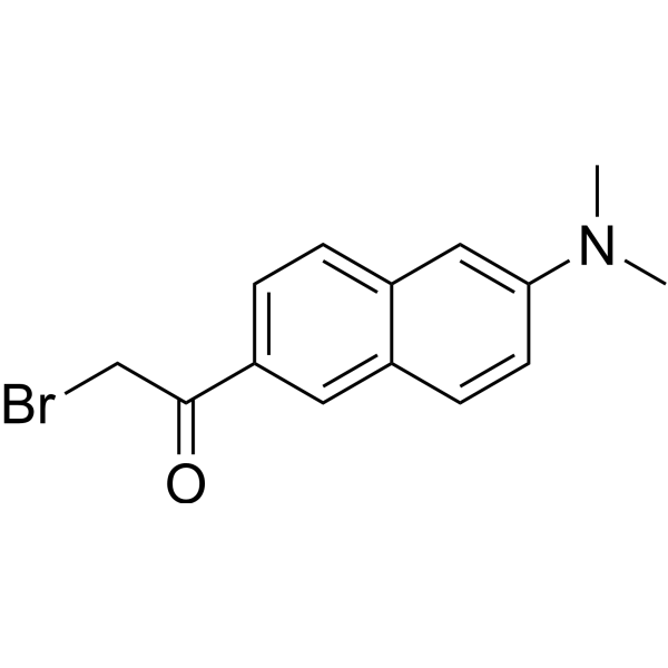
BADAN is a thiol-reactive, environment-sensitive fluorescent probe used for site-specific labeling of protein cysteine residues to detect protein topology, local polarity, protein-lipid interactions, and conformational changes. The bromoacetyl group of BADAN covalently binds to cysteine thiols, and its fluorescence originates from the intramolecular charge transfer (ICT) excited state. The emission peak position varies with the polarity of the surrounding medium: it blue-shifts to approximately 488 nm in nonpolar environments (such as the hydrophobic core of lipid bilayers) and red-shifts to approximately 496-507 nm in polar environments (such as aqueous phases), independent of enzymes, pH, or membrane potential. Ex = 380-402 nm; Em = 400-600 nm .
loading...
    loading...
Cat. No.: HY-D3147
CAS No.: 1965316-23-7
Target:  

Fluorescent Dye

Research Areas:  

Others

RhoVR 1 is a voltage-sensitive probe targeted to the inner mitochondrial membrane. The detection mechanism of RhoVR 1 relies on membrane potential via the photoinduced electron transfer (PeT) mechanism. RhoVR 1 can be used for cell imaging .
loading...
    loading...
Cat. No.: HY-D3127
CAS No.: 455251-97-5
Target:  

Fluorescent Dye

Research Areas:  

Others

DCA-Mln is a Fluorescent probe for ferric iron (Fe³⁺) detection. The probe works via an internal charge transfer (ICT) process from its diethylamino group to its dicyano group, which produces bright pink fluorescence in its unbound state; when Fe³⁺ is present, two molecules of DCA-Mln chelate with one Fe³⁺ ion, with one cyano group of each DCA-Mln participating in the complexation, and this binding triggers a photoinduced electron transfer (PET) process that provides a nonradiative deactivation pathway, resulting in fluorescence quenching; additionally, the probe exhibits a visible color change from purple to peach pink upon Fe³⁺ binding, enabling dual-channel detection. The probe has an excitation wavelength of 570 nm and an emission wavelength of 670 nm, and it shows a rapid response, with fluorescence stabilizing within 15 seconds of Fe³⁺ addition. DCA-Mln is cell-membrane-permeable, nontoxic at the imaging concentration of 10 μM, and can be used for Fe³⁺ detection in actual water samples, Fe³⁺ imaging in living cells, and as an anti-counterfeiting ink[1].
loading...
    loading...
Cat. No.: HY-D3480
BeRST voltage sensitive dye is a silicon-rhodamine-based voltage-sensitive dye that serves as a membrane potential indicator, featuring fast response kinetics, high sensitivity, photostability, and excellent signal-to-noise ratio. BeRST voltage sensitive dye relies on the photoinduced electron transfer (PeT) mechanism to report changes in neuronal membrane potential via fluorescent signals. BeRST voltage sensitive dye localizes to the extracellular surface of neuronal plasma membranes, including cell bodies and neurites. BeRST voltage sensitive dye can be used in studies related to Alzheimer's disease (Ex/Em = 658/683 nm) .
loading...
    loading...
Cat. No.: HY-D3188
CAS No.: 2730023-28-4
gGlu-2OMe SiR600 is a γ-glutamyl transpeptidase (GGT) responsive fluorescent probe. gGlu-2OMe SiR600 can be converted into a highly fluorescent molecule via reaction with GGT, and its initial fluorescence is quenched through a photoinduced electron transfer (PeT) mechanism. gGlu-2OMe SiR600 exhibits fluorescence activation in malignant breast cancer and benign breast fibroadenoma tissues, enabling lesion visualization. gGlu-2OMe SiR600 can be used for research related to breast cancer and fibroadenoma .
loading...
    loading...
Cat. No.: HY-D3602
CAS No.: 2558211-88-2
Target:  

Fluorescent Dye

Research Areas:  

Others

Mito-CS1 is a fluorescent probe for imaging and reversible detection of the exchangeable mitochondrial Cu + pool in living cells. Mito-CS1 combines a Cu +-responsive fluorescent platform with a mitochondria-targeted triphenylphosphonium group, which localizes the probe to mitochondria via a proton gradient. Mito-CS1 contains a thioether-rich receptor for the selective recognition and binding of Cu +, triggering a fluorescence turn-on response by inhibiting photoinduced electron transfer. The apo-form of Mito-CS1 has excitation/emission wavelengths of 555/569 nm, while those of Mito-CS1 after binding to Cu + are 550/558 nm .
loading...
    loading...
Cat. No.: HY-W854727
CAS No.: 151872-44-5
Target:  

Others

Research Areas:  

Others

N-Methylfulleropyrrolidine serves as an electron acceptor molecule characterized by its fullerene core and a nitrogen-free lone pair associated with the pyrrolidine ring. This compound can act as an intermediate in the synthesis of various functionalized fullerenes and plays a significant role in the development of opto-electronic devices. Comprehensive studies have been conducted on the photoinduced energy and electron transfer processes between oligothienylenevinylenes and N-Methylfulleropyrrolidine (MP-C60).
loading...
    loading...
Cat. No.: HY-126926
CAS No.: 36408-16-9
Synonyms: 5-Deazaflavin mononucleotide
Research Areas:  

Others

5-Deaza-FMN (5-Deazaflavin mononucleotide) is a binder of type II isopentenyl diphosphate: dimethylallyl diphosphate isomerase (IDI-2) with a Kd value of 16 μM. 5-Deaza-FMN acts as a radical generator, forming radicals through photoinduced single-electron transfer of tryptophan. 5-Deaza-FMN finds applications in biocatalysis and photocatalysis .
loading...
    loading...
Cat. No.: HY-D3243
CAS No.: 1646537-60-1
Mn1 (M1) is a BODIPY-based Mn 2+-selective "turn-on" Fluorescent sensor and fluorescence modulator. In the absence of Mn 2+, Mn1 undergoes fluorescence quenching via photoinduced electron transfer; binding to Mn 2+ inhibits this mechanism, thereby generating a fluorescence "turn-on" response. Mn1 exhibits Mn 2+-specific cellular fluorescence response .
loading...
    loading...
Cat. No.: HY-D3255
CAS No.: 866534-64-7
CTAP-1 is a monovalent copper ion probe. CTAP-1 binds to monovalent copper Cu (I) to inhibit photoinduced electron transfer, thereby enhancing fluorescence emission. CTAP-1 produces copper-dependent perinuclear staining that colocalizes with mitochondria and the Golgi apparatus. CTAP-1 can be used for imaging Cu (I) pools, excitation/emission Wavelength: 365/480 nm .
loading...
    loading...
Cat. No.: HY-D3250
CAS No.: 2445485-84-5
PYSNO is a lysosome-targeted fluorescent probe based on a pyridazinone skeleton (λem=515-565 nm, λex=405 nm) that can be used to track nitric oxide (NO) production in vivo. PYSNO exhibits a rapid, highly sensitive and highly selective "turn-on" response to endogenous and exogenous NO by blocking photoinduced electron transfer and regulating radiative decay rates. PYSNO enables precise in vivo monitoring in a mouse model of myocardial fibrosis and can be applied to the research of related diseases .
loading...
    loading...
Cat. No.: HY-D3095
CAS No.: 2244914-41-6
Target:  

Fluorescent Dye

Research Areas:  

Others

TPEP-6-NH-MA is a Fluorescent probe that selectively detects cysteine (Cys) and homocysteine (Hcy) without interference from glutathione (GSH) and other amino acids, and can also be used for bioimaging of biothiols in living cells. Its detection mechanism relies on terminating the photoinduced electron transfer (PET) effect via a Michael addition reaction between the maleimide recognition moiety and the thiol group of Cys or Hcy. The excitation wavelength of TPEP-6-NH-MA is Ex = 361 nm .
loading...
    loading...
Cat. No.: HY-D3185
CAS No.: 2243148-62-9
AlDeSense AM is a cell-permeable ALDH1A1-selective fluorescent reporter. AlDeSense AM is oxidized by ALDH1A1, which eliminates photoinduced electron transfer quenching and enhances the fluorescent signal. AlDeSense AM can be used to detect cells with cancer stem cell properties, as well as to monitor the plasticity of cancer stem cells in cell culture systems and animal models. AlDeSense AM is applicable to the study of cancers associated with cancer stem cells, including chronic myeloid leukemia, melanoma, and breast cancer .
loading...
    loading...
Cat. No.: HY-D3431
CFSE-Tz is a fluorescent bioorthogonal probe constructed based on the IEDDA reaction, which is mainly used for visual tracing of amino acid uptake in living cells. The tetrazine group within the CFSE-Tz molecule quenches CFSE fluorescence via photoinduced electron transfer; after the probe specifically undergoes a bioorthogonal reaction with trans-cyclooctene-modified amino acids, the quenching effect is eliminated, and a green fluorescent signal is released, thereby dynamically monitoring the intracellular amino acid uptake process. CFSE-Tz can be applied to in vitro single-cell fluorescence imaging and studies related to amino acid metabolism in tumor cells (Ex = 495 nm, Em = 519 nm) [1] [2].
loading...
    loading...
Cat. No.: HY-D3189
CAS No.: 2370825-44-6
5GluAF-2MeTG is an activatable fluorescent probe targeting the glutamate carboxypeptidase (CP) activity of PSMA (Ex/Em=490/500-600 nm). After being hydrolyzed by PSMA, 5GluAF-2MeTG releases a cell membrane-permeable fluorescent product, and achieves fluorescence activation by disrupting donor-excited photoinduced electron transfer (d-PeT). 5GluAF-2MeTG enables fluorescence imaging of live PSMA-expressing prostate cancer cells in vitro and visualizes the carboxypeptidase activity of PSMA. 5GluAF-2MeTG can be used to detect prostate cancer regions in preclinical excised tissue specimens .
loading...
    loading...
Cat. No.: HY-D3409
CAS No.: 1854026-50-8
Target:  

Fluorescent Dye

Research Areas:  

Others

Si-DMA is a far-red fluorescent singlet oxygen probe (Ex/Em = 640/690 nm). After reacting with singlet oxygen, Si-DMA forms an endoperoxide with a 17-fold fluorescence enhancement. Si-DMA localizes selectively to mitochondria with negligible auto-oxidation. Si-DMA can be used for high spatiotemporal resolution visualization and detection of mitochondrial singlet oxygen in living cells .
loading...
    loading...
  • 1

