



Tricetin
Based on 1 publication(s) in Google Scholar
Tricetin is a potent competitive inhibitor of the Keap1-Nrf2 Protein Protein Interaction (PPI). Tricetin protects against 6-OHDA-induced neurotoxicity in Parkinson's disease model by activating Nrf2/HO-1 signaling pathway and preventing mitochondria-dependent apoptosis pathway.
Nos produits utilisent uniquement pour la recherche. Nous ne vendons pas aux patients.
- Pureté : 99.88%
- CAS No.: 520-31-0
- Formule: C15H10O7
- Masse moléculaire:302.24
-
Stockage:Powder -20°C, 3 years , 4°C, 2 years ; In solvent -80°C, 6 months , -20°C, 1 month

To place orders, for customer services and technical support, please contact: MedChemExpress USA
Tel: 609-228-6898 E-mail: [email protected] [email protected]


-
 Activité biologique
-
Chemical Information
-
Solvant et solubilité
- Protocole
- Pureté et documentation
- Références
-
 Help & FAQs


-

HY-B0015
PaclitaxelAntitumor Agent -
HY-32735
TriptolideImmunosuppressive Agent -
HY-17031
SBE-β-CDExcipient -
HY-15753
EllipticineTopoisomerase Inhibitor -
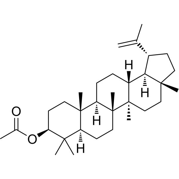
HY-126114
Lupeol acetateAnti-inflammatory Agent -
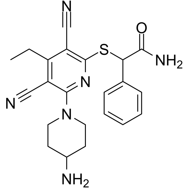
HY-139664
GSK-3685032DNMT1 Inhibitor -
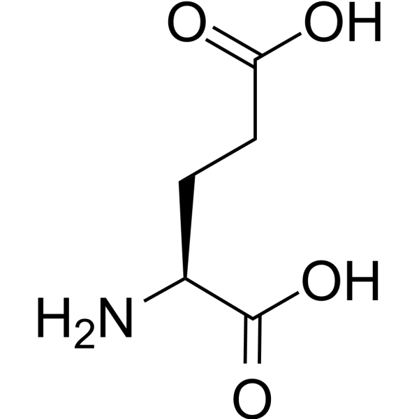
HY-14608
L-Glutamic acidGlutamate Receptors Agonist -
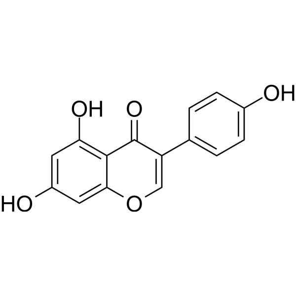
HY-14596
GenisteinAutophagy Inducer -

HY-10182
LaduviglusibGSK-3 Inhibitor -
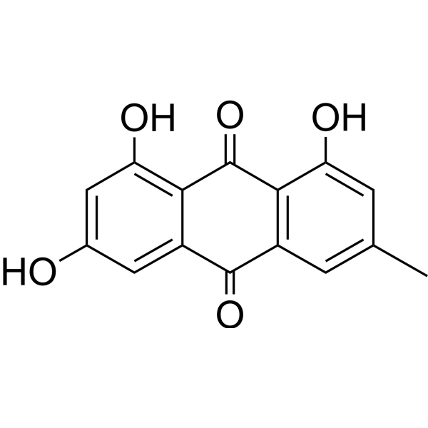
HY-14393
EmodinNatural Anthraquinone -
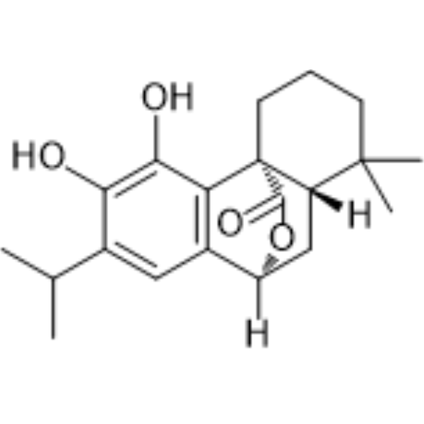
HY-N0643
CarnosolEndogenous Metabolite
Publications Citing Use of MedChemExpress (MCE) Tricetin
More

Activité biologique
Description
Cellular Effect
|
Cell Line

|
Type | Value | Description | References |
|---|---|---|---|---|
| Huh-7 | CC50 |
>50 μM
Compound: 15
|
Cytotoxicity against human Huh7.5.1 cells by MTT assay
Cytotoxicity against human Huh7.5.1 cells by MTT assay
|
[PMID: 22445328] |
| Huh-7 | EC50 |
32.2 μM
Compound: 15
|
Antiviral activity against HCV JFH-1 J399EM infected in Human Huh7.5.1 cells assessed as suppression of viral replication after 72 hrs by EGFP assay
Antiviral activity against HCV JFH-1 J399EM infected in Human Huh7.5.1 cells assessed as suppression of viral replication after 72 hrs by EGFP assay
|
[PMID: 22445328] |
In Vitro
Tricetin is mainly found in natural plants such as Ginkgo biloba L., Carica papaya L. and Murraya exotica L. Tricetin activates the Nrf2/HO-1 pathway to protect cells from oxidative stress. Tricetin possessed the protective effect on dopamine neurons of C. elegans. Tricetin has cytostatic properties and anti-metastatic activity of various solid tumors[1].
Pretreatment with Tricetin (20, 40, and 80 μM; for 4 hours) significantly improves 6-OHDA (200 μM)-induced SH-SY5Y cells viability and suppresses mitochondria-mediated apoptosis[1].
Tricetin (80 μM; for 1, 2 and 4 h) markedly decreased the expressions of p-JNK and p-p38[1].
MedChemExpress (MCE) has not independently confirmed the accuracy of these methods. They are for reference only.
-
Cell Line:SH-SY5Y cells
-
Concentration:20, 40, and 80 μM
-
Incubation Time:Pretreatment for 4 h followed by 6-OHDA (200 μM) for 24 h
-
Result:Significantly increased 6-OHDA-induced SH-SY5Y cells viability.
-
Cell Line:SH-SY5Y cells
-
Concentration:80 μM
-
Incubation Time:1, 2 and 4 h
-
Result:Markedly decreased the expressions of p-JNK and p-p38.
- Neurotoxicity Study
- Detection of Apoptosis
- TUNEL staining for apoptotic DNA fragmentation
- Annexin V plus membrane-impermeant dye apoptosis staining
- Pull-down
- Immunoprecipitation
- Human pluripotent stem cell midbrain dopaminergic neuron differentiation
- Protocol for Bimolecular Fluorescence Complementation (BiFC) Assay
- Apoptosis Solutions
- Co-Immunoprecipitation
- Protocol for Yeast Two-Hybrid (Y2H) Assay
- How to Choose the Right Model Animal


Chemical Information
-
CAS No. 520-31-0
-
Appearance Solid
-
Masse moléculaire 302.24
-
Formule C15H10O7
-
Color Light yellow to brown
-
SMILES
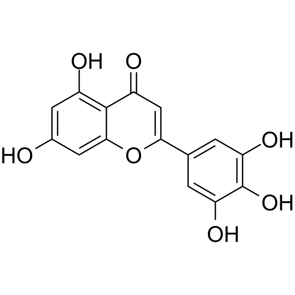
O=C1C=C(C2=CC(O)=C(O)C(O)=C2)OC3=CC(O)=CC(O)=C13
-
Structure Classification
-
Initial Source
-
Livraison
Room temperature in continental US; may vary elsewhere.
-
Stockage
Powder -20°C 3 years 4°C 2 years In solvent -80°C 6 months -20°C 1 month


Publications (1)
-
Journal Impact Factor
-
Most Recent
-
Int Immunopharmacol
Tricetin attenuates atherosclerosis by suppressing macrophage ferroptosis via activation of the NRF2 pathway. [Abstract]2024 Oct 22;143(Pt 2):113418. PMID: 39442188


Solvant et solubilité
In Vitro:
DMSO : 100 mg/mL (330.86 mM; Need ultrasonic; Hygroscopic DMSO has a significant impact on the solubility of product, please use newly opened DMSO)
Please refer to the solubility information to select the appropriate solvent. Once prepared, please aliquot and store the solution to prevent product inactivation from repeated freeze-thaw cycles.
Storage method and period of stock solution: -80°C, 6 months; -20°C, 1 month. When stored at -80°C, please use it within 6 months. When stored at -20°C, please use it within 1 month.
Please refer to the solubility information to select the appropriate solvent. Once prepared, please aliquot and store the solution to prevent product inactivation from repeated freeze-thaw cycles.
Storage method and period of stock solution: -80°C, 6 months; -20°C, 1 month. When stored at -80°C, please use it within 6 months. When stored at -20°C, please use it within 1 month.
Concentration (start) × Volume (start) = Concentration (final) × Volume (final)


Pureté et documentation
-
Fiche technique (275 KB)
-
SDS (394 KB)
- English - EN (394 KB)
- Français - FR (394 KB)
- Deutsch - DE (394 KB)
- Norwegian - NO (394 KB)
- Español - ES (394 KB)
- Swedish - SV (394 KB)
- Italian - IT (394 KB)
- Korean - KR (394 KB)
- Portuguese - PT (394 KB)
-
Instruction de manipulation (2659 KB)
Références


Complete Stock Solution Preparation Table
Please refer to the solubility information to select the appropriate solvent. Once prepared, please aliquot and store the solution to prevent product inactivation from repeated freeze-thaw cycles.
Storage method and period of stock solution: -80°C, 6 months; -20°C, 1 month. When stored at -80°C, please use it within 6 months. When stored at -20°C, please use it within 1 month.
| Optional Solvent | Concentration Solvent Mass | 1 mg | 5 mg | 10 mg | 25 mg |
|---|---|---|---|---|---|
| DMSO | 1 mM | 3.3086 mL | 16.5431 mL | 33.0863 mL | 82.7157 mL |
| 5 mM | 0.6617 mL | 3.3086 mL | 6.6173 mL | 16.5431 mL | |
| 10 mM | 0.3309 mL | 1.6543 mL | 3.3086 mL | 8.2716 mL | |
| 15 mM | 0.2206 mL | 1.1029 mL | 2.2058 mL | 5.5144 mL | |
| 20 mM | 0.1654 mL | 0.8272 mL | 1.6543 mL | 4.1358 mL | |
| 25 mM | 0.1323 mL | 0.6617 mL | 1.3235 mL | 3.3086 mL | |
| 30 mM | 0.1103 mL | 0.5514 mL | 1.1029 mL | 2.7572 mL | |
| 40 mM | 0.0827 mL | 0.4136 mL | 0.8272 mL | 2.0679 mL | |
| 50 mM | 0.0662 mL | 0.3309 mL | 0.6617 mL | 1.6543 mL | |
| 60 mM | 0.0551 mL | 0.2757 mL | 0.5514 mL | 1.3786 mL | |
| 80 mM | 0.0414 mL | 0.2068 mL | 0.4136 mL | 1.0339 mL | |
| 100 mM | 0.0331 mL | 0.1654 mL | 0.3309 mL | 0.8272 mL |


Tricetin Related Classifications
HY-131592 Related Classifications
 Powered by Bioz