



TRK-IN-24
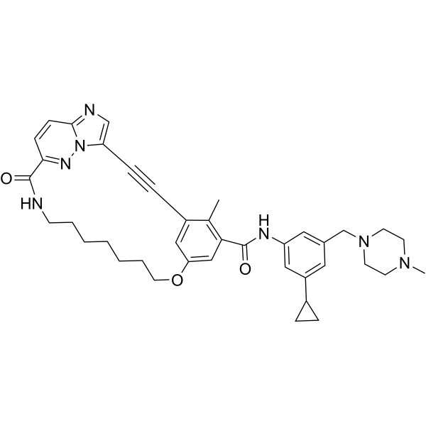
TRK-IN-24 (compound 10g) is a Trk Receptor inhibitor that inhibits TRKA, TRKC, TRKAG595R, TRKAG667C and TRKAF589L IC50s are 5.21, 4.51, 6.77, 1.42 and 6.13 nM respectively. TRK-IN-24 has antitumor efficacy in BaF3-CD74-NTRK1G595R and BaF3-CD74-NTRK1G667C xenograft models. TRK-IN-24 inhibits the proliferation of Ba/F3 cells transfected with single mutants such as SF, GK, and xDFG, with an IC50 of 1.43-47.56 nM.
For research use only. We do not sell to patients.
- CAS No.: 2937544-01-7
- Formula: C39H45N7O3
- Molecular Weight:659.82
-
Storage:
Please store the product under the recommended conditions in the Certificate of Analysis.

To place orders, for customer services and technical support, please contact: MedChemExpress USA
Tel: 609-228-6898 E-mail: [email protected] [email protected]


-
 Biological Activity
-
Chemical Information
- Protocols
- Purity & Documentation
- References
-
 Help & FAQs




Biological Activity
Description
IC50 & Target
IC50: 5.21 nM (TRKA), 4.51 nM (TRKC), 6.77 nM (TRKAG595R), 1.42 nM (TRKAG667C), 6.13 nM (TRKAF589L)[1]
Cellular Effect
|
Cell Line

|
Type | Value | Description | References |
|---|---|---|---|---|
| BaF3 | IC50 |
1.43 nM
Compound: 10g
|
Antiproliferative activity against mouse BaF3 cells harbouring CD74-NTRK1 G667C mutant fusion protein incubated for 3 days by CCK8 method
Antiproliferative activity against mouse BaF3 cells harbouring CD74-NTRK1 G667C mutant fusion protein incubated for 3 days by CCK8 method
|
[PMID: 37676745] |
| BaF3 | IC50 |
1.7 nM
Compound: 10g
|
Antiproliferative activity against mouse BaF3 cells harbouring ETV6-TRKC G696C mutant incubated for 3 days by CCK8 method
Antiproliferative activity against mouse BaF3 cells harbouring ETV6-TRKC G696C mutant incubated for 3 days by CCK8 method
|
[PMID: 37676745] |
| BaF3 | IC50 |
121.09 nM
Compound: 10g
|
Antiproliferative activity against mouse BaF3 cells harbouring CD74-TRKA G595R/G667A mutant incubated for 3 days by CCK8 method
Antiproliferative activity against mouse BaF3 cells harbouring CD74-TRKA G595R/G667A mutant incubated for 3 days by CCK8 method
|
[PMID: 37676745] |
| BaF3 | IC50 |
17.67 nM
Compound: 10g
|
Antiproliferative activity against mouse BaF3 cells harbouring ETV6-NTRK3 fusion protein incubated for 3 days by CCK8 method
Antiproliferative activity against mouse BaF3 cells harbouring ETV6-NTRK3 fusion protein incubated for 3 days by CCK8 method
|
[PMID: 37676745] |
| BaF3 | IC50 |
2.36 nM
Compound: 10g
|
Antiproliferative activity against mouse BaF3 cells harbouring CD74-TRKA G667A mutant incubated for 3 days by CCK8 method
Antiproliferative activity against mouse BaF3 cells harbouring CD74-TRKA G667A mutant incubated for 3 days by CCK8 method
|
[PMID: 37676745] |
| BaF3 | IC50 |
2.37 nM
Compound: 10g
|
Antiproliferative activity against mouse BaF3 cells harbouring CD74-NTRK1 fusion protein incubated for 3 days by CCK8 method
Antiproliferative activity against mouse BaF3 cells harbouring CD74-NTRK1 fusion protein incubated for 3 days by CCK8 method
|
[PMID: 37676745] |
| BaF3 | IC50 |
2.38 nM
Compound: 10g
|
Antiproliferative activity against mouse BaF3 cells harbouring ETV6-TRKC G623E mutant incubated for 3 days by CCK8 method
Antiproliferative activity against mouse BaF3 cells harbouring ETV6-TRKC G623E mutant incubated for 3 days by CCK8 method
|
[PMID: 37676745] |
| BaF3 | IC50 |
2.46 nM
Compound: 10g
|
Antiproliferative activity against mouse BaF3 cells harbouring ETV6-TRKC G696A mutant incubated for 3 days by CCK8 method
Antiproliferative activity against mouse BaF3 cells harbouring ETV6-TRKC G696A mutant incubated for 3 days by CCK8 method
|
[PMID: 37676745] |
| BaF3 | IC50 |
22.82 nM
Compound: 10g
|
Antiproliferative activity against mouse BaF3 cells harbouring ETV6-TRKC V601M mutant incubated for 3 days by CCK8 method
Antiproliferative activity against mouse BaF3 cells harbouring ETV6-TRKC V601M mutant incubated for 3 days by CCK8 method
|
[PMID: 37676745] |
| BaF3 | IC50 |
25.44 nM
Compound: 10g
|
Antiproliferative activity against mouse BaF3 cells harbouring wild type ETV6-TRKB incubated for 3 days by CCK8 method
Antiproliferative activity against mouse BaF3 cells harbouring wild type ETV6-TRKB incubated for 3 days by CCK8 method
|
[PMID: 37676745] |
| BaF3 | IC50 |
26.4 nM
Compound: 10g
|
Antiproliferative activity against mouse BaF3 cells harbouring ETV6-TRKB G709C mutant incubated for 3 days by CCK8 method
Antiproliferative activity against mouse BaF3 cells harbouring ETV6-TRKB G709C mutant incubated for 3 days by CCK8 method
|
[PMID: 37676745] |
| BaF3 | IC50 |
27.07 nM
Compound: 10g
|
Antiproliferative activity against mouse BaF3 cells harbouring CD74-TRKA G6678 mutant incubated for 3 days by CCK8 method
Antiproliferative activity against mouse BaF3 cells harbouring CD74-TRKA G6678 mutant incubated for 3 days by CCK8 method
|
[PMID: 37676745] |
| BaF3 | IC50 |
27.2 nM
Compound: 10g
|
Antiproliferative activity against mouse BaF3 cells harbouring CD74-NTRK1 G595R mutant fusion protein incubated for 3 days by CCK8 method
Antiproliferative activity against mouse BaF3 cells harbouring CD74-NTRK1 G595R mutant fusion protein incubated for 3 days by CCK8 method
|
[PMID: 37676745] |
| BaF3 | IC50 |
315.8 nM
Compound: 10g
|
Antiproliferative activity against mouse BaF3 cells harbouring CD74-TRKA G595R/G667C mutant incubated for 3 days by CCK8 method
Antiproliferative activity against mouse BaF3 cells harbouring CD74-TRKA G595R/G667C mutant incubated for 3 days by CCK8 method
|
[PMID: 37676745] |
| BaF3 | IC50 |
46.15 nM
Compound: 10g
|
Antiproliferative activity against mouse BaF3 cells harbouring ETV6-TRKB G639R mutant incubated for 3 days by CCK8 method
Antiproliferative activity against mouse BaF3 cells harbouring ETV6-TRKB G639R mutant incubated for 3 days by CCK8 method
|
[PMID: 37676745] |
| BaF3 | IC50 |
47.56 nM
Compound: 10g
|
Antiproliferative activity against mouse BaF3 cells harbouring ETV6-TRKB F633L mutant incubated for 3 days by CCK8 method
Antiproliferative activity against mouse BaF3 cells harbouring ETV6-TRKB F633L mutant incubated for 3 days by CCK8 method
|
[PMID: 37676745] |
| BaF3 | IC50 |
5 nM
Compound: 10g
|
Antiproliferative activity against mouse BaF3 cells harbouring CD74-TRKA V573M mutant incubated for 3 days by CCK8 method
Antiproliferative activity against mouse BaF3 cells harbouring CD74-TRKA V573M mutant incubated for 3 days by CCK8 method
|
[PMID: 37676745] |
| BaF3 | IC50 |
5.12 nM
Compound: 10g
|
Antiproliferative activity against mouse BaF3 cells harbouring ETV6-TRKB V617M mutant incubated for 3 days by CCK8 method
Antiproliferative activity against mouse BaF3 cells harbouring ETV6-TRKB V617M mutant incubated for 3 days by CCK8 method
|
[PMID: 37676745] |
| BaF3 | IC50 |
5.56 nM
Compound: 10g
|
Antiproliferative activity against mouse BaF3 cells harbouring ETV6-TRKC G6968 mutant incubated for 3 days by CCK8 method
Antiproliferative activity against mouse BaF3 cells harbouring ETV6-TRKC G6968 mutant incubated for 3 days by CCK8 method
|
[PMID: 37676745] |
| BaF3 | IC50 |
6.13 nM
Compound: 10g
|
Antiproliferative activity against mouse BaF3 cells harbouring TRKA-F589L fusion protein incubated for 3 days by CCK8 method
Antiproliferative activity against mouse BaF3 cells harbouring TRKA-F589L fusion protein incubated for 3 days by CCK8 method
|
[PMID: 37676745] |
| BaF3 | IC50 |
6.42 nM
Compound: 10g
|
Antiproliferative activity against mouse BaF3 cells harbouring ETV6-TRKC G623R mutant incubated for 3 days by CCK8 method
Antiproliferative activity against mouse BaF3 cells harbouring ETV6-TRKC G623R mutant incubated for 3 days by CCK8 method
|
[PMID: 37676745] |
| BaF3 | IC50 |
7.68 nM
Compound: 10g
|
Antiproliferative activity against mouse BaF3 cells harbouring ETV6-TRKC F617L mutant incubated for 3 days by CCK8 method
Antiproliferative activity against mouse BaF3 cells harbouring ETV6-TRKC F617L mutant incubated for 3 days by CCK8 method
|
[PMID: 37676745] |
| BaF3 | IC50 |
8.62 nM
Compound: 10g
|
Antiproliferative activity against mouse BaF3 cells harbouring CD74-NTRKIF 589L mutant fusion protein incubated for 3 days by CCK8 method
Antiproliferative activity against mouse BaF3 cells harbouring CD74-NTRKIF 589L mutant fusion protein incubated for 3 days by CCK8 method
|
[PMID: 37676745] |
| BaF3 | IC50 |
982.75 nM
Compound: 10g
|
Antiproliferative activity against mouse BaF3 cells incubated for 3 days by CCK8 method
Antiproliferative activity against mouse BaF3 cells incubated for 3 days by CCK8 method
|
[PMID: 37676745] |
In Vitro
In Vivo
MedChemExpress (MCE) has not independently confirmed the accuracy of these methods. They are for reference only.


Chemical Information
-
CAS No. 2937544-01-7
-
Molecular Weight 659.82
-
Formula C39H45N7O3
-
SMILES
CC(C(C#CC1=CN=C2C=CC3=NN21)=CC(OCCCCCCCNC3=O)=C4)=C4C(NC5=CC(C6CC6)=CC(CN7CCN(CC7)C)=C5)=O
-
Shipping
Room temperature in continental US; may vary elsewhere.
-
Storage
Please store the product under the recommended conditions in the Certificate of Analysis.


Purity & Documentation
References

Calculators
Concentration (start) × Volume (start) = Concentration (final) × Volume (final)


TRK-IN-24 Related Classifications
HY-156086 Related Classifications
 Powered by Bioz