



Anti-Mouse CCL5 Antibody (R6G9)

Based on 1 Customer Validation
Anti-Mouse CCL5 Antibody (R6G9) is a neutralizing agent that binds to CCL5 to inhibit its chemotactic activity towards CD4+ T cells.Anti-Mouse CCL5 Antibody (R6G9) does not suppress CD4+ T cell migration towards infection-derived liver cells, reduce CD4+ T cell hepatic accumulation, or impair CD4+ T cell migratory capacity.Anti-Mouse CCL5 Antibody (R6G9) serves as a tool to assess CCL5’s role in in vivo target cell killing by LCMV-specific CD8+ effector T cells.Anti-Mouse CCL5 Antibody (R6G9) can be used for the research of malaria infection and lymphocytic choriomeningitis virus infection.
For research use only. We do not sell to patients.
- Purity: 97.24%
-
Storage:
Please store the product under the recommended conditions in the Certificate of Analysis.


To place orders, for customer services and technical support, please contact: MedChemExpress USA
Tel: 609-228-6898 E-mail: [email protected] [email protected]


-
 Biological Activity
-
Chemical Information
- Purity & Documentation
- References
-
 Help & FAQs




Biological Activity
Description
Isotype
Mouse IgG1 kappa
Recommend Isotype Controls
Species Reactivity
Mouse
IC50 & Target
CCL5
In Vitro
Anti-Mouse CCL5 Antibody (R6G9) (10 µg/mL; 2 h pre-incubation) completely blocks the chemotactic response of CD4+ T cells from WSX-1-/- mice infected with Plasmodium berghei NK65 on day 14 to 100 ng/mL recombinant CCL5 in Transwell assays, but fails to inhibit their migration to infected liver tissues from mice in the same infection state[1].
Anti-Mouse CCL5 Antibody (R6G9) (0-60 min after peptide stimulation) enables quantitative detection of pre-stored CCL5 rapidly released by purified p14 effector T cells[2].
MedChemExpress (MCE) has not independently confirmed the accuracy of these methods. They are for reference only.
-
Cell Line:T cells; liver cell suspensions
-
Concentration:10 μg/mL
-
Incubation Time:3 h (T cells 2 h pre‑incubation; liver cell suspensions 1 h pre‑incubation)
-
Result:Failed to inhibit the chemotaxis of splenic CD4⁺ T cells from Plasmodium berghei NK65‑infected (day 14) WSX‑1⁻/⁻ mice toward infected hepatic cell suspensions, despite effectively blocking recombinant CCL5‑induced migration.
In Vivo
Anti-Mouse CCL5 Antibody (R6G9) (100 μg; i.v.; single dose) does not impair the in vivo killing activity of LCMV-specific CD8+ effector T cells[2].
MedChemExpress (MCE) has not independently confirmed the accuracy of these methods. They are for reference only.
-
Animal Model:C57BL/6 IL-27R deficient (WSX-1-/-) (6-10 weeks old; malaria model via intravenous injection of 104 parasitized red blood cells of *Plasmodium berghei* NK65)[1]
-
Dosage:250 μg
-
Administration:i.p.; daily; 7 days starting day 7 post-infection
-
Result:Did not suppress intrahepatic CD4+ T cell accumulation in infected WSX-1-/- mice, with no significant reduction in absolute CD4+ T cell numbers in the liver compared to untreated infected WSX-1-/- mice.
Exhibited unimpaired migration towards infection-derived liver cells in an in vitro transwell assay.
Significantly suppressed migration of infection-derived WSX-1-/- CD4+ T cells towards recombinant CCL5 in an in vitro chemotaxis assay, but did not prevent migration of these cells towards infection-derived WSX-1-/- or WT liver cells.
-
Animal Model:C57BL/6J (B6) mice (sex-matched, 8-10 weeks old, LCMV Armstrong challenge)[2]
-
Dosage:100 μg
-
Administration:i.v.; single dose
-
Result:Did not alter the rapid kinetics of in vivo target cell killing by LCMV-specific CD8+ effector T cells.
Proceeded with the same killing efficiency as in control mice.
Gene ID
Accession
P30882
Conjugated
Unconjugated
Reconsititution
The product can be reconstituted/diluted with sterile PBS or saline.
Format
-
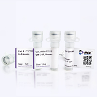
Product Image
Application
in vivo neutralization of CCL5; in vitro neutralization of CCL5; Functional assay; ELISA


Chemical Information
-
Appearance Liquid
-
Color Colorless to light yellow
-
Formulation
Please refer to the lot-specific COA for specific buffer information.
-
Storage
Please store the product under the recommended conditions in the Certificate of Analysis.


Purity & Documentation
-
Data Sheet (263 KB)
-
SDS (251 KB)
- English - EN (251 KB)
- Français - FR (251 KB)
- Deutsch - DE (251 KB)
- Norwegian - NO (251 KB)
- Español - ES (251 KB)
- Swedish - SV (251 KB)
- Italian - IT (251 KB)
- Korean - KR (251 KB)
- Portuguese - PT (251 KB)
-
Inhibitory Antibodies User Guide (603 KB)
References
[1]. Villegas-Mendez A, et al. WSX-1 signalling inhibits CD4⁺ T cell migration to the liver during malaria infection by repressing chemokine-independent pathways. PloS one. 2013;8(11):e78486. [Content Brief]
[2]. Eberlein J, et al. Chemokine Signatures of Pathogen-Specific T Cells I: Effector T Cells. Journal of immunology (Baltimore, Md. : 1950). 2020 Oct 15;205(8):2169-2187. [Content Brief]

Calculators
Concentration (start) × Volume (start) = Concentration (final) × Volume (final)


Anti-Mouse CCL5 Antibody (R6G9) Related Classifications
HY-P991753 Related Classifications
 Powered by Bioz