



BRAF Antibody (YA820)
(Synonyms: BRAF1, RAFB1, BRAF, Serine/threonine-protein kinase B-raf, Proto-oncogene B-Raf, p94, v-Raf murine sarcoma viral oncogene homolog B1)
Based on 1 Customer Validation
BRAF Antibody (YA820) is a Mouse-derived and non-conjugated IgG2b monoclonal antibody, targeting to BRAF.
-
Host:
Mouse
-
Isotype:
IgG
-
Application:
WB, IHC-P, ICC/IF
-
Reactivity :
Human, Mouse, Rat
-
Formulation:
Supplied in 1*PBS (pH7.4), 0.2% BSA and 50% Glycerol. Preservative: 0.05% Sodium Azide.
-
Conjugation:
Non-conjugated


To place orders, for customer services and technical support, please contact: MedChemExpress USA
Tel: 609-228-6898 E-mail: [email protected] [email protected]





Applications
| Application |
WB

WB: Western Blot
|
ICC/IF
ICC/IF: Immunocytochemistry/
Immunofluorescence |
IHC-P
IHC-P: Immunohistochemistry-Paraffin
|
|---|---|---|---|
| Dilution Ratio | 1:500-1:2000 | 1:100-1:500 | 1:100-1:500 |


Product Details
BRAF Antibody (YA820) is a Mouse-derived and non-conjugated IgG2b monoclonal antibody, targeting to BRAF.
-
Host Mouse

-
Clonality Monoclonal

-
Species ReactivityHuman, Mouse, Rat
-
Observed Molecular WeightObserved band size: 84 kDa
Note: Due to possible protein modifications or aggregation, the molecular weight should be confirmed by actual measurement, and the predicted value is for reference only.

-
Calculated Molecular Weight Predicted band size: 84 kDa

Entrez Gene: 673 Human ; 109880 Mouse ; 114486 Rat
SwissProt: P15056 Human ; P28028 Mouse ;
OMIM: 114500 Human
Synthetic peptide corresponding to Human B-Raf.AA range:49-239.
Endogenous
Protein A affinity purified.
Non-conjugated
Unmodified
IgG


Product Properties
-
Appearance
Solution
-
Formulation
Supplied in 1*PBS (pH7.4), 0.2% BSA and 50% Glycerol. Preservative: 0.05% Sodium Azide.
-
Concentration
Batch-dependent, Please check the COA for the concentration of each lot. Check Lot Concentration
-
Storage & Stability
Stored at -20°C for 1 year. Avoid repeated freeze / thaw cycles.
-
Shipping
Shipping with blue ice.


Verification Images
-
 Western blot analysis was performed on protein extracts (25 μg) from K562 (lane 2), HeLa (lane 3) and Ramos (lane 4) using BRAF antibody. Proteins were transferred onto a 0.45 μm PVDF membrane using the Trans-Blot® Turbo™ system for 13 min. The membrane was then blocked with 5% nonfat milk in TBST (HY-K1025) for 1 h at room temperature. The primary antibody (1:1000) and loading control antibody GAPDH Antibody (HRP) (HY-P80954A) (1:5000) were diluted in 5% nonfat milk in TBST and incubated with the membrane overnight at 4°C. After washing, the membrane of primary antibody was incubated with HRP-conjugated goat anti-rabbit/mouse IgG secondary antibody (HY-P8001/HY-P8004) (1:5000) diluted in 5% nonfat milk in TBST for 1 h at room temperature. Protein bands were visualized using an Ultra High Sensitivity ECL detection kit (HY-K1005).
 -
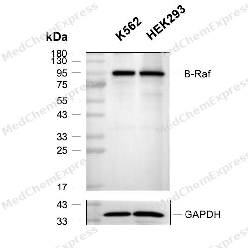 Western blot analysis of extracts from K562(lane 2(20μg) and HEK293(lane 3(20μg) ) using B-Raf (HY-P80036) Mouse mAb. Proteins were transferred to a PVDF membrane and blocked with 5% BSA in TBST for 2 hour at room temperature. The primary antibody (HY-P80036, 1/1000) and Loading control antibody (GAPDH, HY-P80954, 1/3000) was used in 5% BSA in TBST at 4°C overnight. Goat Anti-Mouse IgG-HRP Secondary Antibody (HY-P8004, 1/10,000) was used for 1 hour at room temperature.
 -
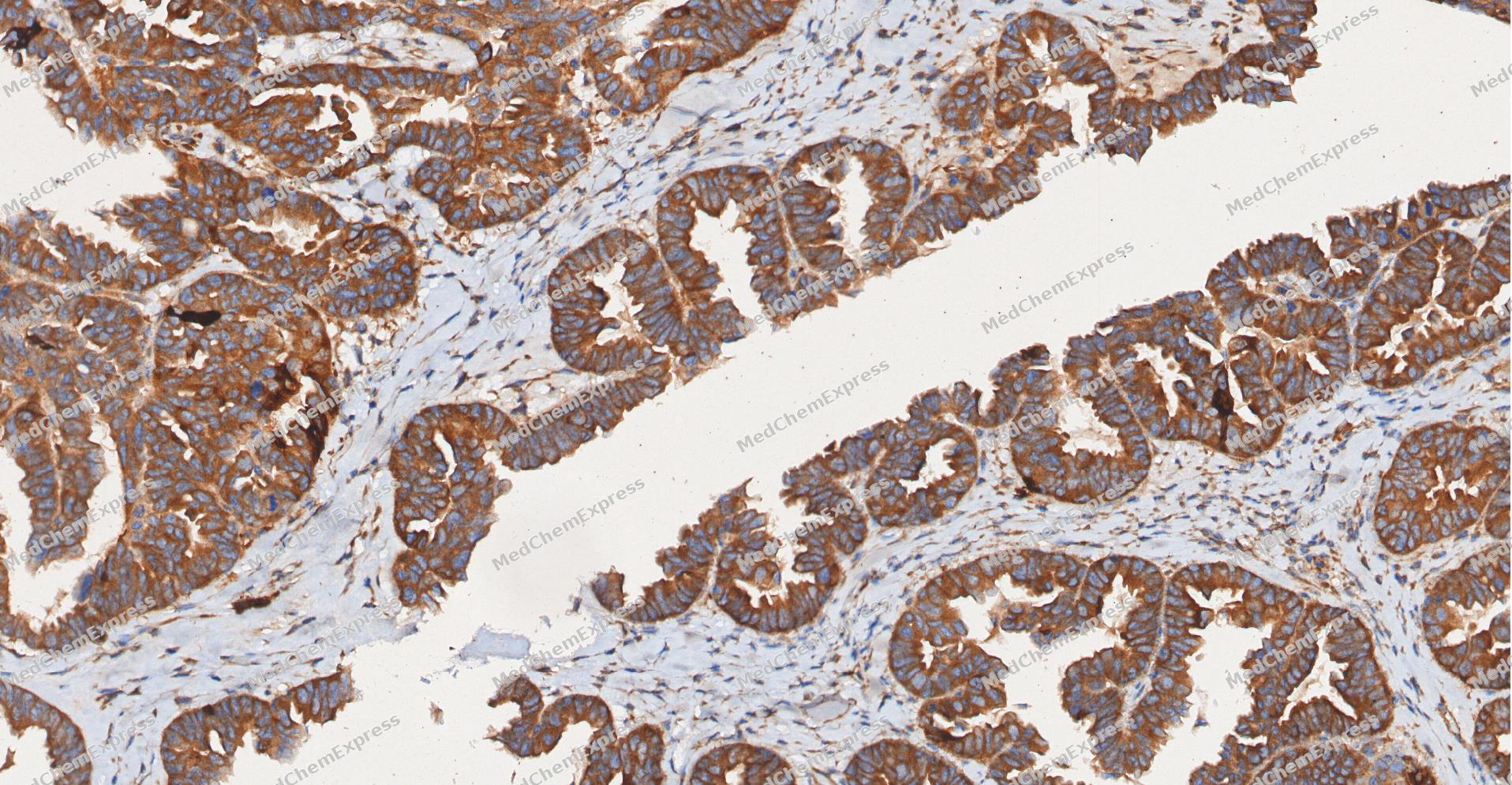 Immunohistochemical analysis of paraffin-embedded human ovarian carcinoma tissue using BRAF antibody was performed. The section was pretreated using high-temperature and high-pressure mediated EDTA antigen retrieval buffer (pH 9.0), for 5 minutes. The tissues were incubated with primary antibody (HY-P80036, 1:100 dilution) at room temperature for 60 minutes. Detection was performed using an HRP conjugated compact polymer system. DAB was used as the chromogen. The tissues were counterstained with hematoxylin and mounted with neutral balsam mounting medium.
 -
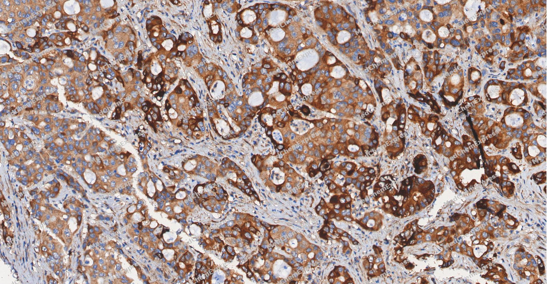 Immunohistochemical analysis of paraffin-embedded human cholangiocarcinoma tissue using BRAF antibody was performed. The section was pretreated using high-temperature and high-pressure mediated EDTA antigen retrieval buffer (pH 9.0), for 5 minutes. The tissues were incubated with primary antibody (HY-P80036, 1:100 dilution) at room temperature for 60 minutes. Detection was performed using an HRP conjugated compact polymer system. DAB was used as the chromogen. The tissues were counterstained with hematoxylin and mounted with neutral balsam mounting medium.
 -
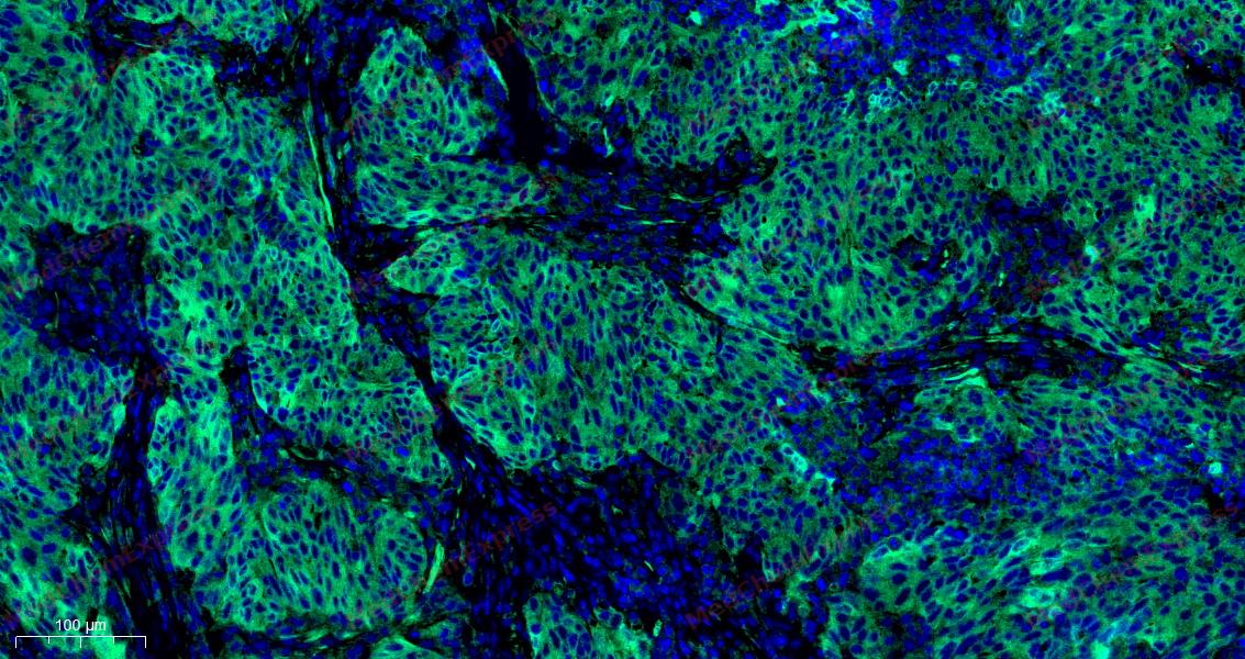 Tyramide signaling amplification based immunofluorescence was performed on paraffin-embedded human ovarian carcinoma (sample 1) tissue using BRAF antibody. Antigen retrieval was performed in EDTA buffer pH 9.0 (95 °C, 20 min) followed by cooling to RT. Then incubated with primary antibody (HY-P80036, 1:100 dilution) at room temperature for 60 minutes and HRP conjugated secondary antibody for 10 minutes. Fluorescence was then developed with TSA520 . The tissues were counterstained with DAPI and mounted with Anti-fade mounting medium.
 -
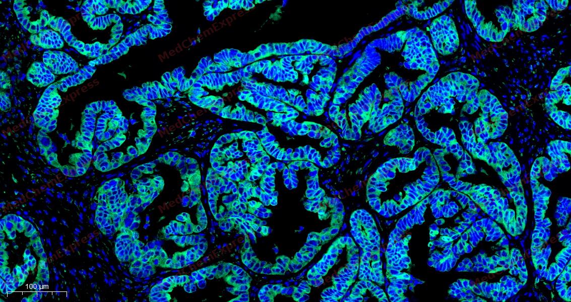 Tyramide signaling amplification based immunofluorescence was performed on paraffin-embedded human ovarian carcinoma (sample 2) tissue using BRAF antibody. Antigen retrieval was performed in EDTA buffer pH 9.0 (95 °C, 20 min) followed by cooling to RT. Then incubated with primary antibody (HY-P80036, 1:100 dilution) at room temperature for 60 minutes and HRP conjugated secondary antibody for 10 minutes. Fluorescence was then developed with TSA520 . The tissues were counterstained with DAPI and mounted with Anti-fade mounting medium.
 -
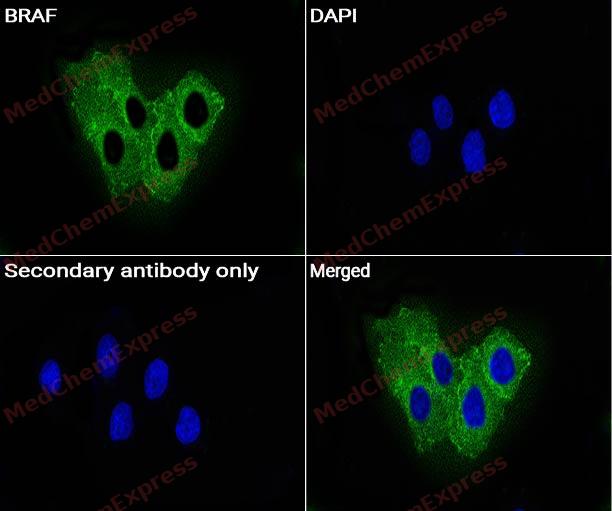 Immunocytochemistry analysis of Hela cells labeling BRAF with BRAF Antibody (HY-P80036) at 1:100 dilution. Cells were fixed in 4% paraformaldehyde for 15 minutes at room temperature, permeabilized with 0.1% Triton X-100 in PBS for 15 minutes at room temperature, then blocked with quick block buffer for 10 minutes at room temperature. Cells were then incubated with BRAF Antibody (HY-P80036) at 1:100 dilution in quick block buffer overnight at 4 ℃. AF488-conjugated Goat Anti-Rabbit IgG H&L(HY-P8002, Green) was used as the secondary antibody at 1/1,000 dilution. PBS instead of the primary antibody was used as the secondary antibody only control. The Nuclear counterstain was DAPI (Blue).
 -
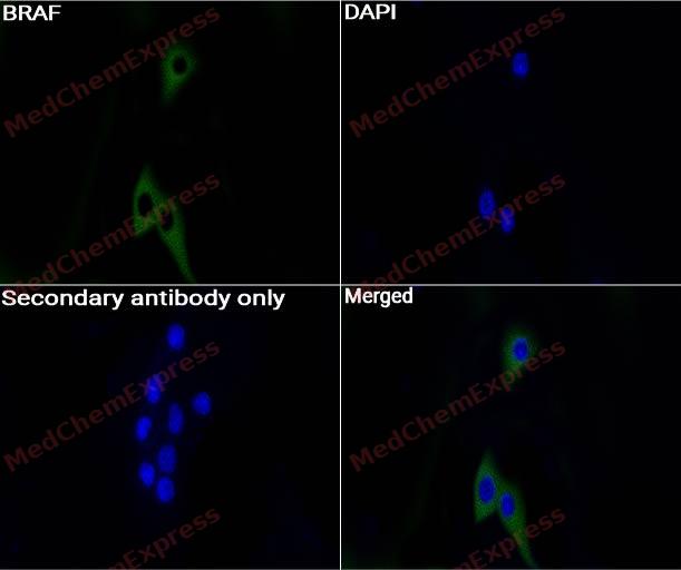 Immunocytochemistry analysis of Hela cells labeling BRAF with BRAFAntibody (HY-P80036) at 1:200 dilution. Cells were fixed in 4% paraformaldehyde for 15 minutes at room temperature, permeabilized with 0.1% Triton X-100 in PBS for 15 minutes at room temperature, then blocked with quick block buffer for 10 minutes at room temperature. Cells were then incubated with BRAF Antibody (HY-P80036) at 1:200 dilution in quick block buffer overnight at 4 ℃. AF488-conjugated Goat Anti-Rabbit IgG H&L(HY-P8002, Green) was used as the secondary antibody at 1/1,000 dilution. PBS instead of the primary antibody was used as the secondary antibody only control. The Nuclear counterstain was DAPI (Blue).


Background
-
Function
B-Raf is a serine/threonine protein kinase encoded by BRAF and functions as a central signal transducer in the RAS-RAF-MEK-ERK signaling cascade that regulates cell proliferation, differentiation, and survival[1][2]. Mechanistically, activated B-Raf phosphorylates MEK, leading to ERK activation and propagation of mitogenic signaling, making B-Raf a critical regulator of MAPK pathway output[2]. Dysregulation of this pathway is strongly linked to oncogenesis, and somatic BRAF mutations are among the most common kinase alterations identified in human cancer, particularly melanoma, colorectal cancer, thyroid cancer, and non-small-cell lung cancer[1][3]. In disease models, the canonical BRAF V600E mutation constitutively activates kinase signaling and promotes tumor cell growth independent of normal upstream regulatory inputs[2][3]. Compared with the related RAF isoforms A-Raf and C-Raf (RAF1), B-Raf displays stronger basal kinase activity and is the most potent activator of MEK-ERK signaling, making it a dominant MAPK pathway driver in many BRAF-mutant tumors[2]. For experimental applications, selective B-Raf inhibitors have been widely used to suppress oncogenic MAPK signaling and to investigate pathway dependence in cancer models[4]. The development of mutation-selective inhibitors, including compounds targeting BRAF V600E, established B-Raf as a validated therapeutic and mechanistic target and continues to support research on signaling regulation, drug resistance, and combination treatment strategies[4][5].
-
Subcellular Localization
Nucleus; Cytoplasm; Cell membrane
-
Expression
Tissue_specificity:brain and testicles -
Subunit
Monomer (PubMed:19710016). Homodimer (PubMed:19710016, PubMed:36402789). Heterodimerizes with RAF1, and the heterodimer possesses a highly increased kinase activity compared to the respective homodimers or monomers (PubMed:19710016).
-
SwissProt ID
-
Synonyms
BRAF1, RAFB1, BRAF, Serine/threonine-protein kinase B-raf, Proto-oncogene B-Raf, p94, v-Raf murine sarcoma viral oncogene homolog B1
-
Research Field
Cell Biology


Documentation
-
Data Sheet (261 KB)
-
SDS (252 KB)
- English - EN (252 KB)
- Français - FR (252 KB)
- Deutsch - DE (252 KB)
- Norwegian - NO (252 KB)
- Español - ES (252 KB)
- Swedish - SV (252 KB)
- Italian - IT (252 KB)
- Korean - KR (252 KB)
- Portuguese - PT (252 KB)
-
User Guide for Antibodies (1077 KB)
References
[1]. Davies H, et al. Mutations of the BRAF gene in human cancer. Nature. 2002 Jun 27;417(6892):949-54. [Content Brief]
[2]. Wan PT, et al. Mechanism of activation of the RAF-ERK signaling pathway by oncogenic mutations of B-RAF. Cell. 2004 Mar 19;116(6):855-67. [Content Brief]
[3]. B-Raf gene information from NCBI.
[4]. Tsai J, et al. Discovery of a selective inhibitor of oncogenic B-Raf kinase with potent antimelanoma activity. Proc Natl Acad Sci U S A. 2008 Feb 26;105(8):3041-6. [Content Brief]
[5]. Bollag G, et al. Vemurafenib: the first drug approved for BRAF-mutant cancer. Nat Rev Drug Discov. 2012 Nov;11(11):873-86. [Content Brief]

 Powered by Bioz
