



FSP27/CIDEC Antibody (YA911)
(Synonyms: Cell Death Activator; Cell death activator CIDE-3; Cell death inducing DFFA like effector protein C; CIDE 3; CIDE C; CIDEC_HUMAN; FLJ20871; FPLD5; FSP27; CIDE-3)
Based on 1 Customer Validation
FSP27/CIDEC Antibody (YA911) is a Mouse-derived and non-conjugated IgG monoclonal antibody, targeting to FSP27/CIDEC.
-
Host:
Mouse
-
Isotype:
IgG
-
Application:
WB, ICC/IF, FC, ELISA
-
Reactivity :
Human, Mouse
-
Formulation:
Supplied in PBS with 0.05% sodium azide.
-
Conjugation:
Non-conjugated


To place orders, for customer services and technical support, please contact: MedChemExpress USA
Tel: 609-228-6898 E-mail: [email protected] [email protected]





Applications
| Application |
WB

WB: Western Blot
|
ICC/IF
ICC/IF: Immunocytochemistry/
Immunofluorescence |
FC
FC: Flow Cytometry
|
ELISA
ELISA: Enzyme Linked Immunosorbent Assay
|
|---|---|---|---|---|
| Dilution Ratio | 1:500-1:2000 | 1:200-1:1000 | 1:200-1:400 | 1:10000 |


Product Details
FSP27/CIDEC Antibody (YA911) is a Mouse-derived and non-conjugated IgG monoclonal antibody, targeting to FSP27/CIDEC.
-
Host Mouse

-
Clonality Monoclonal

-
Species ReactivityHuman, Mouse
-
Observed Molecular WeightObserved band size: 34 kD
Note: Due to possible protein modifications or aggregation, the molecular weight should be confirmed by actual measurement, and the predicted value is for reference only.

Purified recombinant fragment of human CIDEC expressed in E. Coli.
Endogenous
Affinity Purified
Non-conjugated
Unmodified
IgG


Product Properties
-
Appearance
Solution
-
Formulation
Supplied in PBS with 0.05% sodium azide.
-
Concentration
Batch-dependent, Please check the COA for the concentration of each lot. Check Lot Concentration
-
Storage & Stability
Stored at -20°C for 1 year. Avoid repeated freeze / thaw cycles.
-
Envío
Shipping with blue ice.


Verification Images
-
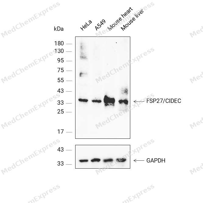 Western blot analysis was performed on protein extracts (25 μg) from HeLa (lane 1), A549 (lane 2), Mouse heart (lane 3), and Mouse liver (lane 4) using FSP27/CIDEC antibody. Proteins were transferred onto a 0.45 μm PVDF membrane using the Trans-Blot® Turbo™ system for 13 min. The membrane was then blocked with 5% nonfat milk in TBST (HY-K1025) for 1 h at room temperature. The primary antibody (1:1000) and loading control antibody GAPDH Antibody (HRP) (HY-P80954A) (1:2500) were diluted in 5% nonfat milk in TBST and incubated with the membrane overnight at 4°C. After washing, the membrane of primary antibody was incubated with HRP-conjugated goat anti-rabbit/mouse IgG secondary antibody (HY-P8001/HY-P8004) (1:5000) diluted in 5% nonfat milk in TBST for 1 h at room temperature. Protein bands were visualized using an Ultra High Sensitivity ECL detection kit (HY-K1005).


Background
-
Function
FSP27/CIDEC is a Lipid transferase specifically expressed in white adipose tissue, which promotes unilocular lipid droplet formation by mediating lipid droplet fusion. Lipid droplet fusion promotes their enlargement, restricting lipolysis and favoring lipid storage. Localizes on the lipid droplet surface, at focal contact sites between lipid droplets, and mediates atypical lipid droplet fusion by undergoing liquid-liquid phase separation (LLPS) and promoting directional net neutral lipid transfer from the smaller to larger lipid droplets. The transfer direction may be driven by the internal pressure difference between the contacting lipid droplet pair. Its role in neutral lipid transfer and lipid droplet enlargement is activated by the interaction with PLIN1. May also act as a CEBPB coactivator in the white adipose tissue to control the expression of a subset of CEBPB downstream target genes, including SOCS1, SOCS3, TGFB1, TGFBR1, ID2 and XDH. When overexpressed in preadipocytes, induces apoptosis or increases cell susceptibility to apoptosis induced by serum deprivation or TGFB treatment[1][2][3][4][5][6].
-
Subcellular Localization
Lipid droplet; Endoplasmic reticulum; Nucleus
-
Expression
Tissue_specificity:It is mainly expressed in adipose tissue, small intestine, heart, colon and stomach, with lower expression levels in brain, kidney and liver. -
Isoforms & Post-Translational Modification
Q96AQ7 has 4 isomers: Q96AQ7-1: 26754 Da (predicted); Q96AQ7-2: 18439 Da (predicted); Q96AQ7-3: 14013 Da (predicted); Q96AQ7-4: 27923 Da (predicted).
Ubiquitinated and targeted to proteasomal degradation, resulting in a short half-life (about 15 minutes in 3T3-L1 cells). Protein stability depends on triaclyglycerol synthesis, fatty acid availability and lipid droplet formation -
Subunit
Homodimer (By similarity). Homooligomer; undergoes liquid-liquid phase separation (LLPS) via its N-terminus, facilitating lipid droplet fusion, occurs at the lipid droplet contact sites (By similarity). Interacts with CIDEA (PubMed:19843876). Interacts with PLIN1 (PubMed:23399566). Interacts with NFAT5; this interaction is direct and retains NFAT5 in the cytoplasm (By similarity). Interacts with CEBPB (By similarity). Interacts with isoform CLSTN3beta of CLSTN3; inhibiting the lipid transferase activity of CIDEC (By similarity)
-
SwissProt ID
-
Synonyms
Cell Death Activator; Cell death activator CIDE-3; Cell death inducing DFFA like effector protein C; CIDE 3; CIDE C; CIDEC_HUMAN; FLJ20871; FPLD5; FSP27; CIDE-3
-
Research Field
Cell Biology


Documentación
-
Ficha de datos (262 KB)
-
SDS (252 KB)
- English - EN (252 KB)
- Français - FR (252 KB)
- Deutsch - DE (252 KB)
- Norwegian - NO (252 KB)
- Español - ES (252 KB)
- Swedish - SV (252 KB)
- Italian - IT (252 KB)
- Korean - KR (252 KB)
- Portuguese - PT (252 KB)
-
User Guide for Antibodies (1077 KB)
References
[1]. Keller P, et al. Fat-specific protein 27 regulates storage of triacylglycerol. J Biol Chem. 2008 May 23;283(21):14355-65. [Content Brief]
[2]. Liu K, et al. Functional analysis of FSP27 protein regions for lipid droplet localization, caspase-dependent apoptosis, and dimerization with CIDEA. Am J Physiol Endocrinol Metab. 2009 Dec;297(6):E1395-413. [Content Brief]
[3]. Rubio-Cabezas O, et al. Partial lipodystrophy and insulin resistant diabetes in a patient with a homozygous nonsense mutation in CIDEC. EMBO Mol Med. 2009 Aug;1(5):280-7. [Content Brief]
[4]. Grahn TH, et al. FSP27 and PLIN1 interaction promotes the formation of large lipid droplets in human adipocytes. Biochem Biophys Res Commun. 2013 Mar 8;432(2):296-301. [Content Brief]
[5]. Wang J, et al. Polybasic RKKR motif in the linker region of lipid droplet (LD)-associated protein CIDEC inhibits LD fusion activity by interacting with acidic phospholipids. J Biol Chem. 2018 Dec 14;293(50):19330-19343. [Content Brief]
[6]. Liang L, et al. Molecular cloning and characterization of CIDE-3, a novel member of the cell-death-inducing DNA-fragmentation-factor (DFF45)-like effector family. Biochem J. 2003 Feb 15;370(Pt 1):195-203. [Content Brief]

 Powered by Bioz
