



CXCR4 Antibody
(Synonyms: C-X-C chemokine receptor type 4; CXC-R4; CXCR-4; Stromal cell-derived factor 1 receptor; SDF-1 receptor; Fusin; Leukocyte-derived seven transmembrane domain receptor; LESTR; CD184 antigen; CXCR4_HUMAN.)
Based on 1 Customer Validation
CXCR4 Antibody is a Rabbit-derived and non-conjugated IgG polyclonal antibody, targeting to CXCR4.
-
Host:
Rabbit
-
Isotype:
IgG
-
Application:
WB, IHC-P, IHC-F, ICC/IF, FC, ELISA
-
Reactivity :
Human, Mouse, Rat
-
Formulation:
Supplied in 0.01M TBS(pH7.4) with 1% BSA, 0.03% Proclin300 and 50% Glycerol.
-
Conjugation:
Non-conjugated


To place orders, for customer services and technical support, please contact: MedChemExpress USA
Tel: 609-228-6898 E-mail: [email protected] [email protected]





Applications
| Application |
WB

WB: Western Blot
|
ELISA
ELISA: Enzyme Linked Immunosorbent Assay
|
IHC-P
IHC-P: Immunohistochemistry-Paraffin
|
IHC-F
IHC-F: Immunohistochemistry-Frozen
|
ICC/IF
ICC/IF: Immunocytochemistry/
Immunofluorescence |
|---|---|---|---|---|---|
| Dilution Ratio | 1:500-2000 | 1:5000-10000 | 1:100-500 | 1:100-500 | 1:100-500 |


Product Details
CXCR4 Antibody is a Rabbit-derived and non-conjugated IgG polyclonal antibody, targeting to CXCR4.
-
Host Rabbit

-
Clonality Polyclonal

-
Species ReactivityHuman, Mouse, Rat Predicted Reactivity: Cow,Rabbit
Note: The predicted reactivity is for reference only and should not be considered a guarantee of product performance.
-
Observed Molecular WeightObserved band size: 45 kDa
Note: Due to possible protein modifications or aggregation, the molecular weight should be confirmed by actual measurement, and the predicted value is for reference only.

-
Calculated Molecular Weight Predicted band size: 40kDa

Entrez Gene: 7852 Human ; 12767 Mouse ; 60628 Rat
SwissProt: P61073 Human ; P70658 Mouse ; O08565 Rat
OMIM: 193670 Human
KLH conjugated synthetic peptide derived from the middle of human CXCR4: 201-294/352
Endogenous
affinity purified
Non-conjugated
Unmodified
IgG


Product Properties
-
Appearance
Solution
-
Formulation
Supplied in 0.01M TBS(pH7.4) with 1% BSA, 0.03% Proclin300 and 50% Glycerol.
-
Concentration
Batch-dependent, Please check the COA for the concentration of each lot. Check Lot Concentration
-
Storage & Stability
Stored at -20°C for 1 year. Avoid repeated freeze / thaw cycles.
-
Shipping
Shipping with blue ice.


Verification Images
-
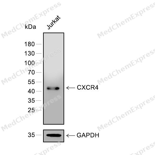 Western blot analysis of extracts from Jurkat (lane 1) using CXCR4 antibody. Proteins were transferred to a PVDF membrane and blocked with 5% nonfat powdered milk in PBST for 2 hour at room temperature. The primary antibody (1/2000) and Loading control antibody (GAPDH, 1/3000) was diluted with 5% nonfat powdered milk in PBST at 4°C overnight. Goat Anti-Rabbit IgG-HRP Secondary Antibody (1/8,000) was incubated for 45min at room temperature.
 -
 Flow cytometric analysis of 1X10⁶ Jurkat cells labeling CXCR4 Antibody (HY-P81089, red). Cells were fixed with 4% paraformaldehyde and permeabilised with 90% methanol. Then stained with the primary antibody at 1μg/Test dilution for an hour at 4℃. AF488-conjugated Goat Anti-Rabbit IgG H&L (HY-P8002) was used as the secondary antibody at 1/1,000 dilution for 30 minutes at 4℃. Rabbit IgG Isotype Control (HY-P80879, blue) was used as the isotype control, cells without incubation with primary antibody were used as the unlabeled control (black).
 -
 Flow cytometric analysis of 1X10⁶ Jurkat cells labeling CXCR4 Antibody (HY-P81089, red). Cells were fixed with 4% paraformaldehyde and permeabilised with 90% methanol. Then stained with the primary antibody at 1μg/Test dilution for an hour at 4℃. AF488-conjugated Goat Anti-Rabbit IgG H&L (HY-P8002) was used as the secondary antibody at 1/1,000 dilution for 30 minutes at 4℃. Rabbit IgG Isotype Control (HY-P80879, blue) was used as the isotype control, cells without incubation with primary antibody were used as the unlabeled control (black).
 -
 Immunocytochemistry analysis of A549 cells labeling CXCR4 with CXCR4 Antibody (HY-P81089) at 1/100 dilution. Cells were fixed in 4% paraformaldehyde for 15 minutes at room temperature, permeabilized with 0.1% Triton X-100 in PBS for 15 minutes at room temperature, then blocked with quick block buffer for 10 minutes at room temperature. Cells were then incubated with CXCR4 Antibody (HY-P81089) at 1/100 dilution in quick block buffer overnight at 4 ℃. AF488-conjugated Goat Anti-Rabbit IgG H&L(HY-P8002, Green) was used as the secondary antibody at 1/1,000 dilution. PBS instead of the primary antibody was used as the secondary antibody only control. The Nuclear counterstain was DAPI (Blue).
 -
 Immunocytochemistry analysis of Hela cells labeling CXCR4 with CXCR4 Antibody (HY-P81089) at 1/100 dilution. Cells were fixed in 4% paraformaldehyde for 15 minutes at room temperature, permeabilized with 0.1% Triton X-100 in PBS for 15 minutes at room temperature, then blocked with quick block buffer for 10 minutes at room temperature. Cells were then incubated with CXCR4 Antibody (HY-P81089) at 1/100 dilution in quick block buffer overnight at 4 ℃. AF488-conjugated Goat Anti-Rabbit IgG H&L(HY-P8002, Green) was used as the secondary antibody at 1/1,000 dilution. PBS instead of the primary antibody was used as the secondary antibody only control. The Nuclear counterstain was DAPI (Blue).


Background
-
Function
CXCR4 is a G protein-coupled chemokine receptor that mediates migration of resting leukocytes and hematopoietic progenitors in response to SDF-1/CXCL12[1]. Mechanistically, CXCR4-CXCL12 signaling supports hematopoiesis and cerebellar development, and CXCR4-deficient mice show hematopoietic and cardiac defects matching SDF-1-deficient mice[1]. In vascular development, mice lacking CXCR4 or PBSF/SDF-1 show defective formation of large vessels supplying the gastrointestinal tract, identifying this axis as a signaling system for organ vascularization[2]. In disease models, CXCR4 and CCR7 are highly expressed in human breast cancer cells, malignant breast tumors, and metastases, linking chemokine receptor programs to tumor cell migration and metastatic spread[3]. Compared with the related ACKR3/CXCR7 receptor, CXCR4 couples to G proteins and directly promotes cell migration, whereas ACKR3 is G protein-independent and scavenges CXCL12 to regulate extracellular chemokine levels[4]. For experimental applications, CXCR4 antagonist structures with IT1t and CVX15 define ligand-binding sites near the extracellular surface, supporting structure-guided inhibitor design[5]. AMD3100 selectively antagonizes CXCR4 and rapidly mobilizes murine and human hematopoietic progenitor cells, especially with G-CSF[6].
-
Subcellular Localization
Cell membrane; Multi-pass membrane protein; Cell junction; Early endosome; Late endosome; Lysosome
-
Expression
Tissue_specificity:This protein is expressed in a variety of tissues, including peripheral blood leukocytes, spleen, thymus, spinal cord, heart, placenta, lung, liver, skeletal muscle, kidney, pancreas, cerebellum, cerebral cortex and medulla oblongata (including microglia and astrocytes) , cerebral microvessels, coronary arteries, and umbilical cord endothelial cells. Isoform 1 was dominant in all tissues examined.
Induction: (Microbial infection) May be down-regulated by Human cytomegalovirus/HHV-5; (Microbial infection) May be down-regulated by HIV-1 tat -
Isoforms & Post-Translational Modification
P61073 has 2 isomers: P61073-1: 39746 Da (predicted); P61073-2: 40221 Da (predicted).
Phosphorylated on agonist stimulation. Rapidly phosphorylated on serine and threonine residues in the C-terminal. Phosphorylation at Ser-324 and Ser-325 leads to recruitment of ITCH, ubiquitination and protein degradation;Ubiquitinated after ligand binding, leading to its degradation (PubMed:28978524). Ubiquitinated by ITCH at the cell membrane on agonist stimulation (PubMed:14602072, PubMed:34927784). The ubiquitin-dependent mechanism, endosomal sorting complex required for transport (ESCRT), then targets CXCR4 for lysosomal degradation. This process is dependent also on prior Ser-/Thr-phosphorylation in the C-terminal of CXCR4. Also binding of ARRB1 to STAM negatively regulates CXCR4 sorting to lysosomes though modulating ubiquitination of SFR5S;Sulfation on Tyr-21 is required for efficient binding of CXCL12/SDF-1alpha and promotes its dimerization. Tyr-7 and Tyr-12 are sulfated in a sequential manner after Tyr-21 is almost fully sulfated, with the binding affinity for CXCL12/SDF-1alpha increasing with the number of sulfotyrosines present. Sulfotyrosines Tyr-7 and Tyr-12 occupy clefts on opposing CXCL12 subunits, thus bridging the CXCL12 dimer interface and promoting CXCL12 dimerization;O- and N-glycosylated. Asn-11 is the principal site of N-glycosylation. There appears to be very little or no glycosylation on Asn-176. N-glycosylation masks coreceptor function in both X4 and R5 laboratory-adapted and primary HIV-1 strains through inhibiting interaction with their Env glycoproteins. The O-glycosylation chondroitin sulfate attachment does not affect interaction with CXCL12/SDF-1alpha nor its coreceptor activity -
Subunit
Monomer. Can form homodimers (PubMed:20929726). Interacts with CD164 (PubMed:17077324). Interacts with ARRB2; the interaction is dependent on the C-terminal phosphorylation of CXCR4 and allows activation of MAPK1 and MAPK3. Interacts with ARR3; the interaction is dependent on the C-terminal phosphorylation of CXCR4 and modulates calcium mobilization (PubMed:20048153). Interacts with RNF113A; the interaction, enhanced by CXCL12, promotes CXCR4 ubiquitination and subsequent degradation (PubMed:28978524). Interacts (via the cytoplasmic C-terminal) with ITCH (via the WW domains I and II); the interaction, enhanced by CXCL12, promotes CXCR4 ubiquitination and leads to its degradation. Interacts with extracellular ubiquitin. Interacts with DBN1; this interaction is enhanced by antigenic stimulation. Following LPS binding, may form a complex with GDF5, HSP90AA1 and HSPA8
-
SwissProt ID
-
Synonyms
C-X-C chemokine receptor type 4; CXC-R4; CXCR-4; Stromal cell-derived factor 1 receptor; SDF-1 receptor; Fusin; Leukocyte-derived seven transmembrane domain receptor; LESTR; CD184 antigen; CXCR4_HUMAN.


Documentation
-
Data Sheet (262 KB)
-
SDS (252 KB)
- English - EN (252 KB)
- Français - FR (252 KB)
- Deutsch - DE (252 KB)
- Norwegian - NO (252 KB)
- Español - ES (252 KB)
- Swedish - SV (252 KB)
- Italian - IT (252 KB)
- Korean - KR (252 KB)
- Portuguese - PT (252 KB)
-
User Guide for Antibodies (1077 KB)
References
[1]. Zou YR, et al. Function of the chemokine receptor CXCR4 in haematopoiesis and in cerebellar development. Nature. 1998 Jun 11;393(6685):595-9. [Content Brief]
[2]. Tachibana K, et al. The chemokine receptor CXCR4 is essential for vascularization of the gastrointestinal tract. Nature. 1998 Jun 11;393(6685):591-4. [Content Brief]
[3]. Müller A, et al. Involvement of chemokine receptors in breast cancer metastasis. Nature. 2001 Mar 1;410(6824):50-6. [Content Brief]
[4]. Schafer CT, et al. Distinct Activation Mechanisms of CXCR4 and ACKR3 Revealed by Single-Molecule Analysis of their Conformational Landscapes. bioRxiv [Preprint]. 2025 Feb 6:2023.10.31.564925. [Content Brief]
[5]. Wu B, et al. Structures of the CXCR4 chemokine GPCR with small-molecule and cyclic peptide antagonists. Science. 2010 Nov 19;330(6007):1066-71. [Content Brief]
[6]. Broxmeyer HE, et al. Rapid mobilization of murine and human hematopoietic stem and progenitor cells with AMD3100, a CXCR4 antagonist. J Exp Med. 2005 Apr 18;201(8):1307-18. [Content Brief]

 Powered by Bioz
