



Iba1 Antibody (YA353)
Based on 3 publication(s) in Google Scholar
Iba1 Antibody (YA353) is a Rabbit-derived and non-conjugated IgG monoclonal antibody, targeting to Iba1.
-
Host:
Rabbit
-
Isotype:
IgG
-
Application:
WB, IHC-P, FC, IP, ICC/IF
-
Reactivity :
Human, Mouse, Rat
-
Formulation:
Supplied in 1*TBS (pH7.4), 0.05% BSA and 40% Glycerol. Preservative: 0.05% Sodium Azide.
-
Conjugation:
Non-conjugated


To place orders, for customer services and technical support, please contact: MedChemExpress USA
Tel: 609-228-6898 E-mail: [email protected] [email protected]



-

HY-P80241
NeuN Antibody (YA272) -
HY-P80140
GFAP Antibody (YA416) -
HY-P80952
AF647-conjugated Goat Anti-Rabbit IgG H&L -
HY-P8001
HRP-conjugated Goat Anti-Rabbit IgG H&L -
HY-P80139
GFAP Antibody (YA755) -
HY-P8002
AF488-conjugated Goat Anti-Rabbit IgG H&L -
HY-P80285
PSD95 Antibody (YA120) -
HY-P80686
GFAP Antibody (YA756) -
HY-P80765
NF-KB p65 Antibody (YA268) -
HY-P80605
CD68 Antibody (YA799) -
HY-P80494
Glucose Transporter GLUT1 Antibody (YA410) -
HY-P80247
NMDAR1 Antibody (YA265)
Publications Citing Use of MedChemExpress (MCE) Iba1 Antibody (YA353)
More

Applications
| Application |
WB

WB: Western Blot
|
ICC/IF
ICC/IF: Immunocytochemistry/
Immunofluorescence |
IHC-P
IHC-P: Immunohistochemistry-Paraffin
|
FC
FC: Flow Cytometry
|
IP
IP: Immunoprecipitation
|
|---|---|---|---|---|---|
| Dilution Ratio | 1:500-1:1000 | 1:50-1:100 | 1:50-1:1000 | 1:50-1:200 | Use at an assay dependent concentration. |


Product Details
Iba1 Antibody (YA353) is a Rabbit-derived and non-conjugated IgG monoclonal antibody, targeting to Iba1.
-
Host Rabbit

-
Clonality Recombinant, Monoclonal

-
Species ReactivityHuman, Mouse, Rat
-
Observed Molecular WeightObserved band size: 17 kDa
Note: Due to possible protein modifications or aggregation, the molecular weight should be confirmed by actual measurement, and the predicted value is for reference only.

-
Calculated Molecular Weight Predicted band size: 17 kDa

Entrez Gene: 199 Human ; 11629 Mouse ; 29427 Rat
SwissProt: P55008 Human ; O70200 Mouse ; P55009 Rat
OMIM: 601833 Human
Synthetic peptide within N-terminal human Iba1.
Endogenous
Protein A affinity purified.
Non-conjugated
Unmodified
IgG


Product Properties
-
Appearance
Liquid
-
Formulation
Supplied in 1*TBS (pH7.4), 0.05% BSA and 40% Glycerol. Preservative: 0.05% Sodium Azide.
-
Concentration
Batch-dependent, Please check the COA for the concentration of each lot. Check Lot Concentration
-
Storage & Stability
Stored at -20°C for 1 year. Avoid repeated freeze / thaw cycles.
-
Shipping
Shipping with blue ice.


Publications (3)
-
Journal Impact Factor
-
Most Recent
-
Int Immunopharmacol
Fibrinogen drives neuroinflammation and neuropathology in perioperative neurocognitive disorders. [Abstract]2026 Jan 1;168(Pt 2):115906. PMID: 41273843


Verification Images
-
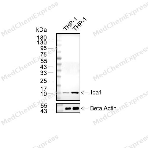 Western blot analysis of extracts from THP-1 (lane 2(20μg) , THP-1 (lane 3(40μg),using Iba1 Antibody (HY-P80501). Proteins were transferred to a PVDF membrane and blocked with 5% BSA in TBST for 2 hour at room temperature. The primary antibody and Loading control antibody (Beta Actin, HY-P80438, 1/3000) was used in 5% BSA in TBST at 4°C overnight. Goat Anti-Mouse/Rabbit IgG-HRP Secondary Antibody (HY-P8004/HY-P8001, 1/10,000) was used for 1 hour at room temperature.
 -
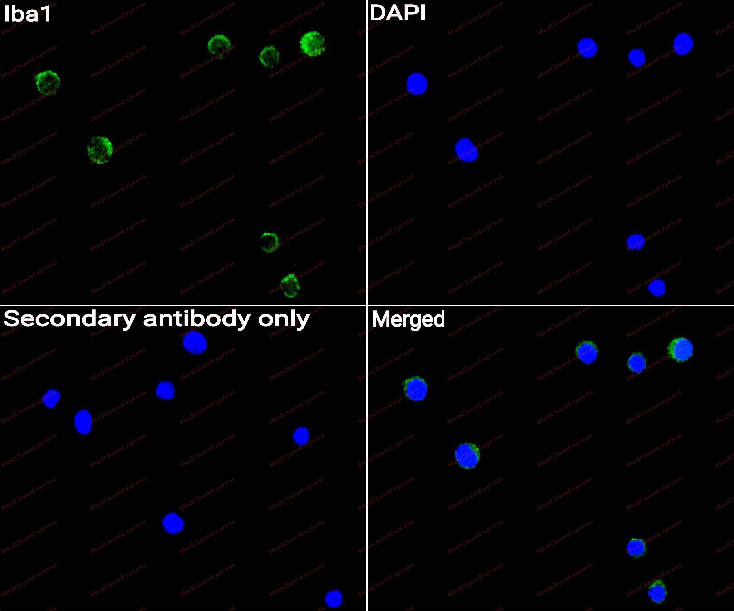 Immunocytochemistry analysis of RAW264.7 cells labeling Iba1 with Iba1 Antibody (HY-P80501) at 1/50 dilution. Cells were fixed in 4% paraformaldehyde for 15 minutes at room temperature, permeabilized with 0.1% Triton X-100 for 10 minutes at room temperature, then blocked with QuickBlock™ Blocking Buffer for Immunol Staining for 10 min at room temperature. Cells were then incubated with Iba1 Antibody (HY-P80501) at 1/50 dilution in QuickBlock™ Blocking Buffer for Immunol Staining at 4 ℃. Alexa Fluor® 488-conjugated AffiniPure Goat Anti-Rabbit IgG H&L(HY-P8002, Green) was used as the secondary antibody at 1/1,000 dilution. PBS instead of the primary antibody was used as the secondary antibody only control. The Nuclear counterstain was DAPI (Blue).
 -
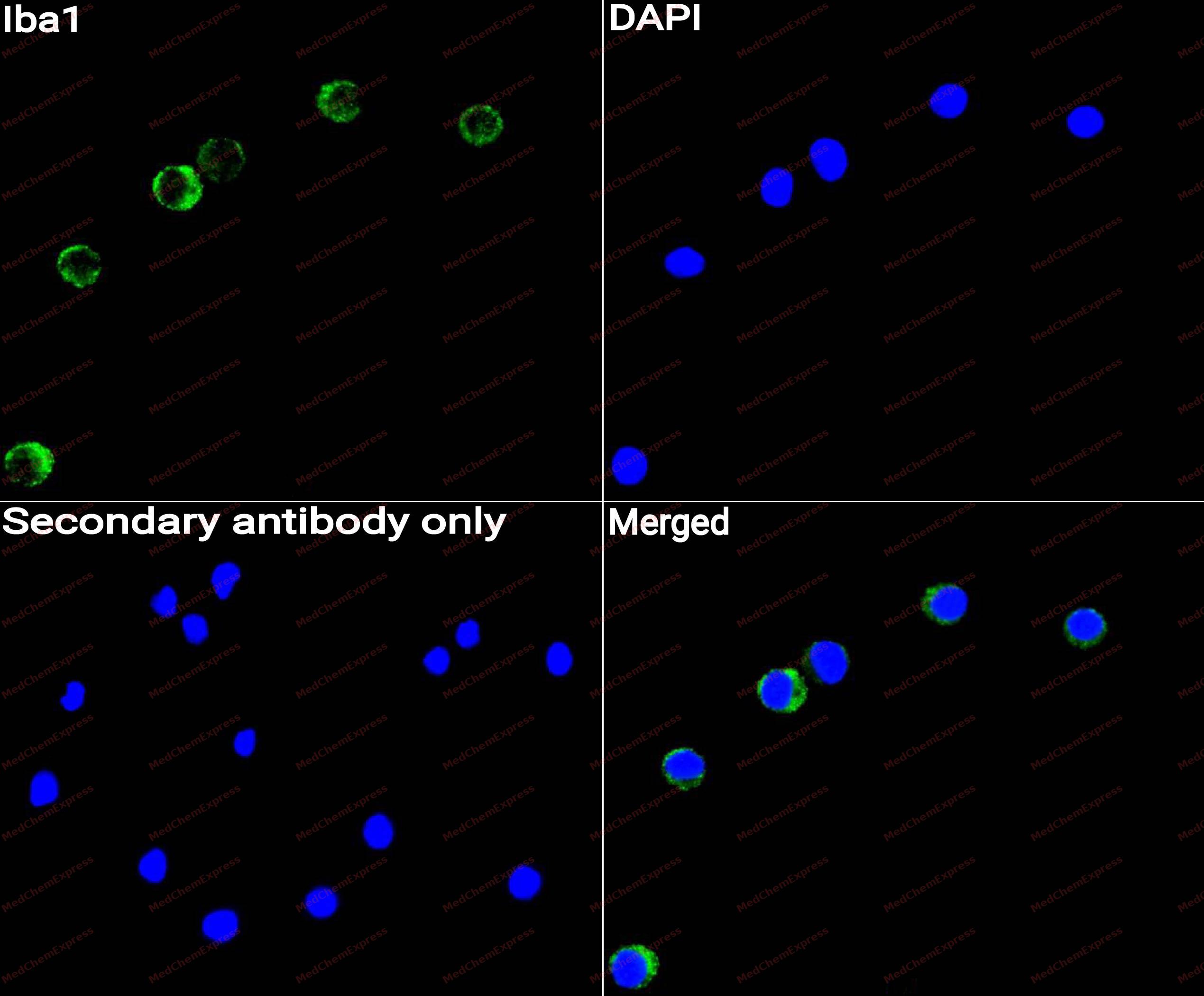 Immunocytochemistry analysis of C6 cells labeling Iba1 with Iba1 Antibody (HY-P80501) at 1/50 dilution. Cells were fixed in 4% paraformaldehyde for 15 minutes at room temperature, permeabilized with 0.1% Triton X-100 for 10 minutes at room temperature, then blocked with QuickBlock™ Blocking Buffer for Immunol Staining for 10 min at room temperature. Cells were then incubated with Iba1 Antibody (HY-P80501)at 1/50 dilution in QuickBlock™ Blocking Buffer for Immunol Staining at 4 ℃. Alexa Fluor® 488-conjugated AffiniPure Goat Anti-Rabbit IgG H&L(HY-P8002,Green) was used as the secondary antibody at 1/1,000 dilution. PBS instead of the primary antibody was used as the secondary antibody only control. The Nuclear counterstain was DAPI (Blue).
 -
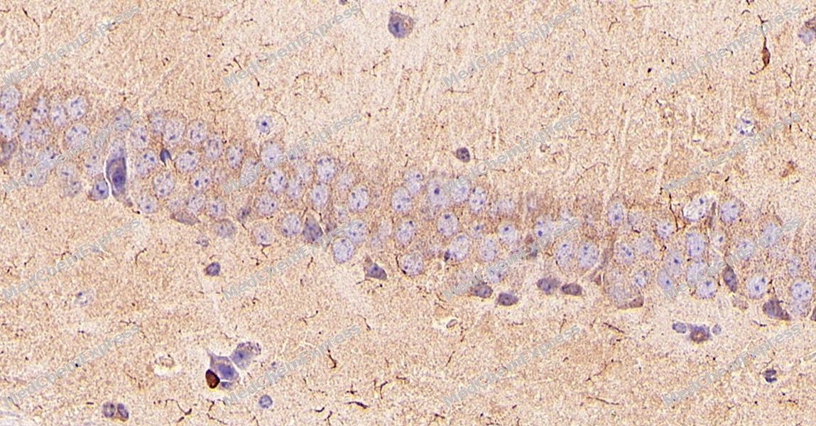 Immunohistochemical analysis of paraffin-embedded Mouse brain tissue using Iba1 Antibody. The section was pre-treated using heat mediated antigen retrieval with Tris-EDTA buffer (pH 9.0) for 8 minutes. The tissues were blocked in QuickBlock for 20 minutes at room temperature, washed with ddH2O and PBS, and then probed with the primary antibody (HY-P80501, 1:1000) in 4℃ overnight. The detection was performed using an HRP conjugated compact polymer system. DAB was used as the chromogen. Tissues were counterstained with hematoxylin and mounted with DPX.
 -
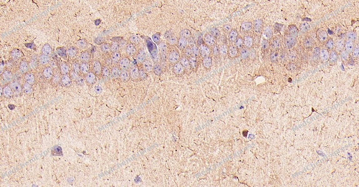 Immunohistochemical analysis of paraffin-embedded Mouse brain tissue using Iba1 Antibody. The section was pre-treated using heat mediated antigen retrieval with Tris-EDTA buffer (pH 9.0) for 8 minutes. The tissues were blocked in QuickBlock for 20 minutes at room temperature, washed with ddH2O and PBS, and then probed with the primary antibody (HY-P80501, 1:1000) in 4℃ overnight. The detection was performed using an HRP conjugated compact polymer system. DAB was used as the chromogen. Tissues were counterstained with hematoxylin and mounted with DPX.
 -
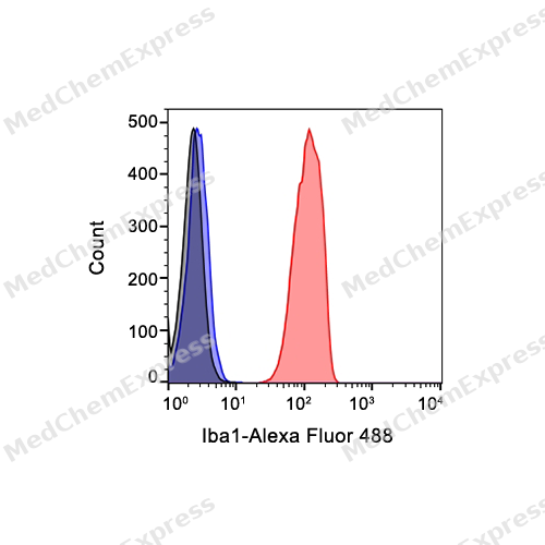 Flow cytometric analysis of 1X10^6 THP-1 cells labeling Iba1 Antibody (HY-P80501, red). Cells were fixed with 4% paraformaldehyde and permeabilised with 90% methanol. Then stained with the primary antibody at 1μg/mL dilution for an hour at 4℃. Alexa Fluor® 488-conjugated AffiniPure Goat Anti-Rabbit IgG H&L (HY-P8002) was used as the secondary antibody at 1/1,000 dilution for 30 minutes at 4℃. Rabbit IgG Isotype Control (HY-P80879, blue) was used as the isotype control, cells without incubation with primary antibody were used as the unlabeled control (black).


Background
-
Function
Iba1 is an Actin-binding protein that enhances membrane ruffling and RAC activation. Enhances the actin-bundling activity of LCP1. Binds calcium. Plays a role in RAC signaling and in phagocytosis. May play a role in macrophage activation and function. Promotes the proliferation of vascular smooth muscle cells and of T-lymphocytes. Enhances lymphocyte migration. Plays a role in vascular inflammation
-
Subcellular Localization
Cytoplasm, cytoskeleton; Cell projection, ruffle membrane; Peripheral membrane protein; Cytoplasmic side; Cell projection, phagocytic cup
-
Expression
Tissue_specificity:It can be detected in both T lymphocytes and peripheral blood mononuclear cells. -
Subunit
Homodimer (Potential). Monomer. Interacts with LCP1
-
SwissProt ID
-
Research Field
Cell Biology
Documentation
-
Data Sheet (234 KB)
-
SDS (251 KB)
- English - EN (251 KB)
- Français - FR (251 KB)
- Deutsch - DE (251 KB)
- Norwegian - NO (251 KB)
- Español - ES (251 KB)
- Swedish - SV (251 KB)
- Italian - IT (251 KB)
- Korean - KR (251 KB)
- Portuguese - PT (251 KB)
-
User Guide for Antibodies (1077 KB)

 Powered by Bioz
