



AF647-conjugated Goat Anti-Rabbit IgG H&L
Based on 1 publication(s) in Google Scholar
AF647-conjugated Goat Anti-Rabbit IgG H&L is an AF647-conjugated,Goat-derived anti-Rabbit antibody. AF647-conjugated Goat Anti-Rabbit IgG H&L can binds to the IgG heavy and light chain of Rabbit antibodies.
-
Host:
Goat
-
Isotype:
IgG
-
Application:
ICC/IF, IF-Tissue, FC
-
Reactivity :
Rabbit
-
Formulation:
Supplied in PBS, pH7.4, containing 1% BSA, 50% glycerol, 0.02% Sodium Azide
-
Conjugation:
AF647


To place orders, for customer services and technical support, please contact: MedChemExpress USA
Tel: 609-228-6898 E-mail: [email protected] [email protected]



-

HY-P81013
AF647-conjugated Goat Anti-Mouse IgG H&L -
HY-P8002
AF488-conjugated Goat Anti-Rabbit IgG H&L -
HY-P8005
AF488-conjugated Goat Anti-Mouse IgG H&L -
HY-P8003
AF594-conjugated Goat Anti-Rabbit IgG H&L -
HY-P8006
AF594-conjugated Goat Anti-Mouse IgG H&L -
HY-P8001
HRP-conjugated Goat Anti-Rabbit IgG H&L -
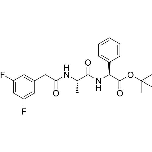
HY-13027
DAPT -
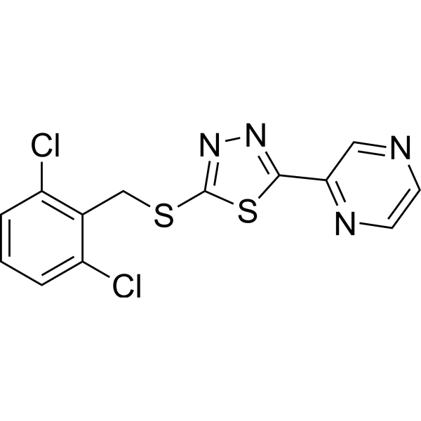
HY-18723
Yoda 1 -
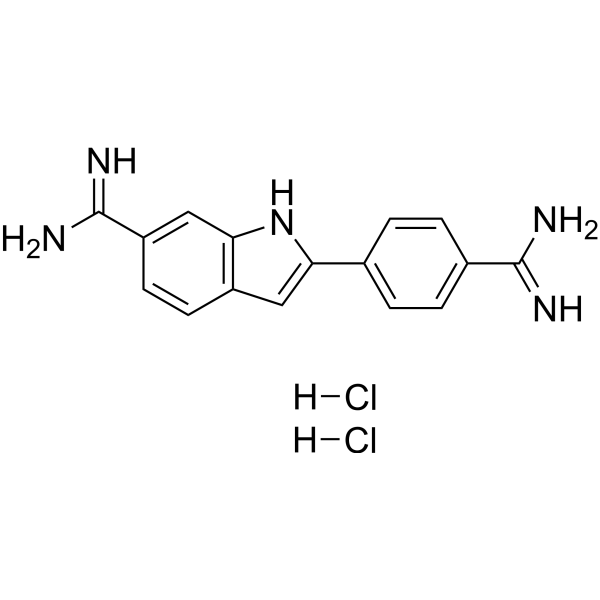
HY-D0814
DAPI dihydrochloride -
HY-P8004
HRP-conjugated Goat Anti-Mouse IgG H&L -
HY-P80501
Iba1 Antibody (YA353) -
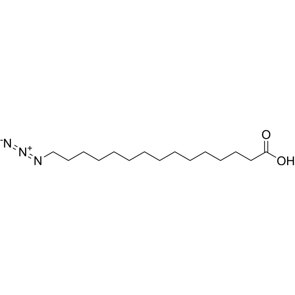
HY-151656
15-Azido-pentadecanoic acid
Publications Citing Use of MedChemExpress (MCE) AF647-conjugated Goat Anti-Rabbit IgG H&L
More

Applications
| Application |
ICC/IF

ICC/IF: Immunocytochemistry/
Immunofluorescence |
IF-Tissue
IF-Tissue: Immunofluorescence-Tissue
|
FC
FC: Flow Cytometry
|
|---|---|---|---|
| Dilution Ratio | 1:100-1000 | 1:100-1000 | 1:1000-4000 |


Product Details
AF647-conjugated Goat Anti-Rabbit IgG H&L is an AF647-conjugated,Goat-derived anti-Rabbit antibody. AF647-conjugated Goat Anti-Rabbit IgG H&L can binds to the IgG heavy and light chain of Rabbit antibodies.
-
Host Goat

-
Clonality Polyclonal

-
Species ReactivityRabbit
Synthetic peptide corresponding to Rabbit IgG.
Affinity Purified
AF647
Unmodified
IgG heavy and light chain
Whole IgG Antibodies
Human, Mouse
IgG


Product Properties
-
Appearance
Solution
-
Formulation
Supplied in PBS, pH7.4, containing 1% BSA, 50% glycerol, 0.02% Sodium Azide
-
Concentration
Batch-dependent, Please check the COA for the concentration of each lot. Check Lot Concentration
-
Storage & Stability
Stored at -20°C for 1 year. Protect from light. Avoid repeated freeze / thaw cycles.
-
Shipping
Shipping with blue ice.


Publications (1)
-
Journal Impact Factor
-
Most Recent


Verification Images
-
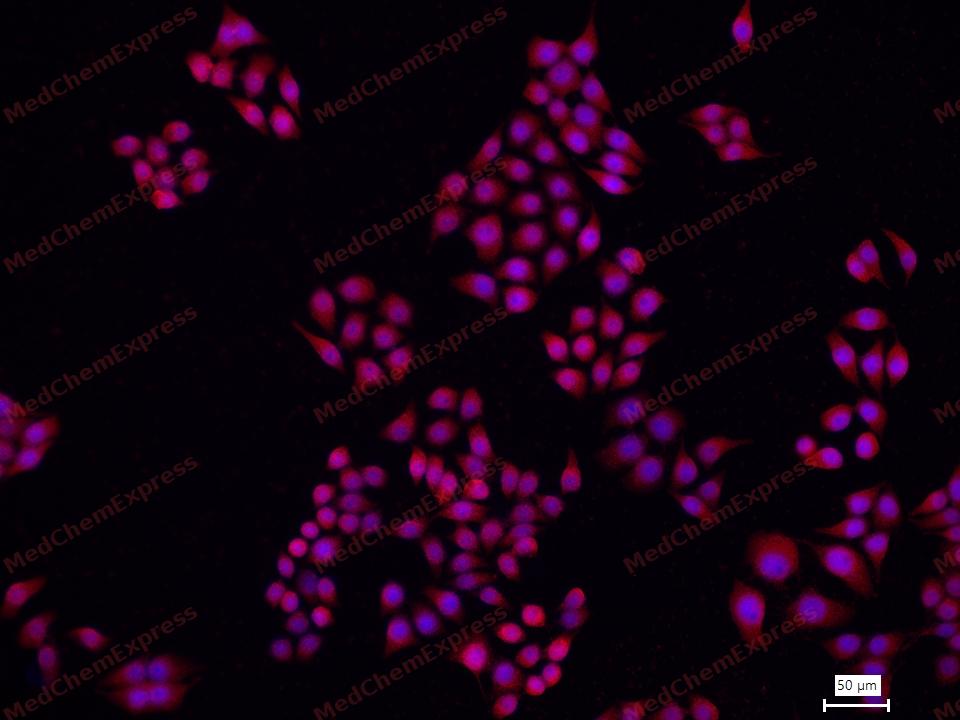 Immunofluorescence analysis of MCF7 cells labeling Cytokeratin 19 with Cytokeratin 19 (HY-P81183) at 1/100 dilution. Cells were fixed in 4% paraformaldehyde for 20 minutes at room temperature, permeabilized with 0.1% Triton X-100 in PBS for 5 minutes at room temperature, then blocked with 1% BSA/10% normal goat serum for 1 hour at room temperature. Cells were then incubated with Cytokeratin 19 (HY-P81183) at 1/100 dilution in 1% BSA in PBST overnight at 4 ℃. Alexa Fluor® 647-conjugated AffiniPure Goat Anti-Rabbit IgG H&L (HY-P80952) was used as the secondary antibody at 1/1,000 dilution. Nuclear DNA was labelled in blue with DAPI.
 -
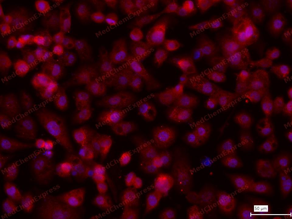 Immunofluorescence analysis of HEC1A cells labeling Cytokeratin 19 with Cytokeratin 19 (HY-P81183) at 1/100 dilution. Cells were fixed in 4% paraformaldehyde for 20 minutes at room temperature, permeabilized with 0.1% Triton X-100 in PBS for 5 minutes at room temperature, then blocked with 1% BSA/10% normal goat serum for 1 hour at room temperature. Cells were then incubated with Cytokeratin 19 (HY-P81183) at 1/100 dilution in 1% BSA in PBST overnight at 4 ℃. Alexa Fluor® 647-conjugated AffiniPure Goat Anti-Rabbit IgG H&L (HY-P80952) was used as the secondary antibody at 1/1,000 dilution. Nuclear DNA was labelled in blue with DAPI.
 -
 Please select the appropriate fluorescent dye based on your fluorescence imaging system. According to the species of the experimental sample you are studying and the host species of the primary antibody, choose the appropriate species of the secondary antibody.


Background
-
Function
Secondary antibodies are antibodies that bind to primary antibodies in immunoassays. They do not directly recognize the target antigen but rather the Fc region of the primary antibody or the entire immunoglobulin (Ig). Secondary antibodies are typically conjugated to fluorescent groups (such as Alexa Fluor), enzymes (such as HRP and alkaline phosphatase), or proteins (such as biotin).
-
Research Field
Immunology
Documentation
-
Data Sheet (230 KB)
-
SDS (252 KB)
- English - EN (252 KB)
- Français - FR (252 KB)
- Deutsch - DE (252 KB)
- Norwegian - NO (252 KB)
- Español - ES (252 KB)
- Swedish - SV (252 KB)
- Italian - IT (252 KB)
- Korean - KR (252 KB)
- Portuguese - PT (252 KB)
-
User Guide for Antibodies (1077 KB)

 Powered by Bioz
