



NF-KB p65 Antibody (YA268)
(Synonyms: NFKB3; RELA; TF65; Transcription factor p65; p65; NFkB)Based on 7 publication(s) in Google Scholar
NF-KB p65 Antibody (YA268) is a Rabbit-derived and non-conjugated IgG monoclonal antibody, targeting to NF-KB p65.
-
Host:
Rabbit
-
Isotype:
IgG
-
Application:
WB, IHC-F, IHC-P, ICC/IF, IP
-
Reactivity :
Human, Mouse
-
Formulation:
Supplied in 50 mM Tris-Glycine (pH 7.4), 0.15 M NaCl, 40% Glycerol and 0.05% BSA. Preservative: 0.01% Sodium azide
-
Conjugation:
Non-conjugated


To place orders, for customer services and technical support, please contact: MedChemExpress USA
Tel: 609-228-6898 E-mail: [email protected] [email protected]



-

HY-P80839
Phospho-NF-KB p65 (Ser536) Antibody -
HY-P80718
IKB alpha Antibody (YA350) -
HY-P81238
Phospho-NF-κB p65 (Ser536) Antibody -
HY-P80246
NLRP3 Antibody (YA266) -
HY-P8001
HRP-conjugated Goat Anti-Rabbit IgG H&L -
HY-13453
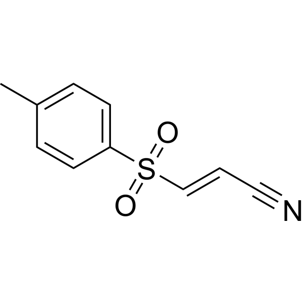
BAY 11-7082 -
HY-P80466
Phospho-IKB alpha (Ser32) Antibody (YA183) -
HY-P80840
Phospho-NF-KB p65 (Thr254) Antibody -
HY-13982
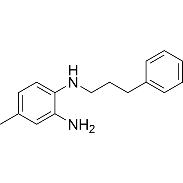
JSH-23 -
HY-12031A
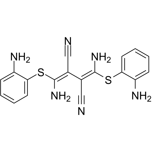
U0126 -
HY-50846
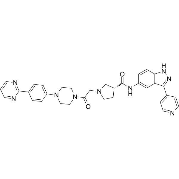
SCH772984 -

HY-14648
Dexamethasone
Publications Citing Use of MedChemExpress (MCE) NF-KB p65 Antibody (YA268)
More- Adv Sci (Weinh). 2025 Feb;12(8):e2405483. [Abstract]
- Eur J Pharmacol. 2025 Jul 5:998:177534. [Abstract]
- Eur J Med Res. 2025 Sep 26;30(1):874. [Abstract]
- Cell Signal. 2026 Aug:144:112541. [Abstract]
- J Inflamm Res. 2026 Jun 18:19:578571. [Abstract]
- Transl Cancer Res. 2024 Jun 30;13(6):2596-2604. [Abstract]
- SSRN. 2025 Mar 6.

 7 Publications Citing Use of MCE NF-KB p65 Antibody (YA268) (7)
-
WB

-
WB

-
WB

-
WB

-
WB


Applications
| Application |
WB

WB: Western Blot
|
IHC-P
IHC-P: Immunohistochemistry-Paraffin
|
IHC-F
IHC-F: Immunohistochemistry-Frozen
|
ICC/IF
ICC/IF: Immunocytochemistry/
Immunofluorescence |
200
|
IP
IP: Immunoprecipitation
|
|---|---|---|---|---|---|---|
| Dilution Ratio | 1:500-1:1000 | 1:50-1:100 | 1:50-1:100 | 1:50-1 | 1:20 |


Product Details
NF-KB p65 Antibody (YA268) is a Rabbit-derived and non-conjugated IgG monoclonal antibody, targeting to NF-KB p65.
-
Host Rabbit

-
Clonality Recombinant,Monoclonal

-
Species ReactivityHuman, Mouse
-
Observed Molecular WeightObserved band size: 65 kDa
Note: Due to possible protein modifications or aggregation, the molecular weight should be confirmed by actual measurement, and the predicted value is for reference only.

-
Calculated Molecular Weight Predicted band size: 60 kDa

Synthetic peptide corresponding to Human NF-kB p65 aa500-551.
Endogenous
affinity purified
Non-conjugated
Unmodified
IgG


Product Properties
-
Appearance
Solution
-
Formulation
Supplied in 50 mM Tris-Glycine (pH 7.4), 0.15 M NaCl, 40% Glycerol and 0.05% BSA. Preservative: 0.01% Sodium azide
-
Concentration
Batch-dependent, Please check the COA for the concentration of each lot. Check Lot Concentration
-
Storage & Stability
Stored at -20°C for 1 year. Avoid repeated freeze / thaw cycles.
-
Shipping
Shipping with blue ice.


Publications (7)
-
Journal Impact Factor
-
Most Recent
-
Adv Sci (Weinh)
Pathophysiology-Directed Engineering of a Combination Nanoanalgesic for Neuropathic Pain. [Abstract]2025 Feb;12(8):e2405483. PMID: 39716944
NF-KB p65 Antibody (YA268) purchased from MedChemExpress. Usage Cited in: Adv Sci (Weinh). 2025 Feb;12(8):e2405483. [Abstract]
Typical Western blotting bands and quantitative analysis of proteins associated with MAPK/NF-κB signaling pathways and macrophage polarization in bone marrow-derived macrophages (BMDMs).
-
Eur J Pharmacol
Chlorogenic acid alleviates DNCB-induced atopic dermatitis by inhibiting the Akt1/NF-κB signaling pathway. [Abstract]2025 Jul 5:998:177534. PMID: 40118327
NF-KB p65 Antibody (YA268) purchased from MedChemExpress. Usage Cited in: Eur J Pharmacol. 2025 Jul 5:998:177534. [Abstract]
Western blot analysis of p-PI3K, PI3K, p-Akt, Akt, p-IκB, IκB-α, p-p65, and p65 proteins. Phosphorylation levels of PI3K in dorsal skin tissues.
NF-KB p65 Antibody (YA268) purchased from MedChemExpress. Usage Cited in: Eur J Pharmacol. 2025 Jul 5:998:177534. [Abstract]
Western blot analysis of p-PI3K, PI3K, p-Akt, Akt, p-IκB, IκB-α, p-p65, and p65 proteins in HaCaT cells.
-
Eur J Med Res
Research on the inhibitory effect of hydrogen gas on ovarian cancer and potential mechanisms. [Abstract]2025 Sep 26;30(1):874. PMID: 41013628
NF-KB p65 Antibody (YA268) purchased from MedChemExpress. Usage Cited in: Eur J Med Res. 2025 Sep 26;30(1):874. [Abstract]
A Western blot analysis and quantification of HIF-1α, P-p65, and p65 protein expression levels in tumor tissues of BALB/c nude mice with SKOV3 cells.
-
Cell Signal
B4GALT1 drives osteoarthritis progression by stabilizing IL-1R1 through N-linked glycosylation. [Abstract]2026 Aug:144:112541. PMID: 42002123
NF-KB p65 Antibody (YA268) purchased from MedChemExpress. Usage Cited in: Cell Signal. 2026 Aug:144:112541. [Abstract]
Western blot analysis was performed to detect p-IRAK1, IRAK1, p-TAK1, TAK1 (the earliest events downstream of IL-1R1 activation) and core inflammatory pathways: p-IκBα, IκBα, p-p65, p65, p-p38 MAPK, p38 MAPK, p-JNK, JNK in human chondrocytes.
-
J Inflamm Res
IL-17RA Promotes Cigarette Smoke-Induced Alveolar Epithelial Cell Pyroptosis in COPD via Dual Activation of the NLRP3/Caspase1/GSDMD and NF-κB/GSDME Pathways. [Abstract]2026 Jun 18:19:578571. PMID: 42339301 -
Transl Cancer Res
Extracellular vesicles isolated from curcumin-medium weakened RKO cell proliferation and migration. [Abstract]2024 Jun 30;13(6):2596-2604. PMID: 38988906
NF-KB p65 Antibody (YA268) purchased from MedChemExpress. Usage Cited in: Transl Cancer Res. 2024 Jun 30;13(6):2596-2604. [Abstract]
Curcumin suppressed the expression of NF-κB p65 in RKOEVs. The expression of NF-κB p65 in RKOEVs, Cur1-RKOEVs and Cur10-RKOEVs was analyzed by western blotting.
-

NF-KB p65 Antibody (YA268) purchased from MedChemExpress. Usage Cited in: SSRN. 2025 Mar 6.
Western blot analysis of p65 phosphorylation and IκBα content restoration in MCF-7 cells. GAPDH was used as a reference.


Verification Images
-
 Western blot analysis of extracts from K562 (lane 2(20μg), RAW264.7 (lane 3(20μg) and Hela (lane 4(20μg) using NF-KB p65 (HY-P80765) Rabbit mAb. Proteins were transferred to a PVDF membrane and blocked with 5% non-fat milk in TBST for 2 hour at room temperature. The primary antibody (1/1000) and Loading control antibody (Beta Actin, HY-P80438, 1/10000) was used in 5% non-fat milk in TBST at 4°C overnight. Goat Anti-Mouse/Rabbit IgG-HRP Secondary Antibody (1/10000) was used for 1 hour at room temperature.
 -
 Immunohistochemical analysis of paraffin-embedded Mouse spleen tissue using NF-KB p65 Antibody. The section was pre-treated using heat mediated antigen retrieval with sodium citrate buffer (pH 6.0) for 8 minutes. The tissues were blocked in QuickBlock for 20 minutes at room temperature, washed with ddH2O and PBS, and then probed with the primary antibody (HY-P80765, 1/100) in 4℃ overnight. The detection was performed using an HRP conjugated compact polymer system. DAB was used as the chromogen. Tissues were counterstained with hematoxylin and mounted with DPX.
 -
 Immunohistochemical analysis of paraffin-embedded Mouse spleen tissue using NF-KB p65 Antibody. The section was pre-treated using heat mediated antigen retrieval with sodium citrate buffer (pH 6.0) for 8 minutes. The tissues were blocked in QuickBlock for 20 minutes at room temperature, washed with ddH2O and PBS, and then probed with the primary antibody (HY-P80765, 1/100) in 4℃ overnight. The detection was performed using an HRP conjugated compact polymer system. DAB was used as the chromogen. Tissues were counterstained with hematoxylin and mounted with DPX.
 -
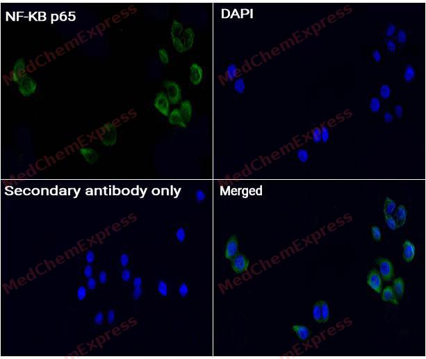 Immunocytochemistry analysis of Hela cells labeling NF-KB p65 with NF-KB p65 antibody (HY-P80765) at 1/50 dilution. Cells were fixed in 4% paraformaldehyde for 15 minutes at room temperature, permeabilized with 0.1% Triton X-100 for 10 minutes at room temperature, then blocked with QuickBlock™ Blocking Buffer for Immunol Staining for 10 min at room temperature. Cells were then incubated with NF-KB p65 antibody (HY-P80765) at 1/50 dilution in QuickBlock™ Blocking Buffer for Immunol Staining at 4 ℃. Alexa Fluor® 488-conjugated AffiniPure Goat Anti-Rabbit IgG H&L(HY-P8002, Green) was used as the secondary antibody at 1/1,000 dilution. PBS instead of the primary antibody was used as the secondary antibody only control. The Nuclear counterstain was DAPI (Blue).
 -
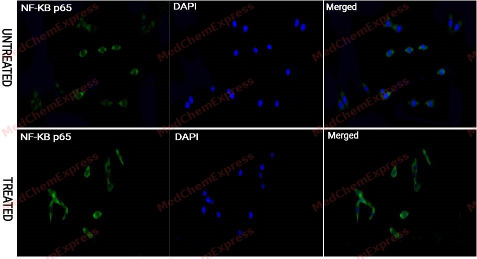 Immunocytochemistry analysis of NIH/3T3 cells labeling NF-KB p65 with NF-KB p65 antibody (HY-P80765) at 1/50 dilution. Confocal image showing the signal translocated from the cytoplasm into the nucleus in NIH/3T3 cells after the treatment with TNF-alpha (50 ng/ml) for 20 min. Cells were fixed in 4% paraformaldehyde for 15 minutes at room temperature, permeabilized with 0.1% Triton X-100 for 10 minutes at room temperature, then blocked with QuickBlock™ Blocking Buffer for Immunol Staining for 10 min at room temperature. Cells were then incubated with NF-KB p65 antibody (HY-P80765) at 1/200 dilution in QuickBlock™ Blocking Buffer for Immunol Staining at 4 ℃. Alexa Fluor® 488-conjugated AffiniPure Goat Anti-Rabbit IgG H&L(HY-P8002, Green) was used as the secondary antibody at 1/1,000 dilution. PBS instead of the primary antibody was used as the secondary antibody only control. The Nuclear counterstain was DAPI (Blue).


Background
-
Function
NF-kappa-B is a pleiotropic transcription factor present in almost all cell types and is the endpoint of a series of signal transduction events that are initiated by a vast array of stimuli related to many biological processes such as inflammation, immunity, differentiation, cell growth, tumorigenesis and apoptosis. NF-kappa-B is a homo- or heterodimeric complex formed by the Rel-like domain-containing proteins RELA/p65, RELB, NFKB1/p105, NFKB1/p50, REL and NFKB2/p52. The heterodimeric RELA-NFKB1 complex appears to be most abundant one. The dimers bind at kappa-B sites in the DNA of their target genes and the individual dimers have distinct preferences for different kappa-B sites that they can bind with distinguishable affinity and specificity. Different dimer combinations act as transcriptional activators or repressors, respectively. The NF-kappa-B heterodimeric RELA-NFKB1 and RELA-REL complexes, for instance, function as transcriptional activators. NF-kappa-B is controlled by various mechanisms of post-translational modification and subcellular compartmentalization as well as by interactions with other cofactors or corepressors. NF-kappa-B complexes are held in the cytoplasm in an inactive state complexed with members of the NF-kappa-B inhibitor (I-kappa-B) family. In a conventional activation pathway, I-kappa-B is phosphorylated by I-kappa-B kinases (IKKs) in response to different activators, subsequently degraded thus liberating the active NF-kappa-B complex which translocates to the nucleus. The inhibitory effect of I-kappa-B on NF-kappa-B through retention in the cytoplasm is exerted primarily through the interaction with RELA. RELA shows a weak DNA-binding site which could contribute directly to DNA binding in the NF-kappa-B complex. Besides its activity as a direct transcriptional activator, it is also able to modulate promoters accessibility to transcription factors and thereby indirectly regulate gene expression. Associates with chromatin at the NF-kappa-B promoter region via association with DDX1. Essential for cytokine gene expression in T-cells. The NF-kappa-B homodimeric RELA-RELA complex appears to be involved in invasin-mediated activation of IL-8 expression. Key transcription factor regulating the IFN response during SARS-CoV-2 infection
-
Subcellular Localization
Nucleus; Cytoplasm
-
Isoforms & Post-Translational Modification
NF-κB has 4 isomers: Q04206-1: 60219 Da (predicted); Q04206-2: 58807 Da (predicted); Q04206-3: 59068 Da (predicted); Q04206-4: 59910 Da (predicted).
Ubiquitinated by RNF182, leading to its proteasomal degradation (PubMed:31432514). Degradation is required for termination of NF-kappa-B response. Polyubiquitinated via 'Lys-29'-linked ubiquitin; leading to lysosomal degradation (PubMed:21518757);Monomethylated at Lys-310 by SETD6 (PubMed:21515635). Monomethylation at Lys-310 is recognized by the ANK repeats of EHMT1 and promotes the formation of repressed chromatin at target genes, leading to down-regulation of NF-kappa-B transcription factor activity. Phosphorylation at Ser-311 disrupts the interaction with EHMT1 without preventing monomethylation at Lys-310 and relieves the repression of target genes (By similarity);Phosphorylation at Ser-311 disrupts the interaction with EHMT1 and promotes transcription factor activity (By similarity). Phosphorylation on Ser-536 stimulates acetylation on Lys-310 and interaction with CBP; the phosphorylated and acetylated forms show enhanced transcriptional activity. Phosphorylation at Ser-276 by RPS6KA4 and RPS6KA5 promotes its transactivation and transcriptional activities;Phosphorylation at Ser-75 by herpes simplex virus 1/HHV-1 inhibits NF-kappa-B activation;Reversibly acetylated; the acetylation seems to be mediated by CBP, the deacetylation by HDAC3 and SIRT2. Acetylation at Lys-122 enhances DNA binding and impairs association with NFKBIA. Acetylation at Lys-310 is required for full transcriptional activity in the absence of effects on DNA binding and NFKBIA association. Acetylation at Lys-310 promotes interaction with BRD4. Acetylation can also lower DNA-binding and results in nuclear export. Interaction with BRMS1 promotes deacetylation of Lys-310. Lys-310 is deacetylated by SIRT2;S-nitrosylation of Cys-38 inactivates the enzyme activity;Sulfhydration at Cys-38 mediates the anti-apoptotic activity by promoting the interaction with RPS3 and activating the transcription factor activity;Sumoylation by PIAS3 negatively regulates DNA-bound activated NF-kappa-B;Proteolytically cleaved within a conserved N-terminus region required for base-specific contact with DNA in a CPEN1-mediated manner, and hence inhibits NF-kappa-B transcriptional activity (PubMed:18212740) -
Subunit
Component of the NF-κB p65-p50 complex. Interacts with NFKBIA. Interacts with NFKBIE. Interacts with NFKBIZ. Interacts with EHMT1 (via ANK repeats). Part of a 70-90 kDa complex at least consisting of CHUK, IKBKB, NFKBIA, RELA, ELP1 and MAP3K14. Interacts with HDAC3; HDAC3 mediates the deacetylation of RELA. Interacts with HDAC1; the interaction requires non-phosphorylated RELA. Interacts with CBP; the interaction requires phosphorylated RELA. Interacts (phosphorylated at 'Thr-254') with PIN1; the interaction inhibits p65 binding to NFKBIA. Interacts with SOCS1. Interacts with UXT. Interacts with MTDH and PHF11. Interacts with ARRB2. Interacts with NFKBIA (when phosphorylated), the interaction is direct; phosphorylated NFKBIA is part of a SCF(BTRC)-like complex lacking CUL1. Interacts with RNF25. Interacts (via C-terminus) with DDX1. Interacts with UFL1 and COMMD1. Interacts with BRMS1; this promotes deacetylation of 'Lys-310'. Directly interacts with MEN1; this interaction represses NFKB-mediated transactivation. Interacts with AKIP1, which promotes the phosphorylation and nuclear retention of RELA. Interacts (via the RHD) with GFI1; the interaction, after bacterial lipopolysaccharide (LPS) stimulation, inhibits the transcriptional activity by interfering with the DNA-binding activity to target gene promoter DNA. Interacts (when acetylated at Lys-310) with BRD4; leading to activation of the NF-κB pathway. Interacts with MEFV. Interacts (via N-terminus) with CPEN1. Interacts with FOXP3. Interacts with CDK5RAP3. Interacts with DHX9; this interaction is direct and activates NF-κB-mediated transcription. Interacts with LRRC25. Interacts with ZBTB7A; involved in the control by RELA of the accessibility of target gene promoters. Directly interacts with DDX3X; this interaction may trap RELA in the cytoplasm, impairing nuclear relocalization upon TNF activating signals.
-
SwissProt ID
-
Synonyms
NFKB3; RELA; TF65; Transcription factor p65; p65; NFkB
-
Research Field
Cell Biology
Documentation
-
Data Sheet (234 KB)
-
SDS (251 KB)
- English - EN (251 KB)
- Français - FR (251 KB)
- Deutsch - DE (251 KB)
- Norwegian - NO (251 KB)
- Español - ES (251 KB)
- Swedish - SV (251 KB)
- Italian - IT (251 KB)
- Korean - KR (251 KB)
- Portuguese - PT (251 KB)
-
User Guide for Antibodies (1077 KB)

 Powered by Bioz
