



MLKL Antibody (YA289)
Based on 2 publication(s) in Google Scholar
MLKL Antibody (YA289) is a Rabbit-derived and non-conjugated IgG monoclonal antibody, targeting to MLKL.
-
Host:
Rabbit
-
Isotype:
IgG
-
Application:
WB, IHC-P
-
Reactivity :
Human, Mouse
-
Formulation:
Supplied in 1*TBS (pH7.4), 0.05% BSA and 40% Glycerol. Preservative: 0.05% Sodium Azide.
-
Conjugation:
Non-conjugated


To place orders, for customer services and technical support, please contact: MedChemExpress USA
Tel: 609-228-6898 E-mail: [email protected] [email protected]



-

HY-P80468
Phospho-MLKL (Ser345) Antibody (YA174) -
HY-P81878
Phospho-MLKL (Ser358) Antibody (YA1623) -
HY-P86351
RIP3 Antibody (YA6043) -
HY-P80246
NLRP3 Antibody (YA266) -
HY-P80704
HIF1 alpha Antibody -
HY-P81539
Phospho-RIP(S166) Antibody (YA1284) -
HY-P87753
Phospho-MLKL (Ser345) Antibody (YA7438) -
HY-P80046
Caspase-3 Antibody (YA557)
Publications Citing Use of MedChemExpress (MCE) MLKL Antibody (YA289)
More

Applications
| Application |
WB

WB: Western Blot
|
IHC-P
IHC-P: Immunohistochemistry-Paraffin
|
|---|---|---|
| Dilution Ratio | 1:1000 | 1:50 |


Product Details
MLKL Antibody (YA289) is a Rabbit-derived and non-conjugated IgG monoclonal antibody, targeting to MLKL.
-
Host Rabbit

-
Clonality Recombinant, Monoclonal

-
Species ReactivityHuman, Mouse
-
Observed Molecular WeightObserved band size: 54 kDa
Note: Due to possible protein modifications or aggregation, the molecular weight should be confirmed by actual measurement, and the predicted value is for reference only.

-
Calculated Molecular Weight Predicted band size: 54 kDa;

Entrez Gene: 197259 Human ; 74568 Mouse ;
SwissProt: Q8NB16 Human ; Q9D2Y4 Mouse ;
OMIM: 615153 Human
Synthetic peptide corresponding to Human MLKL.AA range:333-471.
Endogenous
Protein A affinity purified.
Non-conjugated
Unmodified
IgG


Product Properties
-
Appearance
Liquid
-
Formulation
Supplied in 1*TBS (pH7.4), 0.05% BSA and 40% Glycerol. Preservative: 0.05% Sodium Azide.
-
Concentration
Batch-dependent, Please check the COA for the concentration of each lot. Check Lot Concentration
-
Storage & Stability
Stored at -20°C for 1 year. Avoid repeated freeze / thaw cycles.
-
Shipping
Shipping with blue ice.


Publications (2)
-
Journal Impact Factor
-
Most Recent
-
ACS Chem Neurosci
Calycosin Inhibit PANoptosis and Alleviate Brain Damage: A Bioinformatics and Experimental Verification Approach. [Abstract]2025 Apr 16;16(8):1550-1564. PMID: 40156525


Verification Images
-
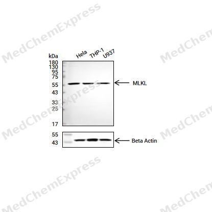 Western blot analysis of extracts from Hela (lane 2(20μg) , THP-1(lane 3(20μg) and U937(lane 4(20μg) using MLKL(HY-P80227) Rabbit mAb. Proteins were transferred to a PVDF membrane and blocked with 5% non-fat milk in TBST for 2 hour at room temperature. The primary antibody (1/1000) and Loading control antibody (Beta Actin, HY-P80438, 1/10000) was used in 5% non-fat milk in TBST at 4°C overnight. Goat Anti-Rabbit/Mouse IgG-HRP Secondary Antibody (1/10000) was used for 1 hour at room temperature.
 -
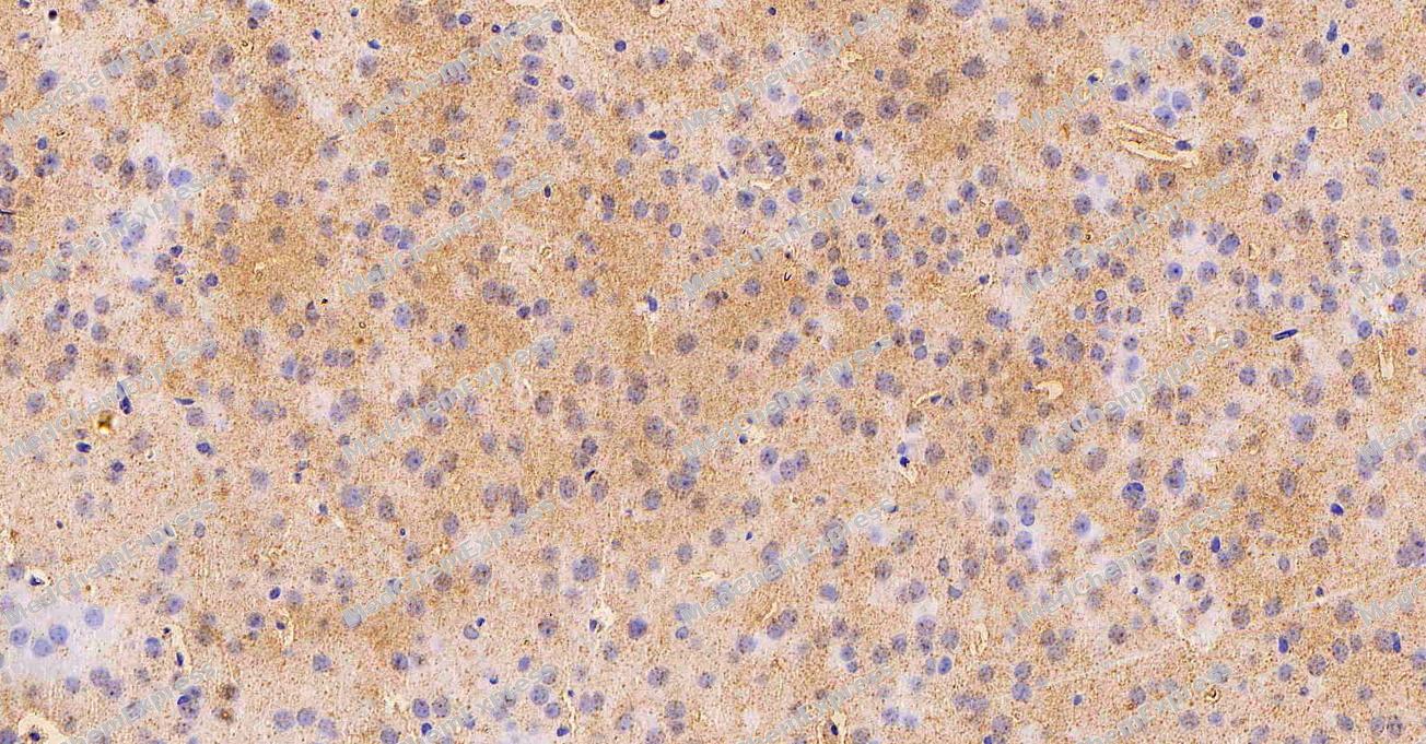 Immunohistochemical analysis of paraffin-embedded Mouse brain tissue using MLKL Antibody. The section was pre-treated using heat mediated antigen retrieval with Tris-EDTA buffer (pH 9.0) for 8 minutes. The tissues were blocked in QuickBlock for 20 minutes at room temperature, washed with ddH2O and PBS, and then probed with the primary antibody (HY-P80227, 1/1000) in 4℃ overnight. The detection was performed using an HRP conjugated compact polymer system. DAB was used as the chromogen. Tissues were counterstained with hematoxylin and mounted with DPX.
 -
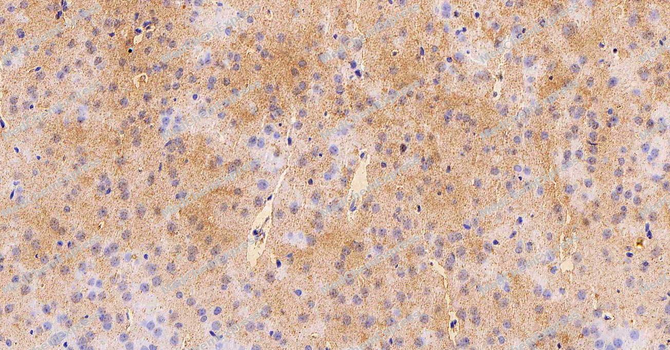 Immunohistochemical analysis of paraffin-embedded Mouse brain tissue using MLKL Antibody. The section was pre-treated using heat mediated antigen retrieval with Tris-EDTA buffer (pH 9.0) for 8 minutes. The tissues were blocked in QuickBlock for 20 minutes at room temperature, washed with ddH2O and PBS, and then probed with the primary antibody (HY-P80227, 1/1000) in 4℃ overnight. The detection was performed using an HRP conjugated compact polymer system. DAB was used as the chromogen. Tissues were counterstained with hematoxylin and mounted with DPX.


Background
-
Function
MLKL is a Pseudokinase that plays a key role in TNF-induced necroptosis, a programmed cell death process. Does not have protein kinase activity. Activated following phosphorylation by RIPK3, leading to homotrimerization, localization to the plasma membrane and execution of programmed necrosis characterized by calcium influx and plasma membrane damage. In addition to TNF-induced necroptosis, necroptosis can also take place in the nucleus in response to orthomyxoviruses infection: following activation by ZBP1, MLKL is phosphorylated by RIPK3 in the nucleus, triggering disruption of the nuclear envelope and leakage of cellular DNA into the cytosol.following ZBP1 activation, which senses double-stranded Z-RNA structures, nuclear RIPK3 catalyzes phosphorylation and activation of MLKL, promoting disruption of the nuclear envelope and leakage of cellular DNA into the cytosol. Binds to highly phosphorylated inositol phosphates such as inositolhexakisphosphate (InsP6) which is essential for its necroptotic function[1][2][3][4][5].
-
Subcellular Localization
Cytoplasm; Cell membrane; Nucleus
-
Subunit
Homooligomer (PubMed:29883610). Homotrimer; forms homotrimers on necroptosis induction (PubMed:24316671). Upon TNF-induced necrosis, forms in complex with PGAM5, RIPK1 and RIPK3 (PubMed:22265414). Within this complex, may play a role in the proper targeting of RIPK1-RIPK3 to its downstream effector PGAM5 (PubMed:22265414). Interacts with RIPK3; the interaction is direct and promotes its phosphorylation and subsequent activation (PubMed:22265413, PubMed:22421439, PubMed:29883609)
-
SwissProt ID
-
Research Field
Signal Transduction
Documentation
-
Data Sheet (235 KB)
-
SDS (313 KB)
- English - EN (313 KB)
- Français - FR (313 KB)
- Deutsch - DE (313 KB)
- Norwegian - NO (313 KB)
- Español - ES (313 KB)
- Swedish - SV (313 KB)
- Italian - IT (313 KB)
- Korean - KR (313 KB)
- Portuguese - PT (313 KB)
-
User Guide for Antibodies (1077 KB)

 Powered by Bioz
