



RhoA Antibody (YA093)
(Synonyms: ARH12, ARHA, RHO12, RHOA, Transforming protein RhoA, Rho cDNA clone 12, h12)
Based on 1 Customer Validation
RhoA Antibody (YA093) is a Rabbit-derived and non-conjugated IgG monoclonal antibody, targeting to RhoA.
-
Host:
Rabbit
-
Isotype:
IgG
-
Application:
WB, ICC/IF, FC
-
Reactivity :
Human, Mouse
-
Formulation:
Supplied in 1*TBS (pH7.4), 0.05% BSA and 40% Glycerol. Preservative: 0.05% Sodium Azide.
-
Conjugation:
Non-conjugated


To place orders, for customer services and technical support, please contact: MedChemExpress USA
Tel: 609-228-6898 E-mail: [email protected] [email protected]





Applications
| Application |
WB

WB: Western Blot
|
ICC/IF
ICC/IF: Immunocytochemistry/
Immunofluorescence |
FC
FC: Flow Cytometry
|
|---|---|---|---|
| Dilution Ratio | 1:1000-1:2000 | 1:50-1:200 | 1:50-1:100 |


Product Details
RhoA Antibody (YA093) is a Rabbit-derived and non-conjugated IgG monoclonal antibody, targeting to RhoA.
-
Host Rabbit

-
Clonality Recombinant,Monoclonal

-
Species ReactivityHuman, Mouse
-
Observed Molecular WeightObserved band size: 22 kDa
Note: Due to possible protein modifications or aggregation, the molecular weight should be confirmed by actual measurement, and the predicted value is for reference only.

-
Calculated Molecular Weight Predicted band size: 22 kDa

Entrez Gene: 387 Human ; 11848 Mouse ; 117273 Rat
SwissProt: P61586 Human ; Q9QUI0 Mouse ; P61589 Rat
OMIM: 618727 Human
Synthetic peptide corresponding to Human.AA range:144-193.
Endogenous
Protein A affinity purified.
Non-conjugated
Unmodified
IgG


Product Properties
-
Appearance
Solution
-
Formulation
Supplied in 1*TBS (pH7.4), 0.05% BSA and 40% Glycerol. Preservative: 0.05% Sodium Azide.
-
Concentration
Batch-dependent, Please check the COA for the concentration of each lot. Check Lot Concentration
-
Storage & Stability
Stored at -20°C for 1 year. Avoid repeated freeze / thaw cycles.
-
Shipping
Shipping with blue ice.


Verification Images
-
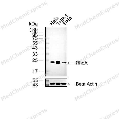 Western blot analysis of extracts from Hela (lane 2(20μg) , THP-1 (lane 3(20μg) , SiHa (lane 4(20μg) , C2C12 (lane 4(20μg),using RhoA Antibody (HY-P80311). Proteins were transferred to a PVDF membrane and blocked with 5% BSA in TBST for 2 hour at room temperature. The primary antibody and Loading control antibody (Beta Actin, HY-P80438, 1/3000) was used in 5% BSA in TBST at 4°C overnight. Goat Anti-Mouse/Rabbit IgG-HRP Secondary Antibody (HY-P8004/HY-P8001, 1/10,000) was used for 1 hour at room temperature.
 -
 Immunocytochemistry analysis of Hela cells labeling RhoA with RhoA Antibody (HY-P80311) at 1/100 dilution. Cells were fixed in 4% paraformaldehyde for 15 minutes at room temperature, permeabilized with 0.1% Triton X-100 in PBS for 15 minutes at room temperature, then blocked with quick block buffer for 10 minutes at room temperature. Cells were then incubated with RhoA Antibody (HY-P80311) at 1/100 dilution in quick block buffer overnight at 4 ℃. AF488-conjugated Goat Anti-Rabbit IgG H&L(HY-P8002, Green) was used as the secondary antibody at 1/1,000 dilution. PBS instead of the primary antibody was used as the secondary antibody only control. The Nuclear counterstain was DAPI (Blue).
 -
 Immunocytochemistry analysis of U2OS cells labeling RhoA with RhoA Antibody (HY-P80311) at 1/100 dilution. Cells were fixed in 4% paraformaldehyde for 15 minutes at room temperature, permeabilized with 0.1% Triton X-100 in PBS for 15 minutes at room temperature, then blocked with quick block buffer for 10 minutes at room temperature. Cells were then incubated with RhoA Antibody (HY-P80311) at 1/100 dilution in quick block buffer overnight at 4 ℃. AF488-conjugated Goat Anti-Rabbit IgG H&L(HY-P8002, Green) was used as the secondary antibody at 1/1,000 dilution. PBS instead of the primary antibody was used as the secondary antibody only control. The Nuclear counterstain was DAPI (Blue).
 -
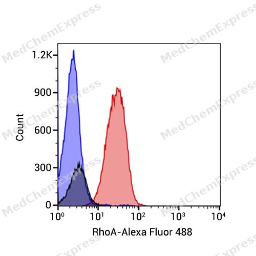 Flow cytometric analysis of 1X10^6 Jurkat cells labeling RhoA Antibody(HY-P80311, red). Cells were fixed with 4% paraformaldehyde and permeabilised with 90% methanol. Then stained with the primary antibody at 1/150 dilution for an hour at 4℃. Alexa Fluor® 488-conjugated AffiniPure Goat Anti-Rabbit IgG H&L (HY-P8002) was used as the secondary antibody at 1/1,000 dilution for 30 minutes at 4℃. Rabbit IgG Isotype Control (HY-P80879, blue) was used as the isotype control, cells without incubation with primary antibody were used as the unlabeled control (black).


Background
-
Function
RhoA is a Small GTPase which cycles between an active GTP-bound and an inactive GDP-bound state. Mainly associated with cytoskeleton organization, in active state binds to a variety of effector proteins to regulate cellular responses such as cytoskeletal dynamics, cell migration and cell cycle. Regulates a signal transduction pathway linking plasma membrane receptors to the assembly of focal adhesions and actin stress fibers. Involved in a microtubule-dependent signal that is required for the myosin contractile ring formation during cell cycle cytokinesis. Plays an essential role in cleavage furrow formation. Required for the apical junction formation of keratinocyte cell-cell adhesion. Essential for the SPATA13-mediated regulation of cell migration and adhesion assembly and disassembly. The MEMO1-RHOA-DIAPH1 signaling pathway plays an important role in ERBB2-dependent stabilization of microtubules at the cell cortex. It controls the localization of APC and CLASP2 to the cell membrane, via the regulation of GSK3B activity. In turn, membrane-bound APC allows the localization of the MACF1 to the cell membrane, which is required for microtubule capture and stabilization. Regulates KCNA2 potassium channel activity by reducing its location at the cell surface in response to CHRM1 activation; promotes KCNA2 endocytosis. Acts as an allosteric activator of guanine nucleotide exchange factor ECT2 by binding in its activated GTP-bound form to the PH domain of ECT2 which stimulates the release of PH inhibition and promotes the binding of substrate RHOA to the ECT2 catalytic center. May be an activator of PLCE1. In neurons, involved in the inhibition of the initial spine growth. Upon activation by CaMKII, modulates dendritic spine structural plasticity by relaying CaMKII transient activation to synapse-specific, long-term signaling. Acts as a regulator of platelet alpha-granule release during activation and aggregation of platelets. When activated by DAAM1 may signal centrosome maturation and chromosomal segregation during cell division. May also be involved in contractile ring formation during cytokinesis; (Microbial infection) Serves as a target for the yopT cysteine peptidase from Yersinia pestis, vector of the plague[1][2][3][4][5][6][7][8][9][10][11][12][13][14].
-
Subcellular Localization
Cell membrane; Lipid-anchor; Cytoplasmic side; Cytoplasm, cytoskeleton; Cleavage furrow; Cytoplasm, cell cortex; Midbody; Cell projection, lamellipodium; Cell projection, dendrite; Nucleus; Cytoplasm
-
Subunit
Interacts with ARHGEF28 (By similarity). Interacts (via GTP-bound form) with RIPOR1 (via N-terminus); this interaction links RHOA to STK24 and STK26 kinases (PubMed:27807006). Interacts with RIPOR2 (via active GTP- or inactive GDP-bound forms) isoform 1 and isoform 2; these interactions are direct, block the loading of GTP to RHOA and decrease upon chemokine CCL19 stimulation in primary T lymphocytes (PubMed:25588844). Binds PRKCL1, ROCK1 and ROCK2 (PubMed:10388627, PubMed:8617235, PubMed:8641286). Interacts with ARHGEF2, ARHGEF3, NET1 and RTKN (PubMed:10940294, PubMed:12221096, PubMed:9857026). Interacts with PLCE1 and AKAP13 (PubMed:11696353, PubMed:12900402). Interacts with DIAPH1 (PubMed:23325789). Interacts (in the constitutively activated, GTP-bound form) with DGKQ (PubMed:10066731). Interacts with RACK1; enhances RHOA activation (PubMed:20499158). Interacts with PKP4; the interaction is detected at the midbody (PubMed:17115030). Interacts (GTP-bound form preferentially) with PKN2; the interaction stimulates autophosphorylation and phosphorylation of PKN2 (PubMed:20974804, PubMed:9121475). Interacts with ARHGDIA; this interaction inactivates and stabilizes RHOA (PubMed:20400958). Interacts with ARHGDIB. Interacts (GTP-bound form) with KCNA2 (via cytoplasmic N-terminal domain) (PubMed:9635436). Interacts (GTP-bound form) with ECT2; the interaction results in allosteric activation of ECT2 (PubMed:31888991). Interacts with RAP1GDS1; the interaction is direct and in a 1:1 stoichiometry (PubMed:12551911, PubMed:20709748, PubMed:28630045, PubMed:30190425)
-
SwissProt ID
-
Synonyms
ARH12, ARHA, RHO12, RHOA, Transforming protein RhoA, Rho cDNA clone 12, h12
-
Research Field
Signal Transduction


Documentation
-
Data Sheet (235 KB)
-
SDS (251 KB)
- English - EN (251 KB)
- Français - FR (251 KB)
- Deutsch - DE (251 KB)
- Norwegian - NO (251 KB)
- Español - ES (251 KB)
- Swedish - SV (251 KB)
- Italian - IT (251 KB)
- Korean - KR (251 KB)
- Portuguese - PT (251 KB)
-
User Guide for Antibodies (1077 KB)
References
[1]. Wei J, et al. A new mechanism of RhoA ubiquitination and degradation: roles of SCF(FBXL19) E3 ligase and Erk2. Biochim Biophys Acta. 2013 Dec;1833(12):2757-2764. [Content Brief]
[2]. Vabres P, et al. Postzygotic inactivating mutations of RHOA cause a mosaic neuroectodermal syndrome. Nat Genet. 2019 Oct;51(10):1438-1441. [Content Brief]
[3]. Quilliam LA, et al. Isolation of a NCK-associated kinase, PRK2, an SH3-binding protein and potential effector of Rho protein signaling. J Biol Chem. 1996 Nov 15;271(46):28772-6. [Content Brief]
[4]. Vincent S, et al. The PRK2 kinase is a potential effector target of both Rho and Rac GTPases and regulates actin cytoskeletal organization. Mol Cell Biol. 1997 Apr;17(4):2247-56. [Content Brief]
[5]. Wing MR, et al. Direct activation of phospholipase C-epsilon by Rho. J Biol Chem. 2003 Oct 17;278(42):41253-8. [Content Brief]
[6]. Kamijo K, et al. Dissecting the role of Rho-mediated signaling in contractile ring formation. Mol Biol Cell. 2006 Jan;17(1):43-55. [Content Brief]
[7]. Wallace SW, et al. The Rho target PRK2 regulates apical junction formation in human bronchial epithelial cells. Mol Cell Biol. 2011 Jan;31(1):81-91. [Content Brief]
[8]. Dubash AD, et al. The GEF Bcr activates RhoA/MAL signaling to promote keratinocyte differentiation via desmoglein-1. J Cell Biol. 2013 Aug 19;202(4):653-66. [Content Brief]
[9]. Bristow JM, et al. The Rho-family GEF Asef2 activates Rac to modulate adhesion and actin dynamics and thereby regulate cell migration. J Cell Sci. 2009 Dec 15;122(Pt 24):4535-46. [Content Brief]
[10]. Zaoui K, et al. ErbB2 receptor controls microtubule capture by recruiting ACF7 to the plasma membrane of migrating cells. Proc Natl Acad Sci U S A. 2010 Oct 26;107(43):18517-22. [Content Brief]
[11]. Stirling L, et al. Dual roles for RHOA/RHO-kinase in the regulated trafficking of a voltage-sensitive potassium channel. Mol Biol Cell. 2009 Jun;20(12):2991-3002. [Content Brief]
[12]. Cachero TG, et al. The small GTP-binding protein RhoA regulates a delayed rectifier potassium channel. Cell. 1998 Jun 12;93(6):1077-85. [Content Brief]
[13]. Chen M, et al. Structure and regulation of human epithelial cell transforming 2 protein. Proc Natl Acad Sci U S A. 2020 Jan 14;117(2):1027-1035. [Content Brief]
[14]. Yüce O, et al. An ECT2-centralspindlin complex regulates the localization and function of RhoA. J Cell Biol. 2005 Aug 15;170(4):571-82. [Content Brief]

 Powered by Bioz
