



KCC-07
Based on 2 publication(s) in Google Scholar
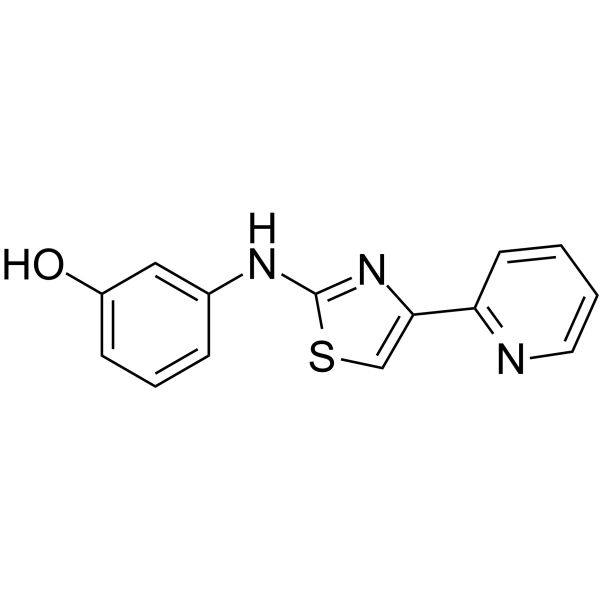
KCC-07 is a potent and brain-penetrant MBD2 (methyl-CpG-binding domain protein 2) inhibitor. KCC-07 prevents binding of MBD2 to methylated DNA and activates brain specific angiogenesis inhibitor 1 (BAI1) inducing anti-proliferative BAI1/p53/p21 signaling. KCC-07 is a TRACER (transcriptional regulation via active control of epigenetic reprogramming) that can stimulate ER transcriptional activity. KCC-07 can be used for the stduy of breast cancer.
For research use only. We do not sell to patients.
- Purity : 99.95%
- CAS No.: 315702-75-1
- Formula: C14H11N3OS
- Molecular Weight:269.32
-
Storage:
4°C, protect from light
* In solvent : -80°C, 6 months; -20°C, 1 month (protect from light)

To place orders, for customer services and technical support, please contact: MedChemExpress USA
Tel: 609-228-6898 E-mail: [email protected] [email protected]


-
 Biological Activity
-
Chemical Information
-
Solvent & Solubility
- Protocols
- Purity & Documentation
- References
-
 Help & FAQs


-
HY-13030
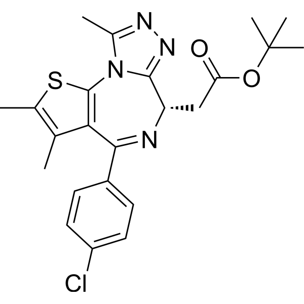
(+)-JQ-1BET Inhibitor -
HY-B0245
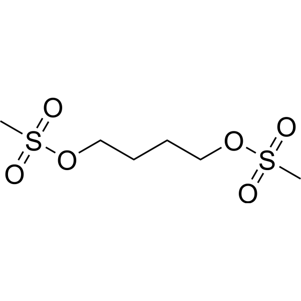
BusulfanAlkylating Antineoplastic Agent -
HY-13669
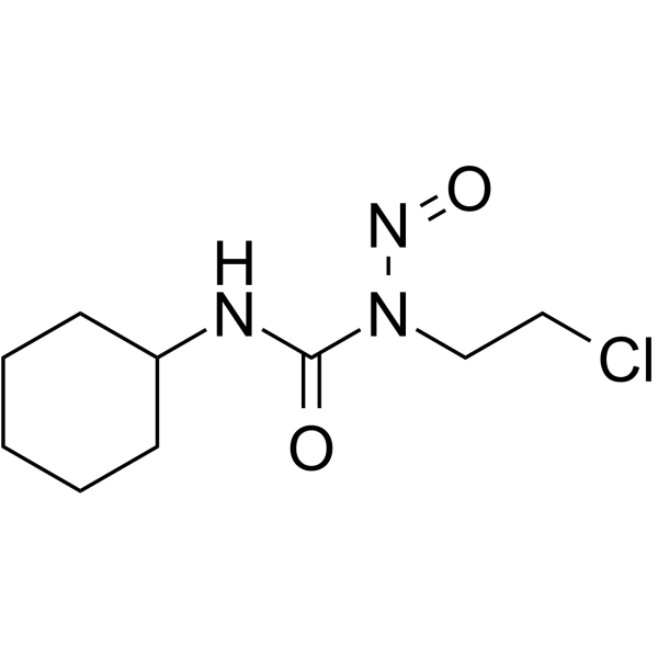
LomustineDNA Alkylator -
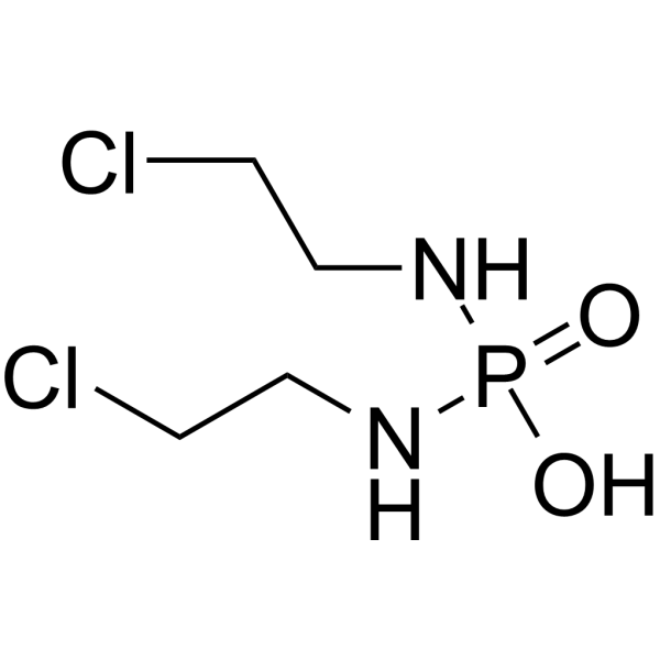
HY-14798
Palifosfamide -

HY-B0245R
Busulfan (Standard)Alkylating Antineoplastic Agent -
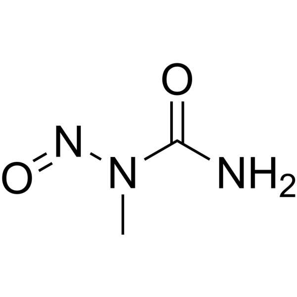
HY-34758
N-Nitroso-N-methylureaCarcinogen Agent -
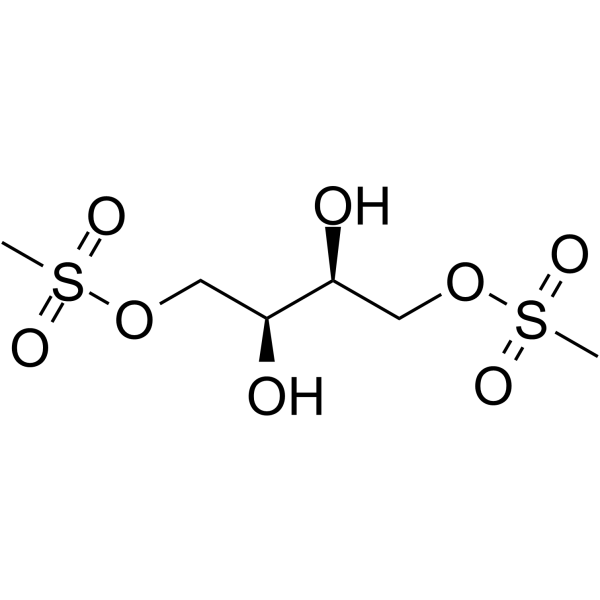
HY-16503
TreosulfanAlkylating Cytotoxic Agent -
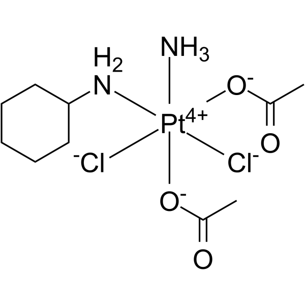
HY-17576
SatraplatinDNA Alkylator -
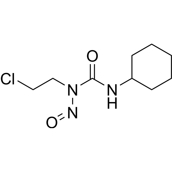
HY-13669R
Lomustine (Standard)DNA Alkylator -
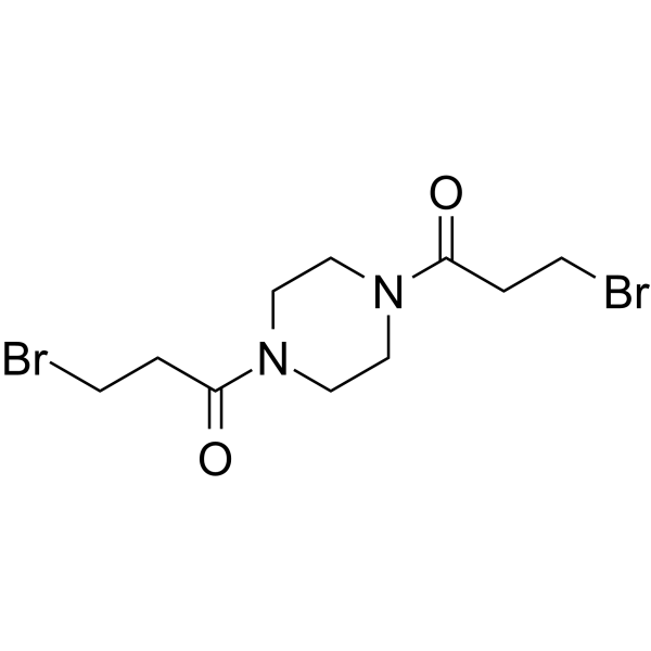
HY-16398
PipobromanAlkylating Agent -
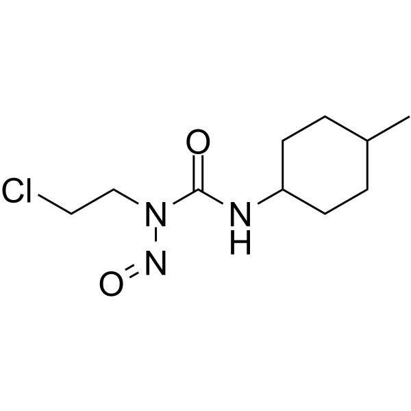
HY-13747
SemustineAntitumor Alkylating Agent -
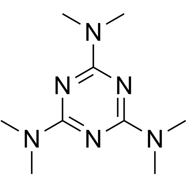
HY-B0181
AltretamineDNA Alkylator/Crosslinker
Publications Citing Use of MedChemExpress (MCE) KCC-07
More

Biological Activity
Description
IC50 & Target
MBD2 (methyl-CpG-binding domain protein 2)[1]
In Vitro
KCC-07 (10 μM; 72 hours; MB cells) treatment clearly inhibited MB cell growth in vitro, consistent with induction of anti-proliferative BAI1/p53/p21 signaling[1].
KCC-07 (10 μM; 48 hours; MB cells) treatment largely abrogates MBD2 binding to the ADGRB1 promoter and restores BAI1 mRNA and protein expression in BAI1-silent MB cells[1].
MedChemExpress (MCE) has not independently confirmed the accuracy of these methods. They are for reference only. Further protocols information, click here.
-
Cell Line:Medulloblastomas (MB) cells
-
Concentration:10 μM
-
Incubation Time:72 hours
-
Result:Clearly inhibited MB cell growth in vitro.
-
Cell Line:Medulloblastomas (MB) cells
-
Concentration:10 μM
-
Incubation Time:48 hours
-
Result:Largely abrogated MBD2 binding to the ADGRB1 promoter in BAI1-silent MB cells.
In Vivo
MedChemExpress (MCE) has not independently confirmed the accuracy of these methods. They are for reference only.
-
Animal Model:Outbred athymic nude mice (females; 8-10 weeks old) injected with MB cells[1]
-
Dosage:100 mg/kg
-
Administration:Intraperitoneal injection; 5 days/week
-
Result:Significantly extended the survival of MB xenografts in vivo.


Chemical Information
-
CAS No. 315702-75-1
-
Appearance Solid
-
Molecular Weight 269.32
-
Formula C14H11N3OS
-
Color White to yellow
-
SMILES
OC1=CC=CC(NC2=NC(C3=NC=CC=C3)=CS2)=C1
-
Shipping
Room temperature in continental US; may vary elsewhere.
-
Storage
4°C, protect from light
* In solvent : -80°C, 6 months; -20°C, 1 month (protect from light)


Publications (2)
-
Journal Impact Factor
-
Most Recent
-
Redox Biol
Methylation reader MBD2-mediated GPX4 transcriptional repression drives ovarian granulosa cell ferroptosis in PCOS. [Abstract]2026 Mar:90:104034. PMID: 41576700 -
Cancer Biol Ther
MBD2 suppresses SFRP1 expression and promotes colorectal cancer development by blocking MED19 binding to its methylated promoter. [Abstract]2026 Dec 31;27(1):2667568. PMID: 42084802


Solvent & Solubility
In Vitro:
DMSO : 125 mg/mL (464.13 mM; Need ultrasonic; Hygroscopic DMSO has a significant impact on the solubility of product, please use newly opened DMSO)
Please refer to the solubility information to select the appropriate solvent. Once prepared, please aliquot and store the solution to prevent product inactivation from repeated freeze-thaw cycles.
Storage method and period of stock solution: -80°C, 6 months; -20°C, 1 month (protect from light). When stored at -80°C, please use it within 6 months. When stored at -20°C, please use it within 1 month.
Please refer to the solubility information to select the appropriate solvent. Once prepared, please aliquot and store the solution to prevent product inactivation from repeated freeze-thaw cycles.
Storage method and period of stock solution: -80°C, 6 months; -20°C, 1 month (protect from light). When stored at -80°C, please use it within 6 months. When stored at -20°C, please use it within 1 month.
Concentration (start) × Volume (start) = Concentration (final) × Volume (final)
In Vivo:
Select the appropriate dissolution method based on your experimental animal and administration route.
- For the following dissolution methods, please ensure to first prepare a clear stock solution using an In Vitro approach and then sequentially add co-solvents:
- To ensure reliable experimental results, the clarified stock solution can be appropriately stored based on storage conditions. As for the working solution for In Vivo experiments, it is recommended to prepare freshly and use it on the same day.
- The percentages shown for the solvents indicate their volumetric ratio in the final prepared solution. If precipitation or phase separation occurs during preparation, heat and/or sonication can be used to aid dissolution.
Add each solvent one by one: 10% DMSO 40% PEG300 5% Tween-80 45% Saline
Solubility: ≥ 2.08 mg/mL (7.72 mM); Clear solution
This protocol yields a clear solution of ≥ 2.08 mg/mL (saturation unknown).
Taking 1 mL working solution as an example, add 100 μL DMSO stock solution (20.8 mg/mL) to 400 μL PEG300, and mix evenly; then add 50 μL Tween-80 and mix evenly; then add 450 μL Saline to adjust the volume to 1 mL.
Preparation of Saline: Dissolve 0.9 g sodium chloride in ddH₂O and dilute to 100 mL to obtain a clear Saline solution.
Add each solvent one by one: 10% DMSO 90% (20% SBE-β-CD in Saline)
Solubility: ≥ 2.08 mg/mL (7.72 mM); Clear solution
This protocol yields a clear solution of ≥ 2.08 mg/mL (saturation unknown).
Taking 1 mL working solution as an example, add 100 μL DMSO stock solution (20.8 mg/mL) to 900 μL 20% SBE-β-CD in Saline, and mix evenly.
Preparation of 20% SBE-β-CD in Saline (4°C, storage for one week): 2 g SBE-β-CD powder is dissolved in 10 mL Saline, completely dissolve until clear.
For the following dissolution methods, please prepare the working solution directly:
It is recommended to prepare fresh solutions and use them promptly within a short period of time.
The percentages shown for the solvents indicate their volumetric ratio in the final prepared solution. If precipitation or phase separation occurs during preparation, heat and/or sonication can be used to aid dissolution.
Add each solvent one by one: 50% PEG300 50% Saline
Solubility: 16.67 mg/mL (61.90 mM); Clear solution; Need ultrasonic
In Vivo Dissolution Calculator
Please enter the basic information of animal experiments:
-
-
-
-
Recommended: Prepare an additional quantity of animals to account for potential losses during experiments.
Please enter your animal formula composition:
-
%DMSO +
Recommended: Keep the proportion of DMSO in working solution below 2% if your animal is weak.
-
%+
-
+%Tween-80 + +
-
%Saline +
The co-solvents required include: DMSO, . All of co-solvents are available by MedChemExpress (MCE). , Tween 80. All of co-solvents are available by MedChemExpress (MCE).
Working solution concentration: 0.22 mg/mL
Method for preparing stock solution: mg drug dissolved in μL DMSO. Stock solution concentration: mg/mL. * In solvent : -80°C, 6 months; -20°C, 1 month (protect from light)
1. Take μL DMSO stock solution;
2. Add μL .
μL , mix evenly;
3. Then add μL Tween 80, mix evenly;
4. Then add μL
Please ensure that the stock solution in the first step is dissolved to a clear state, and add co-solvents in sequence. You can use ultrasonic heating (ultrasonic cleaner, recommended frequency 20-40 kHz), vortexing, etc. to assist dissolution.

Protocols
-
Dual Luciferin reporter gene assay
Luciferin reporter gene assay is a reporting system to detect the activity of Firefly Luciferase using luciferin as a substrate, which is often used in the research of miRNA target gene verification and promoter transcriptive activity regulation. Dual luciferase usually refers to Firefly luciferase and Renilla luciferase.
-
Human pluripotent stem cell endothelial-cell differentiation
Human pluripotent stem cell endothelial differentiation is based on stepwise developmental patterning: early activation of WNT/GSK3β inhibition promotes mesodermal or vascular progenitor entry, followed by endothelial specification using VEGF-related signaling, BMP4, FGF2, Notch modulation, or cAMP depending on the published protocol. Endothelial differentiation is read out by acquisition of CD31, CD34, VE-cadherin/CD144, KDR/VEGFR2, vWF, Tie2, NOS3, acetylated LDL uptake, tube/network formation, barrier function, and in vivo vessel-forming capacity where tested.
-
Vascular/Branching Fractal Analysis
Vascular/branching fractal analysis quantifies the geometric complexity of vessel trees or vascular networks from segmented 2D images, commonly by converting vessels into binary and/or skeletonized maps and estimating fractal dimension using box-counting or related approaches. Fractal dimension is interpreted as an image-derived readout of vascular branching complexity, space filling, or density, and has been applied to retinal photographs, fluorescein angiography, OCT angiography, capillary perfusion maps, and in vitro Matrigel angiogenesis networks. The assay readout is generated from vessel-positive pixels after image preprocessing, vessel segmentation, binarization, and optional skeletonization; reported outputs include fractal dimension, vessel density, branchpoint density, endpoint density, vessel length density, tortuosity, and generation-based branching metrics when VESGEN-style analysis is used. The biological interpretation is limited to quantitative vascular patterning and s
-
Endothelial Tube Formation Assay
Endothelial tube formation assay evaluates the ability of endothelial cells to attach, migrate, align, and organize into capillary-like networks when cultured on gelled basement membrane extract or Matrigel; the readout is the morphology and quantity of tube-like networks, which reflects an in vitro endothelial morphogenesis step related to angiogenesis. Basement membrane extract/Matrigel provides laminin-rich extracellular matrix cues that support endothelial differentiation into capillary-like structures, but it can contain biologically active growth factors, so growth-factor-reduced matrix is preferred when testing defined angiogenic stimulators or inhibitors.
-
Breast Cancer Modeling
Breast cancer is a heterogeneous cancer, and it has been distinguished into four subtypes: luminal A, luminal B, HER2-positive and basal-like. Molecular mutations, epigenetic alterations, hormone exposure and immune microenvironment are related to the progression of breast cancer.
-
Endothelial Cell Migration/Angiogenic Sprouting Assay
Endothelial cell migration and angiogenic sprouting assays are in vitro (and partially ex vivo-adapted) functional models that quantify the ability of endothelial cells to undergo coordinated migration, extracellular matrix invasion, and multicellular organization into capillary-like sprouts in response to pro-angiogenic stimuli such as VEGF, bFGF, or conditioned microenvironments. These assays are used to model early angiogenic events including tip-cell formation, directional migration, and lumen-like sprout extension, which collectively reflect angiogenic activation and vascular morphogenesis processes observed in vivo.
-
Cell Viability Determination by MTT Colorimetric Assay
The following protocol uses the MTT colorimetric assay as a classic literature-established method for assessing cell viability/metabolic activity in cultured mammalian cells. MTT[3-(4,5-dimethylthiazol-2-yl)-2,5-diphenyltetrazolium bromide] is reduced by metabolically active cells to a colored formazan product; the amount of formazan is quantified spectrophotometrically and provides an indirect measure of metabolically active viable cells. Importantly, MTT reduction reflects cellular oxidoreductase/metabolic activity rather than an absolute direct count of living cells, so changes in cellular metabolism can alter the signal independently of cell number.


Purity & Documentation
-
Data Sheet (277 KB)
-
SDS (393 KB)
- English - EN (393 KB)
- Français - FR (393 KB)
- Deutsch - DE (393 KB)
- Norwegian - NO (393 KB)
- Español - ES (393 KB)
- Swedish - SV (393 KB)
- Italian - IT (393 KB)
- Korean - KR (393 KB)
- Portuguese - PT (393 KB)
-
Handling Instructions (2659 KB)
References
[1]. Dan Zhu, et al. BAI1 Suppresses Medulloblastoma Formation by Protecting p53 From Mdm2-Mediated Degradation. Cancer Cell. 2018 Jun 11;33(6):1004-1016.e5. [Content Brief]
[2]. Stieger CE, et al. Targeted Transcriptional Repression by Induced Proximity. bioRxiv [Preprint]. 2025 Oct 22:2025.10.22.680877. [Content Brief]


Complete Stock Solution Preparation Table
Please refer to the solubility information to select the appropriate solvent. Once prepared, please aliquot and store the solution to prevent product inactivation from repeated freeze-thaw cycles.
Storage method and period of stock solution: -80°C, 6 months; -20°C, 1 month (protect from light). When stored at -80°C, please use it within 6 months. When stored at -20°C, please use it within 1 month.
| Optional Solvent | Concentration Solvent Mass | 1 mg | 5 mg | 10 mg | 25 mg |
|---|---|---|---|---|---|
| DMSO | 1 mM | 3.7131 mL | 18.5653 mL | 37.1306 mL | 92.8264 mL |
| 5 mM | 0.7426 mL | 3.7131 mL | 7.4261 mL | 18.5653 mL | |
| 10 mM | 0.3713 mL | 1.8565 mL | 3.7131 mL | 9.2826 mL | |
| 15 mM | 0.2475 mL | 1.2377 mL | 2.4754 mL | 6.1884 mL | |
| 20 mM | 0.1857 mL | 0.9283 mL | 1.8565 mL | 4.6413 mL | |
| 25 mM | 0.1485 mL | 0.7426 mL | 1.4852 mL | 3.7131 mL | |
| 30 mM | 0.1238 mL | 0.6188 mL | 1.2377 mL | 3.0942 mL | |
| 40 mM | 0.0928 mL | 0.4641 mL | 0.9283 mL | 2.3207 mL | |
| 50 mM | 0.0743 mL | 0.3713 mL | 0.7426 mL | 1.8565 mL | |
| 60 mM | 0.0619 mL | 0.3094 mL | 0.6188 mL | 1.5471 mL | |
| 80 mM | 0.0464 mL | 0.2321 mL | 0.4641 mL | 1.1603 mL | |
| 100 mM | 0.0371 mL | 0.1857 mL | 0.3713 mL | 0.9283 mL |


KCC-07 Related Classifications
HY-131031 Related Classifications
 Powered by Bioz