



KT195
KT195 is a serine hydrolase inhibitor that targets ABHD6 (IC50 = 10 nM) and ABHD2, with no significant activity against DAGLβ. KT195 inhibits endoplasmic reticulum calcium release and mitochondrial calcium uptake by targeting ABHD2, thereby blocking A23187 (HY-N6687) and H2O2-induced necrotic cell death and apoptosis. By inhibiting ABHD6 activity, KT195 significantly induces 2-AG accumulation in Neuro2A cells and reduces IL-1β secretion in lipopolysaccharide-treated macrophages. KT195 has been used as a negative control probe for DAGLβ and can be applied in studies of ABHD6 and ABHD2 in calcium signaling, lipid metabolism, neuronal function, and inflammatory.
For research use only. We do not sell to patients.
- CAS No.: 1402612-58-1
- Formula: C27H26N4O2
- Molecular Weight:438.52
-
Storage:
Please store the product under the recommended conditions in the Certificate of Analysis.


To place orders, for customer services and technical support, please contact: MedChemExpress USA
Tel: 609-228-6898 E-mail: [email protected] [email protected]


-
 Biological Activity
-
Chemical Information
- Protocols
- Purity & Documentation
- References
-
 Help & FAQs


-
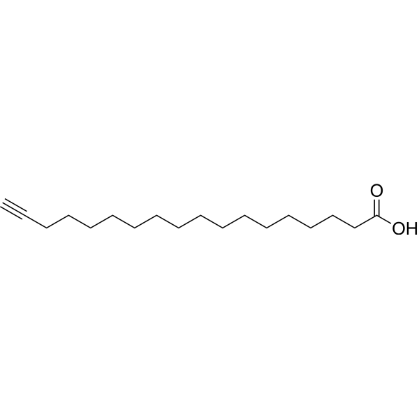
HY-101016
17-ODYACYP450 ω-Hydroxylase Inhibitor -
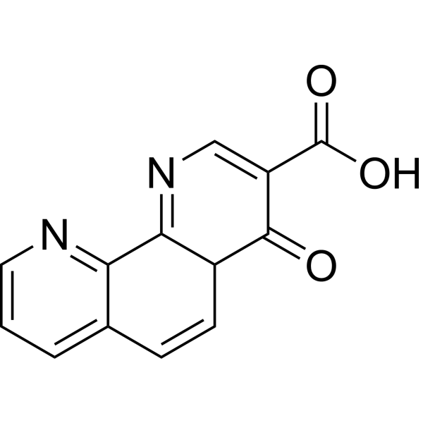
HY-121172
1,4-DPCAProlyl-4-hydroxylase Inhibitor -
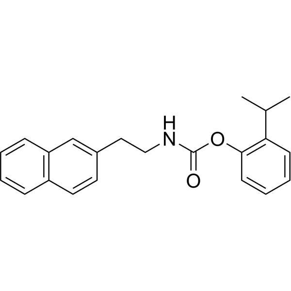
HY-107582
JW480KIAA1363/AADACL1 Inhibitor -

HY-101016R
17-ODYA (Standard)CYP450 ω-Hydroxylase Inhibitor -

HY-107582R
JW480 (Standard)Reference Standard -

HY-125775
12(S)-Hydroxy-16-heptadecynoic acidP450 ω-Hydroxylase Inhibitor -
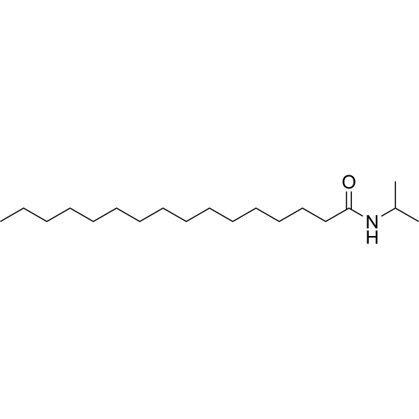
HY-103324
PalmitoylisopropylamideAnandamide Metabolism Inhibitor


Biological Activity
Description
|
ABHD6 10 nM (IC50) |
ABHD2 |
In Vitro
KT195 (0.2-2 μM; pretreated for 30 min) significantly inhibits 50 μMH2O2-induced apoptosis in MLF cells, partially inhibits 1 mM H2O2-induced [3H]arachidonic acid release and LDH release, and inhibits MPTP formation; in IMLF α⁻/⁻ cells, it concentration-dependently inhibits A23187-induced [3H]arachidonic acid release and LDH release (IC50 ≈ 0.7 μM); but it shows no inhibitory effect on 250 nM Staurosporine (HY-15141)-induced apoptosis[1][2].
KT195-alkyne (0.2-10 μM; 1 h) labels ABHD2 and ABHD6 in a concentration-dependent manner in the IMLF cell membrane proteome[1].
KT195-alkyne (30 min) inhibits A23187 (HY-N6687)-induced necrotic cell death in IMLF cells, with an IC50 of approximately 1.5 μM, comparable to unmodified KT195 (IC50 0.7 μM)[1].
KT195 (10 μM) competitively blocks the binding of KT195-alkyne to ABHD2 in IMLF cells, indicating that ABHD2 is a target of KT195[1].
KT195 (2 μM; pretreated for 30 min + co-incubated with A23187 for 30 min) still inhibits A23187-induced [3H]arachidonic acid release and LDH release in ABHD6-knockdown IMLF α⁻/⁻ cells, indicating that its anti-necrotic effect is independent of ABHD6[2].
KT195 (0.025-2 μM) completely blocks HT-01 probe binding to ABHD6 in IMLF α⁻/⁻ cells and concentration-dependently inhibits HT-01 probe binding to ABHD6 in the IMLF α⁻/⁻ membrane proteome, with an IC50 of approximately 25 nM[2].
KT195 (pretreated for 30 min + co-incubated with A23187 for 30 min) completely blocks mitochondrial calcium uptake in IMLF α⁻/⁻ cells, but only partially reduces cytosolic calcium elevation[2].
KT195 (2 μM; pretreated for 30 min + co-incubated with 20 μMThapsigargin for 60 min) shows no significant inhibitory effect on Thapsigargin-induced [3H]arachidonic acid release and LDH release in IMLF α⁻/⁻ cells[2].
KT195 (2 μMor 10 μM; pretreated for 30 min then washed out) fails to inhibit [3H]arachidonic acid release and LDH release in IMLF α⁻/⁻ cells when stimulated with A23187 30 min after washout, indicating that its inhibitory effect is reversible or requires continuous target occupancy[2].
KT195 (0.0032-10 μM; 4 h) shows potent inhibitory activity against ABHD6 in recombinant proteomes, tissue proteomes, Neuro2A cells, and PC3 cells (ex vivo IC50 = 10 nM; in situ IC50 ≈ 1 nM), but shows no activity against DAGLβ or other detected serine hydrolases (DAGLβ IC50 > 10 μM); it does not affect SAG, 2-AG, or arachidonic acid levels in Neuro2A and PC3 cells, but induces significant accumulation of 2-AG[3].
KT195 (2 μM; pretreated for 30 min) fully binds and inhibits ABHD6 in rat primary cortical neurons, but shows no significant inhibitory effect on NMDA (100 μM, 15 min)-induced excitotoxic cell death and mitochondrial calcium elevation[4].
MedChemExpress (MCE) has not independently confirmed the accuracy of these methods. They are for reference only.
-
Cell Line:MLF cells
-
Concentration:2 μM
-
Incubation Time:30 min pretreatment + 6 h stimulation (with 50 μMH2O2)
-
Result:Significantly inhibited apoptosis (decreased Annexin V-positive cells) induced by 50 μMH2O2, but showed no inhibition against 250 nM staurosporine-induced apoptosis.
-
Cell Line:IMLF α⁻/⁻ cells
-
Concentration:0.2, 1 and 2 μM
-
Incubation Time:30 min pretreatment + 30 min stimulation (with A23187)
-
Result:Significantly inhibited [3H]arachidonic acid release and LDH release (necrotic cell death) induced by A23187 (1 μg/ml), with an IC50 of ~0.7 μM(determined from concentration-response curve).
-
Cell Line:ABHD6-knockdown IMLF α⁻/⁻ cells and MLF α+/+ /α⁻/⁻ cells
-
Concentration:2 μM
-
Incubation Time:30 min pretreatment + 30 min stimulation (with A23187) for ABHD6-knockdown cells; 30 min pretreatment + 18 h stimulation (with H2O2) for MLF cells
-
Result:Still inhibited A23187-induced [3H]arachidonic acid release and LDH release in ABHD6-knockdown IMLF cells; partially inhibited H2O2-induced [3H]arachidonic acid release (~36–44% in MLF α+/+ ; ~55% in MLF α⁻/⁻) and LDH release (~45–50%) in MLF cells, and inhibited MPTP formation (calcein fluorescence quenching). These results indicate that its anti-necrotic effect is independent of ABHD6.
-
Cell Line:IMLF α⁻/⁻ cells
-
Concentration:2 μM or 10 μM
-
Incubation Time:30 min pretreatment + 30 min stimulation (with A23187)
-
Result:Failed to inhibit [3H]arachidonic acid release or LDH release induced by A23187 (1 μg/ml) when cells were washed to remove the inhibitor after pretreatment, indicating that its inhibitory effect requires continuous target occupancy.
-
Cell Line:IMLF α⁻/⁻ cells
-
Concentration:2 μM
-
Incubation Time:30 min pretreatment + 60 min stimulation (with Thapsigargin)
-
Result:Showed no significant inhibition of [3H]arachidonic acid release or LDH release induced by Thapsigargin (20 μM).
-
Cell Line:Rat primary cortical neurons
-
Concentration:2 μM
-
Incubation Time:30 min pretreatment + 15 min NMDA stimulation + 24 h recovery
-
Result:Showed no significant inhibition of NMDA (100 μM)-induced excitotoxic cell death (LDH release).
In Vivo
MedChemExpress (MCE) has not independently confirmed the accuracy of these methods. They are for reference only.
-
Animal Model:Female C57BL/6 mice (6-8 weeks old) were intraperitoneally injected with 4% (w/v) thioglycollate medium (2.5 mL per mouse) to induce peritoneal macrophage recruitment, and compound treatment was administered 4 days later[3].
-
Dosage:0.1, 0.5, 1, 5, 10 mg/kg (dose-response study); 5 mg/kg (time-course, metabolomics, and cytokine studies); 2 or 5 mg/kg (ABPP-MudPIT study)
-
Administration:Intraperitoneal injection (i.p.); single dose
-
Result:Showed no inhibitory activity against DAGLβ at any dose tested (0.1-10 mg/kg) as measured by gel-based competitive ABPP with the HT-01 probe.
Showed dose-dependent inhibition of ABHD6 at ≥0.5 mg/kg, with complete inhibition observed at 5 mg/kg.
Showed no inhibition of DAGLβ across the entire time course (1-16 h) at 5 mg/kg.
ABPP-MudPIT analysis at 5 mg/kg for 4 h confirmed inhibition of ABHD6 but not DAGLβ; also inhibited off-targets CES3, CES2G, and PLA2G15 (activity reduced by ~50-80%).
At 5 mg/kg for 4 h, showed no significant effects on 2-AG, SAG (C18:0/C20:4 DAG), arachidonic acid, PGE₂, or PGD₂ levels in peritoneal macrophages.
At 5 mg/kg for 2 h, macrophages were isolated and stimulated with LPS (5 μg/mL, 90 min); showed no significant inhibition of TNF-α release.


Chemical Information
-
CAS No. 1402612-58-1
-
Molecular Weight 438.52
-
Formula C27H26N4O2
-
SMILES
O=C(N1CCCCC1C2=CC=CC=C2)N3N=NC(C4=CC=C(C5=CC=C(OC)C=C5)C=C4)=C3
-
Shipping
Room temperature in continental US; may vary elsewhere.
-
Storage
Please store the product under the recommended conditions in the Certificate of Analysis.


Purity & Documentation
References
[1]. Yun B, et al. Regulation of calcium release from the endoplasmic reticulum by the serine hydrolase ABHD2. Biochem Biophys Res Commun. 2017 Sep 2;490(4):1226-1231. [Content Brief]
[2]. Yun B, et al. Serine hydrolase inhibitors block necrotic cell death by preventing calcium overload of the mitochondria and permeability transition pore formation. J Biol Chem. 2014 Jan 17;289(3):1491-504. [Content Brief]
[3]. Hsu KL, et al. DAGLβ inhibition perturbs a lipid network involved in macrophage inflammatory responses. Nat Chem Biol. 2012 Dec;8(12):999-1007. [Content Brief]
[4]. Manterola A, et al. Re-examining the potential of targeting ABHD6 in multiple sclerosis: Efficacy of systemic and peripherally restricted inhibitors in experimental autoimmune encephalomyelitis. Neuropharmacology. 2018 Oct;141:181-191. [Content Brief]

Calculators
Concentration (start) × Volume (start) = Concentration (final) × Volume (final)


KT195 Related Classifications
HY-18542 Related Classifications
 Powered by Bioz
Keywords
- KT195
- 1402612-58-1
- KT 195
- KT-195
- Lipase
- ABHD2
- ABHD6
- DAGLβ
- DAGLα
- CES3
- CES2G
- PLA2G15
- MLF
- IMLF
- MLF α?/?
- ABHD2 KD IMLF
- ABHD6 KD IMLF
- Rat cortical neurons
- Neuro2A
- PC3
- ER Ca2? release
- Mitochondrial Ca2? uptake
- MPTP
- Necrosis
- Apoptosis
- Arachidonic acid release
- LDH release
- Caspase activation
- Annexin V
- Calcein quenching
- Rhod-2
- Fura Red
- G-CEPIAer
- CEPIA2mt
- 2-AG
- SAG
- PGE2
- PGD2
- TNF-α
- Excitotoxicity
- NMDA excitotoxicity
- In situ IC50
- ABPP-SILAC
- Metabolomics
- ABPP
- Click chemistry
- KT195-alkyne
- Streptavidin pull-down
- LC-MS/MS
- HT-01
- FP-Rh
- Gel-based ABPP
- ABPP-MudPIT
- A23187
- H2O2
- Staurosporine
- Thapsigargin
- NMDA
- Thioglycollate-elicited peritoneal macrophages
- Inhibitor
- inhibitor
- inhibit