



Fluoflavine

Based on 1 Customer Validation
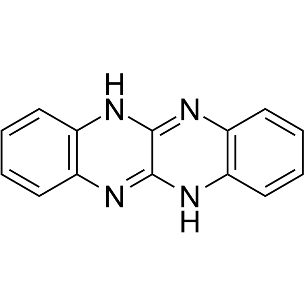
Fluoflavine (ML-090) is a selective NOX1 inhibitor and reactive oxygen species inhibitor. Fluoflavine reduces reactive oxygen species production, NOX1-mediated downstream signaling events, and oxygen-glucose deprivation-induced retinal ganglion cell death. Fluoflavine inhibits NADPH oxidase activity and pathological retinal neovascularization induced by oxygen-induced retinopathy in the retinas of ischemic mice. Fluoflavine can be used in studies related to retinal ischemia-reperfusion injury and proliferative retinopathy.
Nos produits utilisent uniquement pour la recherche. Nous ne vendons pas aux patients.
- Pureté : 98.47%
- CAS No.: 531-46-4
- Formule: C14H10N4
- Masse moléculaire:234.26
-
Stockage:Powder -20°C, 3 years ; In solvent -80°C, 6 months , -20°C, 1 month

To place orders, for customer services and technical support, please contact: MedChemExpress USA
Tel: 609-228-6898 E-mail: [email protected] [email protected]


-
 Activité biologique
-
Chemical Information
- Protocole
- Pureté et documentation
- Références
-
 Help & FAQs


-
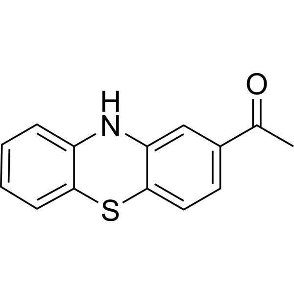
HY-12805
ML171NADPH Oxidase Inhibitor -
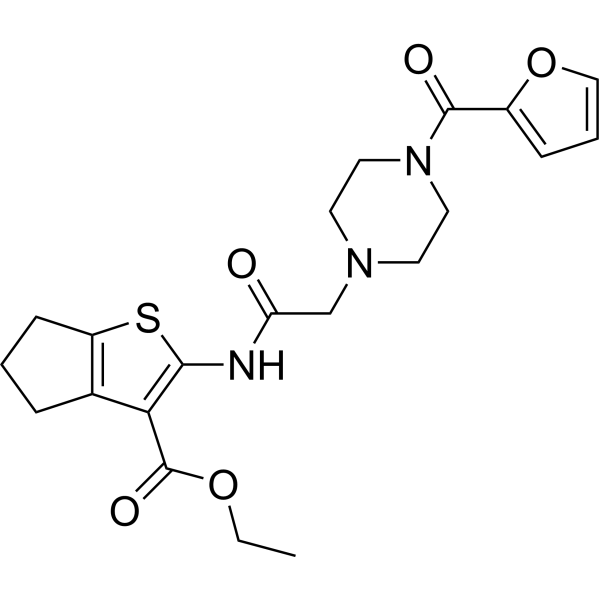
HY-100111
GLX351322NADPH Oxidase 4 Inhibitor -
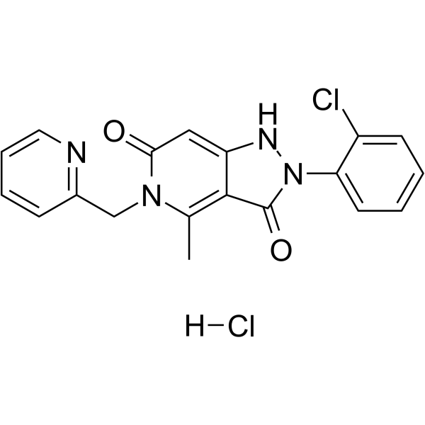
HY-101499A
GKT136901 hydrochlorideNOX1/4 Inhibitor -
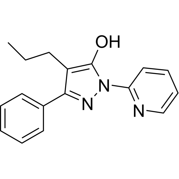
HY-120801A
APX-115 free basePan-Nox Inhibitor -
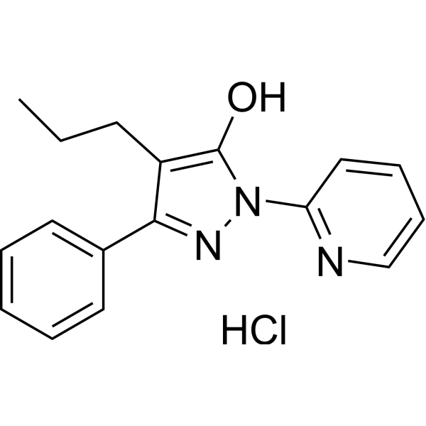
HY-120801
APX-115Pan-Nox Inhibitor -

HY-12805R
ML171 (Standard)NADPH Oxidase Inhibitor -
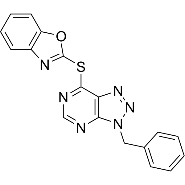
HY-12804
VAS2870Nox Inhibitor -
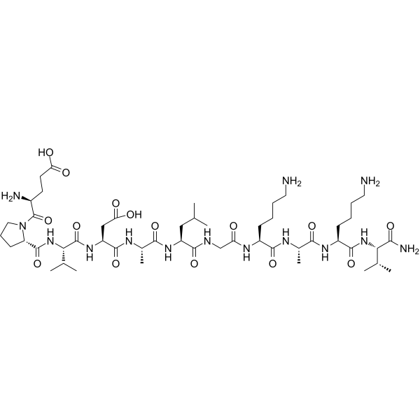
HY-P1435
NoxA1dsNox1 Inhibitor -
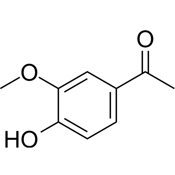
HY-N0088
ApocyninNADPH-Oxidase Inhibitor -

HY-100111R
GLX351322 (Standard)NADPH Oxidase 4 Inhibitor -

HY-101499AR
GKT136901 hydrochloride (Standard)NOX1/4 Inhibitor -
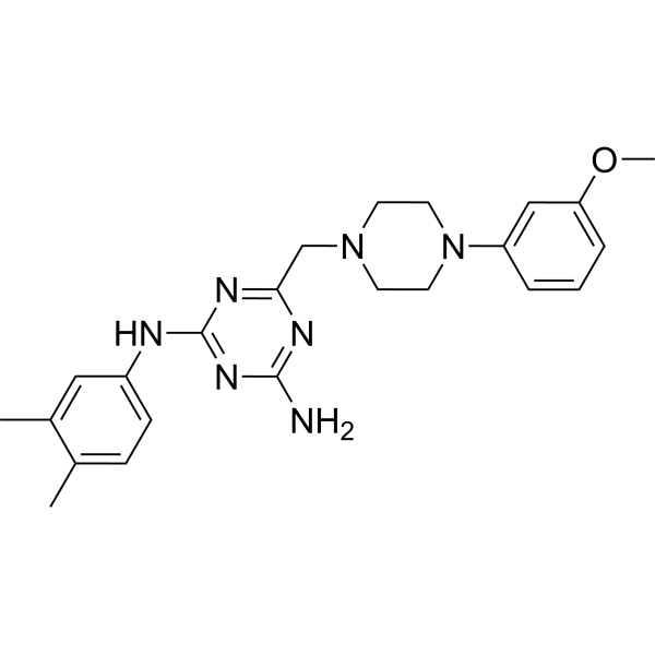
HY-153977
GLX481304Nox2/4 Inhibitor


Activité biologique
Description
IC50 & Target
|
NOX1 |
In Vitro
Fluoflavine (ML-090) (0.09 μM; during OGD and 2 hours of reoxygenation) significantly reduces OGD-induced superoxide production in primary RGCs, with efficacy comparable to a nonselective NAD(P)H oxidase inhibitor[1].
Fluoflavine (0.09 μM; during 4 hours of OGD and 24 hours of reoxygenation) potently protects primary from OGD-induced apoptotic and necrotic death, with efficacy comparable to nonselective NAD(P)H oxidase inhibitors[1].
Fluoflavine (10 μM; 30 min preincubation, 2 h IL-33 treatment) attenuates IL-33-induced barrier disruption in HRMVECs, as demonstrated by reduced FITC-dextran flux[2].
Fluoflavine (10 μM; 30 min preincubation, 30 min IL-33 treatment) reduces IL-33-induced ZO-1 serine/threonine phosphorylation in HRMVECs[2].
Fluoflavine (10 μM; 30 min preincubation, 30 min IL-33 treatment) attenuates IL-33-induced PKCδ phosphorylation in HRMVECs[2].
Fluoflavine (10 μM; 30 min preincubation, 2 h IL-33 treatment) reverses IL-33-induced tight junction disruption in HRMVECs, as shown by restored ZO-1 localization at cell junctions[2].
Fluoflavine (10 μM; 30 min preincubation, 24 h IL-33 treatment) reduces IL-33-induced proliferation of HRMVECs[2].
MedChemExpress (MCE) has not independently confirmed the accuracy of these methods. They are for reference only.
-
Cell Line:human retinal microvascular endothelial cells (HRMVECs)
-
Concentration:10 μM
-
Incubation Time:30 min (preincubation); 30 min (IL-33 treatment)
-
Result:Attenuated IL-33-induced ZO-1 serine/threonine phosphorylation in HRMVECs.\nEffectively reduced IL-33-induced PKCd serine phosphorylation (at Ser643/676) in HRMVECs.
-
Cell Line:human retinal microvascular endothelial cells (HRMVECs)
-
Concentration:10 μM
-
Incubation Time:30 min (preincubation); 2 h (IL-33 treatment)
-
Result:Restored IL-33-induced tight junction breakdown, as evidenced by increased ZO-1 fluorescence intensity at cell junctions compared to IL-33-only treated cells.
-
Cell Line:human retinal microvascular endothelial cells (HRMVECs)
-
Concentration:10 μM
-
Incubation Time:30 min (preincubation); 24 h (IL-33 treatment)
-
Result:Inhibited IL-33-induced HRMVEC proliferation, as measured by reduced absorbance in the MTT assay.


Chemical Information
-
CAS No. 531-46-4
-
Appearance Solid
-
Masse moléculaire 234.26
-
Formule C14H10N4
-
Color Light yellow to yellow
-
SMILES
C1(N2)=NC3=C(C=CC=C3)NC1=NC4=C2C=CC=C4
-
Synonyms
ML-090
-
Livraison
Room temperature in continental US; may vary elsewhere.
-
Stockage
Powder -20°C 3 years In solvent -80°C 6 months -20°C 1 month


Pureté et documentation
-
Fiche technique (274 KB)
-
SDS (251 KB)
- English - EN (251 KB)
- Français - FR (251 KB)
- Deutsch - DE (251 KB)
- Norwegian - NO (251 KB)
- Español - ES (251 KB)
- Swedish - SV (251 KB)
- Italian - IT (251 KB)
- Korean - KR (251 KB)
- Portuguese - PT (251 KB)
-
Instruction de manipulation (2659 KB)
Références
[1]. Dvoriantchikova G, et al. Neuronal NAD(P)H oxidases contribute to ROS production and mediate RGC death after ischemia. Invest Ophthalmol Vis Sci. 2012;53(6):2823-2830. Published 2012 May 14. [Content Brief]
[2]. Kaur G, et al. NADPH oxidase 1-PKCδ-dependent ZO-1 phosphorylation mediates IL-33-induced inner blood-retinal barrier disruption in proliferative retinopathies. Am J Physiol Cell Physiol. 2025;329(5):C1577-C1592. [Content Brief]

Calculators
Concentration (start) × Volume (start) = Concentration (final) × Volume (final)


Fluoflavine Related Classifications
HY-126195 Related Classifications
 Powered by Bioz