



Emrusolmin

Based on 1 Customer Validation
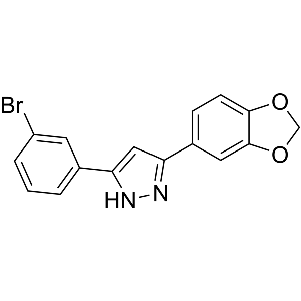
Emrusolmin (Anle138b), an oligomeric aggregation inhibitor, blocks the formation of pathological aggregates of prion protein (PrPSc) and of α-synuclein (α-syn). Emrusolmin strongly inhibits oligomer accumulation, neuronal degeneration, and disease progression in vivo. Emrusolmin has low toxicity and an excellent oral bioavailability and blood-brain-barrier penetration. Emrusolmin blocks Aβ channels and rescues disease phenotypes in a mouse model for amyloid pathology.
Nos produits utilisent uniquement pour la recherche. Nous ne vendons pas aux patients.
- Pureté : 99.93%
- CAS No.: 882697-00-9
- Formule: C16H11BrN2O2
- Masse moléculaire:343.17
-
Stockage:Powder -20°C, 3 years , 4°C, 2 years ; In solvent -80°C, 2 years , -20°C, 1 year

To place orders, for customer services and technical support, please contact: MedChemExpress USA
Tel: 609-228-6898 E-mail: [email protected] [email protected]


-
 Activité biologique
-
Chemical Information
-
Solvant et solubilité
- Protocole
- Pureté et documentation
- Références
-
 Help & FAQs


-
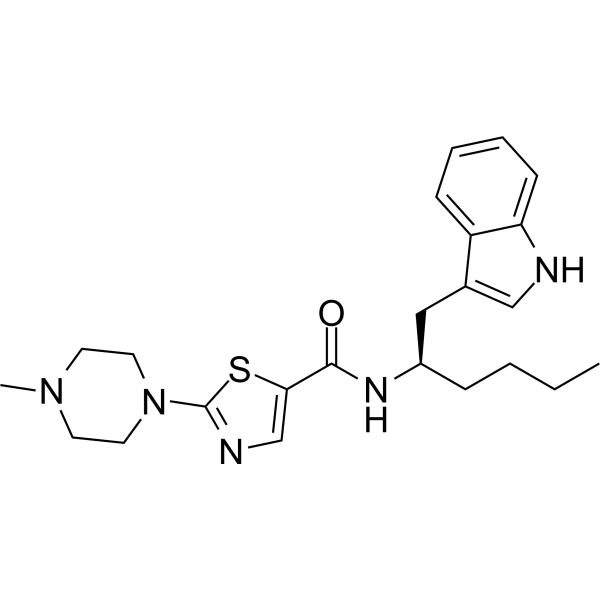
HY-145580
Minzasolminα-Synuclein Oligomerization Inhibitor -
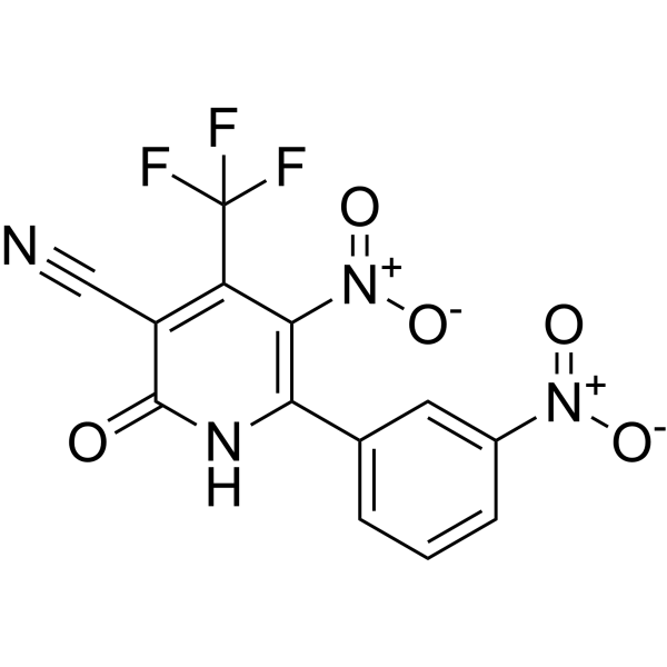
HY-124876
SynuClean-Dα-synuclein Aggregation Inhibitor -

HY-10219
RapamycinmTOR Inhibitor -
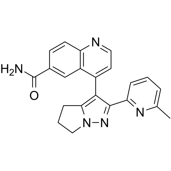
HY-13226
GalunisertibTGFβRI Inhibitor -

HY-10227
BortezomibProteasome Inhibitor -
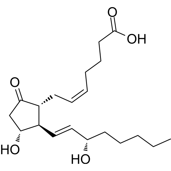
HY-101952
Prostaglandin E2Prostaglandin Receptor Agonist -

HY-B0015
PaclitaxelAntitumor Agent -
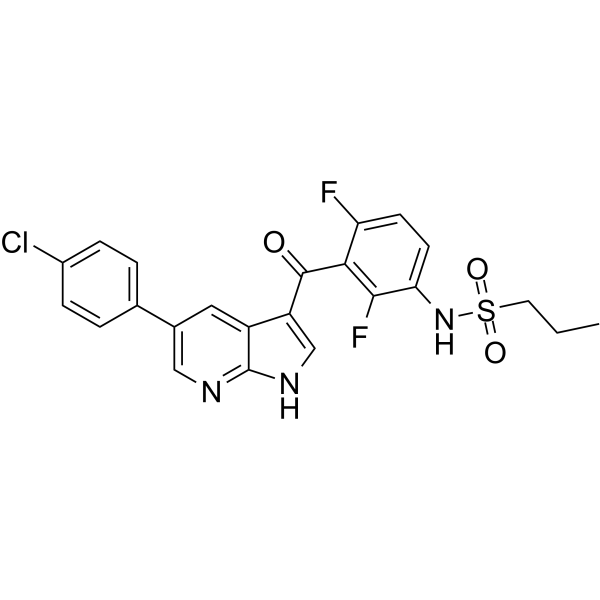
HY-12057
VemurafenibB-Raf Inhibitor -
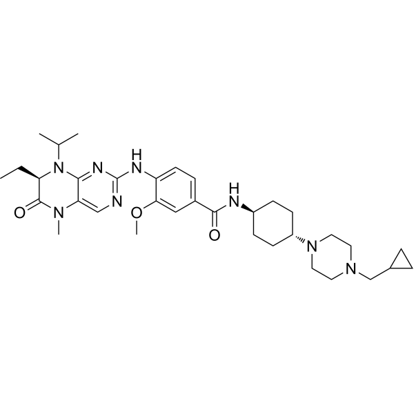
HY-12137
VolasertibPLK1 Inhibitor -
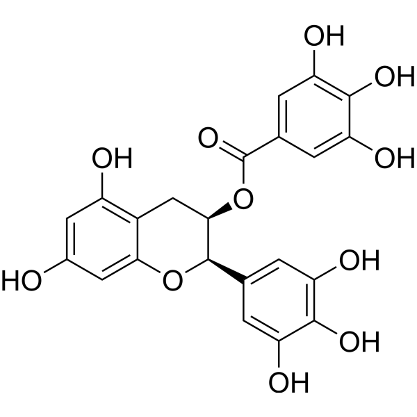
HY-13653
(-)-Epigallocatechin GallateAnticancer Agent -
HY-16106
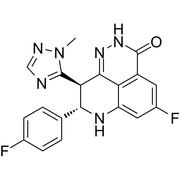
TalazoparibPARP Inhibitor -
HY-112251
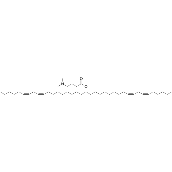
D-Lin-MC3-DMAsiRNA Delivery Vehicle


Activité biologique
Description
In Vitro
Oligomeric aggregates are presumed to be the key neurotoxic agent. Emrusolmin blocksthe formation of pathological aggregates of prion protein and of α-synuclein, which is deposited in Parkinson’s disease and other synucleinopathies such as dementia with Lewy bodies and multiple system atrophy. Emrusolmin strongly inhibits all prion strains tested including BSE-derived and human prions. Emrusolmin shows structure-dependent binding to pathological aggregates and strongly inhibits formation of pathological oligomers both for prion protein and α-synuclein[1].
MedChemExpress (MCE) has not independently confirmed the accuracy of these methods. They are for reference only. Further protocols information, click here.
In Vivo
Emrusolmin (0.6-2 g/kg; p.o.) modulates α-synuclein oligomerization[3].
MedChemExpress (MCE) has not independently confirmed the accuracy of these methods. They are for reference only.
-
Animal Model:Two-month-old PLP-hαSyn mice[3]
-
Dosage:0.6 and 2 g/kg
-
Administration:Oral
-
Result:Prevented motor deficits and neurodegeneration in the PLP-hαSyn mice.
Essai clinique
| NCT Number | Sponsor | Condition | Start Date |
Phase

|
|---|---|---|---|---|
| NCT01329991 | Plexxikon| | 2011-05 | PHASE1 |


Chemical Information
-
CAS No. 882697-00-9
-
Appearance Solid
-
Masse moléculaire 343.17
-
Formule C16H11BrN2O2
-
Color Off-white to light yellow
-
SMILES
BrC1=CC(C2=CC(C3=CC=C(OCO4)C4=C3)=NN2)=CC=C1
-
Synonyms
Anle138b
-
Livraison
Room temperature in continental US; may vary elsewhere.
-
Stockage
Powder -20°C 3 years 4°C 2 years In solvent -80°C 2 years -20°C 1 year


Solvant et solubilité
In Vitro:
DMSO : 87.5 mg/mL (254.98 mM; Need ultrasonic; Hygroscopic DMSO has a significant impact on the solubility of product, please use newly opened DMSO)
Please refer to the solubility information to select the appropriate solvent. Once prepared, please aliquot and store the solution to prevent product inactivation from repeated freeze-thaw cycles.
Storage method and period of stock solution: -80°C, 2 years; -20°C, 1 year. When stored at -80°C, please use it within 2 years. When stored at -20°C, please use it within 1 year.
Please refer to the solubility information to select the appropriate solvent. Once prepared, please aliquot and store the solution to prevent product inactivation from repeated freeze-thaw cycles.
Storage method and period of stock solution: -80°C, 2 years; -20°C, 1 year. When stored at -80°C, please use it within 2 years. When stored at -20°C, please use it within 1 year.
Concentration (start) × Volume (start) = Concentration (final) × Volume (final)
In Vivo:
Select the appropriate dissolution method based on your experimental animal and administration route.
- For the following dissolution methods, please ensure to first prepare a clear stock solution using an In Vitro approach and then sequentially add co-solvents:
- To ensure reliable experimental results, the clarified stock solution can be appropriately stored based on storage conditions. As for the working solution for In Vivo experiments, it is recommended to prepare freshly and use it on the same day.
- The percentages shown for the solvents indicate their volumetric ratio in the final prepared solution. If precipitation or phase separation occurs during preparation, heat and/or sonication can be used to aid dissolution.
Add each solvent one by one: 10% DMSO 40% PEG300 5% Tween-80 45% Saline
Solubility: ≥ 2.5 mg/mL (7.29 mM); Clear solution
This protocol yields a clear solution of ≥ 2.5 mg/mL (saturation unknown).
Taking 1 mL working solution as an example, add 100 μL DMSO stock solution (25.0 mg/mL) to 400 μL PEG300, and mix evenly; then add 50 μL Tween-80 and mix evenly; then add 450 μL Saline to adjust the volume to 1 mL.
Preparation of Saline: Dissolve 0.9 g sodium chloride in ddH₂O and dilute to 100 mL to obtain a clear Saline solution.
In Vivo Dissolution Calculator
Please enter the basic information of animal experiments:
-
-
-
-
Recommended: Prepare an additional quantity of animals to account for potential losses during experiments.
Please enter your animal formula composition:
-
%DMSO +
Recommended: Keep the proportion of DMSO in working solution below 2% if your animal is weak.
-
%+
-
+%Tween-80 + +
-
%Saline +
The co-solvents required include: DMSO, . All of co-solvents are available by MedChemExpress (MCE). , Tween 80. All of co-solvents are available by MedChemExpress (MCE).
Working solution concentration: 0.22 mg/mL
Method for preparing stock solution: mg drug dissolved in μL DMSO. Stock solution concentration: mg/mL.
1. Take μL DMSO stock solution;
2. Add μL .
μL , mix evenly;
3. Then add μL Tween 80, mix evenly;
4. Then add μL
Please ensure that the stock solution in the first step is dissolved to a clear state, and add co-solvents in sequence. You can use ultrasonic heating (ultrasonic cleaner, recommended frequency 20-40 kHz), vortexing, etc. to assist dissolution.

Protocole
-
Protocol for Pharmacokinetic Study
Pharmacokinetic studies quantify how an organism handles a drug over time through absorption, distribution, metabolism, and excretion, and the core experimental readout is the concentration-time profile of parent drug and, when relevant, metabolites in biological matrices such as plasma, whole blood, urine, bile, or tissue. Pharmacokinetic analysis links dose, route, exposure, clearance, half-life, distribution, bioavailability, and systemic exposure to drug efficacy and toxicity hypotheses rather than measuring a signaling pathway directly. The literature links pharmacokinetics to drug-development phenotypes by showing that drug metabolism and pharmacokinetics influence compound progression, exposure-response interpretation, safety margins, dosing strategy, and failure risk during discovery and development. DMPK science contributes to compound optimization by integrating physicochemical properties, in vitro metabolism, transporter behavior, in vivo exposure, and pharmacodynamic contex
-
Protocol for Hematoxylin-Eosin (H&E) Staining
Hematoxylin-eosin staining is a routine histological method that stains nuclei mainly blue-purple with hemalum and stains cytoplasm, extracellular matrix, and many stromal components pink with eosin, allowing tissue architecture, cell morphology, necrosis, inflammation, fibrosis, tumor growth pattern, and treatment-associated injury to be evaluated by light microscopy. In cancer cells, primary neurons, mouse tumor models, intestinal organoids, inflammatory macrophage preparations, and drug-screening tissues, H&E is a morphology assay rather than a molecular assay; it should be interpreted with complementary molecular or immunostaining assays when the biological question concerns specific proteins, RNA levels, ferroptosis, mitophagy, or immune phenotypes.
-
Amyloid: Congo Red Amyloid Staining
Congo red amyloid staining is a histochemical method used to detect extracellular amyloid deposits in tissue sections based on the affinity of Congo red dye for β-pleated sheet-rich protein aggregates. When bound to amyloid, Congo red produces characteristic apple-green birefringence under polarized light microscopy, which is widely regarded as a diagnostic feature of amyloid deposition in histopathology. The diagnostic principle relies on the combination of dye binding (congophilia) and optical anisotropy under polarized illumination, which distinguishes amyloid from most non-amyloid eosinophilic extracellular deposits in routine histological evaluation. Amyloid identification by Congo red staining remains a cornerstone in diagnostic pathology despite the availability of adjunct methods such as immunohistochemistry and mass spectrometry, particularly because of its ability to localize deposits directly within tissue architecture. The specificity of Congo red-positive deposits is incre
-
Alzheimer’s Disease Modeling
Alzheimer’s Disease (AD) is a neurodegenerative disorder characterized by a progressive decline in cognitive functions and loss of specific types of neurons and synapses. Alzheimer's symptoms can be simulated in mice by injecting drugs (such as Aβ) or genetically modified.
-
How to Choose the Right Model Animal
Choosing the right model animal is a validity-driven decision in which the species, strain, sex, age, genetic background, disease-induction method, outcome measures, and welfare burden must match the scientific question rather than laboratory tradition or convenience. A model should be selected by judging face validity, construct validity, and predictive validity: whether it resembles the human phenotype, whether it reproduces relevant mechanisms, and whether results are likely to predict human biology or treatment response. Animal studies often fail to translate because of species differences, weak disease resemblance, poor experimental design, inadequate reporting, publication bias, and underuse of randomization, blinding, and sample-size justification. Unresolved questions include how to rank competing models objectively, how much human-disease complexity must be reproduced for a given objective, and when non-animal systems such as organoids, ex vivo tissue, or computational models


Pureté et documentation
-
Fiche technique (275 KB)
-
SDS (393 KB)
- English - EN (393 KB)
- Français - FR (393 KB)
- Deutsch - DE (393 KB)
- Norwegian - NO (393 KB)
- Español - ES (393 KB)
- Swedish - SV (393 KB)
- Italian - IT (393 KB)
- Korean - KR (393 KB)
- Portuguese - PT (393 KB)
-
Instruction de manipulation (2659 KB)
Références
[1]. Wagner J, et al. Anle138b: a novel oligomer modulator for disease-modifying therapy of neurodegenerative diseases such as prion and Parkinson's disease. Acta Neuropathol. 2013 Jun;125(6):795-813. [Content Brief]
[2]. Martinez Hernandez A, et al. The diphenylpyrazole compound anle138b blocks Aβ channels and rescues disease phenotypes in a mouse model for amyloid pathology. EMBO Mol Med. 2018;10(1):32-47. [Content Brief]
[3]. Heras-Garvin A, et al. Anle138b modulates α-synuclein oligomerization and prevents motor decline and neurodegeneration in a mouse model of multiple system atrophy. Mov Disord. 2019;34(2):255-263. [Content Brief]


Complete Stock Solution Preparation Table
Please refer to the solubility information to select the appropriate solvent. Once prepared, please aliquot and store the solution to prevent product inactivation from repeated freeze-thaw cycles.
Storage method and period of stock solution: -80°C, 2 years; -20°C, 1 year. When stored at -80°C, please use it within 2 years. When stored at -20°C, please use it within 1 year.
| Optional Solvent | Concentration Solvent Mass | 1 mg | 5 mg | 10 mg | 25 mg |
|---|---|---|---|---|---|
| DMSO | 1 mM | 2.9140 mL | 14.5700 mL | 29.1401 mL | 72.8502 mL |
| 5 mM | 0.5828 mL | 2.9140 mL | 5.8280 mL | 14.5700 mL | |
| 10 mM | 0.2914 mL | 1.4570 mL | 2.9140 mL | 7.2850 mL | |
| 15 mM | 0.1943 mL | 0.9713 mL | 1.9427 mL | 4.8567 mL | |
| 20 mM | 0.1457 mL | 0.7285 mL | 1.4570 mL | 3.6425 mL | |
| 25 mM | 0.1166 mL | 0.5828 mL | 1.1656 mL | 2.9140 mL | |
| 30 mM | 0.0971 mL | 0.4857 mL | 0.9713 mL | 2.4283 mL | |
| 40 mM | 0.0729 mL | 0.3643 mL | 0.7285 mL | 1.8213 mL | |
| 50 mM | 0.0583 mL | 0.2914 mL | 0.5828 mL | 1.4570 mL | |
| 60 mM | 0.0486 mL | 0.2428 mL | 0.4857 mL | 1.2142 mL | |
| 80 mM | 0.0364 mL | 0.1821 mL | 0.3643 mL | 0.9106 mL | |
| 100 mM | 0.0291 mL | 0.1457 mL | 0.2914 mL | 0.7285 mL |


Emrusolmin Related Classifications
HY-101855 Related Classifications
 Powered by Bioz