



DCOIT

Based on 1 Customer Validation
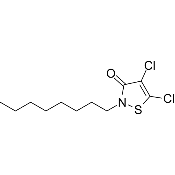
DCOIT (Kathon 930) is an isothiazolinone biocide and environmental pollutant that directly binds to G protein α subunits, with KD values of 0.92 mM and 0.4 mM for Gαs and Gαi, respectively. Binding of DCOIT to Gαi enhances the interaction between Gαi and mitochondrial calcium uniporter (MCU), alters the subcellular distribution of intracellular Ca2+, and activates PKC/MEK/ERK signaling. DCOIT upregulates CYP19a transcription and aromatase activity, thereby disrupting steroid hormone homeostasis. DCOIT is used in studies of reproductive endocrine disruption and environmental toxicology.
For research use only. We do not sell to patients.
- Purity : 99.87%
- CAS No.: 64359-81-5
- Formula: C11H17Cl2NOS
- Molecular Weight:282.23
-
Storage:Powder -20°C, 3 years , 4°C, 2 years ; In solvent -80°C, 6 months , -20°C, 1 month

To place orders, for customer services and technical support, please contact: MedChemExpress USA
Tel: 609-228-6898 E-mail: [email protected] [email protected]


-
 Biological Activity
-
Chemical Information
-
Solvent & Solubility
- Protocols
- Purity & Documentation
- References
-
 Help & FAQs


-
HY-W588722
ICG-DBCOFluorescent Click Chemistry Reagent -
HY-148835
AzGGKAmino Acid -

HY-W041308R
DCOIT (Standard)Reference Standard -

HY-I0144
ThiazolidineHeterocyclic Compound -
HY-P0009A
Cetrorelix acetateGnRH Receptor Antagonist -
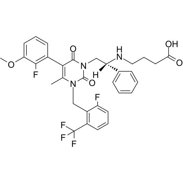
HY-14789
(R)-ElagolixGnRH Receptor Antagonist -
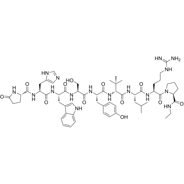
HY-P0051
LecirelinGnRH Analogue -
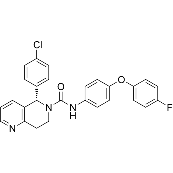
HY-130249
BAY-298LH-R Antagonist -
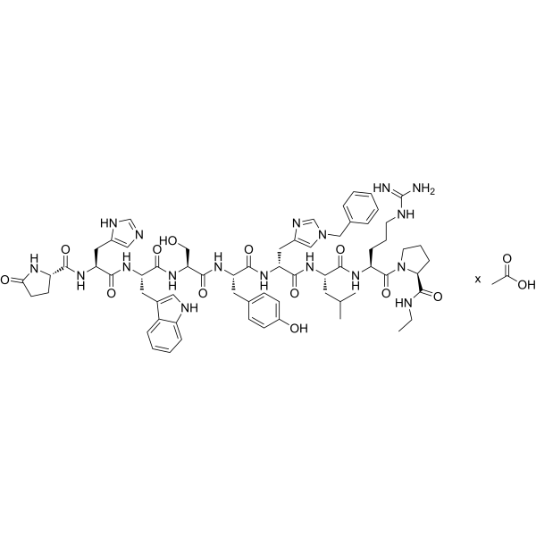
HY-P0056A
Histrelin acetateGnRH Receptor Agonist -
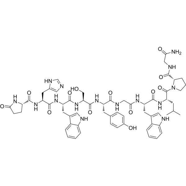
HY-P0243
Luteinizing Hormone Releasing Hormone (LH-RH), salmonLuteinizing Hormone-releasing Hormone -
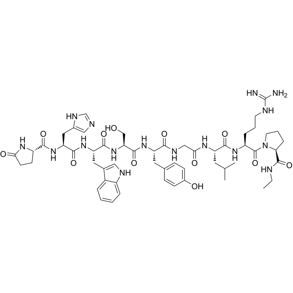
HY-P0053
FertirelinGnRH Analog -
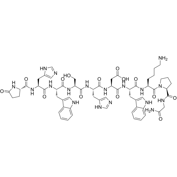
HY-P1808
LGnRH-III, lampreyGnRH Agonist


Biological Activity
Description
IC50 & Target
[1]|
Gαs 0.92 mM (Kd) |
Gαi 0.4 mM (Kd) |
In Vitro
DCOIT (2-4 mg/L) concentration-dependently reduces its binding to the DCOIT matrix in pull-down assays with recombinant Gαs and Gαi proteins; it decreases the GTPase activity of recombinant Gαi, while having no significant effect on the GTPase activity of Gαs[1].
DCOIT (1.6 μM; 24 h) does not alter total MEK and ERK protein levels in H295R cells, but increases p-MEK and p-ERK levels by 1.7-fold and 1.9-fold, respectively, and ELISA results further confirm the activation of MAPK signaling[1].
DCOIT (1.6 μM; 24 h) upregulates CYP19a transcription and increases aromatase activity in H295R cells, promoting the conversion of testosterone to estradiol and elevating the E2/T ratio[1].
DCOIT (1.6 μM; 24 h; co-exposure with PMA (HY-18739) or U0126 (HY-12031A)) induces changes in CYP19a transcription, aromatase activity, E2 levels, and the E2/T ratio in H295R cells that are enhanced by the MAPK activator PMA and antagonized by the MEK inhibitor U0126[1].
DCOIT (1.6 μM; 24 h; co-exposure with PTX or GTP)-induced MEK/ERK phosphorylation in H295R cells is alleviated by the Gαi inhibitor PTX and reversed by co-exposure with GTP; GTP also restores E2 levels and the E2/T ratio to near baseline[1].
DCOIT (1.6 μM; 24 h) elevates PLC, IP3, DAG, and PKC levels in H295R cells[1].
DCOIT (0.10-12.80 μM; 24 h) exerts a concentration-dependent effect on H295R cell viability, in which 0.10-1.60 μM does not reduce cell viability, 1.60 μM is the maximum no-observed-effect concentration (NOEC), and 3.20, 6.40, and 12.80 μM significantly reduce cell viability[1].
DCOIT (1.6 μM; 24 h; co-treatment with 12.5 μM Progesterone (HY-N0437)) further elevates p-MEK and p-ERK levels in H295R cells; it upregulates CYP19a gene transcription but does not further enhance aromatase activity or alterations in hormone homeostasis[1].
MedChemExpress (MCE) has not independently confirmed the accuracy of these methods. They are for reference only.
-
Cell Line:H295R
-
Concentration:1.6 μM
-
Incubation Time:24 h
-
Result:Left total MEK abundance unchanged.
Left total ERK abundance unchanged.
Increased p-MEK by 1.7-fold.
Increased p-ERK by 1.9-fold.
-
Cell Line:H295R
-
Concentration:1.6 μM
-
Incubation Time:24 h
-
Result:Increased p-MEK content.
Increased p-ERK content.
-
Cell Line:H295R
-
Concentration:1.6 μM
-
Incubation Time:24 h
-
Result:Increased CYP19a gene transcription.
-
Cell Line:H295R
-
Concentration:1.6 μM + 10 nM PMA
-
Incubation Time:24 h
-
Result:Further increased CYP19a gene transcription relative to DCOIT alone.
-
Cell Line:H295R
-
Concentration:1.6 μM + 10 μM U0126
-
Incubation Time:24 h
-
Result:Antagonized the DCOIT-induced increase in CYP19a gene transcription.
-
Cell Line:H295R
-
Concentration:1.6 μM + 20 μM NF449
-
Incubation Time:24 h
-
Result:NF449 did not appreciably alter the DCOIT-induced increases in p-MEK and p-ERK.
-
Cell Line:H295R
-
Concentration:1.6 μM + 12.5 μM progesterone
-
Incubation Time:24 h
-
Result:Further increased p-MEK.
Further increased p-ERK relative to DCOIT alone.
-
Cell Line:H295R
-
Concentration:1.6 μM + 12.5 μM progesterone
-
Incubation Time:24 h
-
Result:Further increased CYP19a gene transcription relative to DCOIT alone.
In Vivo
DCOIT (1-10 μg/L; life-cycle exposure, from embryo to sexual maturity) disrupts GnRHR-mediated gonadotropin signaling in marine medaka; 10 μg/L increases GnRH in female brain by 2.2-fold and FSH in blood by 1.5-fold, while 3 μg/L increases LH and FSH in male blood by 2.1-fold and 1.4-fold, respectively[1].
DCOIT (1-10 μg/L; life-cycle exposure, from embryo to sexual maturity) alters the transcription of HPG axis-related genes in marine medaka; 10 μg/L increases the E2/T ratio by 1.9-fold in male fish and elevates testicular CYP19a transcription by 3.0-fold[1].
DCOIT (1-10 μg/L; life-cycle exposure, from embryo to sexual maturity) alters gametogenesis and reproductive performance in marine medaka; 1 and 3 μg/L increase the mean egg production by 1.4-fold and 1.8-fold, respectively, whereas 10 μg/L slightly inhibits cumulative egg production and decreases the gonadosomatic index in both males and females[1].
MedChemExpress (MCE) has not independently confirmed the accuracy of these methods. They are for reference only.
-
Animal Model:Oryzias melastigma[1]
-
Dosage:1 μg/L, 3 μg/L, 10 μg/L
-
Administration:waterborne; static life-cycle exposure from embryo to sexual maturation
-
Result:Increased brain GnRH concentration by 2.2-fold and blood FSH concentration by 1.5-fold in female fish at 10 μg/L, accompanied by upregulation of sGnRH, mGnRH, GnRHR, and FSHβ gene transcription in the brain.
Increased blood LH by 2.1-fold and FSH by 1.4-fold in male fish at 3 μg/L, with concurrent upregulation of FSHβ, GTHα, and LHβ gene transcription in the brain.
Increased the testicular E2/T ratio by 1.9-fold and elevated testicular CYP19a transcription by 3.0-fold in male fish at 10 μg/L.
Promoted ovarian oogenic progression, with a lower proportion of primary oocytes and higher proportions of cortical-alveolar, early vitellogenic, and late/mature oocytes.
Blocked spermatogenesis in the testis at 10 μg/L, reducing the proportion of mature sperm and increasing the proportion of spermatocytes.
Increased average fecundity 1.4-fold at 1 μg/L and 1.8-fold at 3 μg/L, while mildly suppressing cumulative fecundity at 10 μg/L.
Decreased both female and male gonadosomatic index values.
Skewed the sex ratio toward female dominance at 10 μg/L.
Caused increased mortality and malformation, delayed hatching, faster heartbeat, and longer body length in offspring in a concentration-dependent manner.
Altered the brain proteome at 10 μg/L, with 848 differentially abundant proteins in females and 892 in males, including G protein subunits (Gαi, Gαo, Gαq), Ca2+ signaling proteins, and MAPK pathway proteins (PKC, MEK).
Significantly decreased mitochondrial calcium uniporter protein in the male brain at 10 μg/L.
Was detected in brain tissue after 10 μg/L life-cycle exposure.


Chemical Information
-
CAS No. 64359-81-5
-
Appearance Solid
-
Molecular Weight 282.23
-
Formula C11H17Cl2NOS
-
Color White to off-white
-
SMILES
O=C1N(CCCCCCCC)SC(Cl)=C1Cl
-
Synonyms
Kathon 930
-
Shipping
Room temperature in continental US; may vary elsewhere.
-
Storage
Powder -20°C 3 years 4°C 2 years In solvent -80°C 6 months -20°C 1 month


Solvent & Solubility
In Vitro:
DMSO : 133.33 mg/mL (472.42 mM; Need ultrasonic; Hygroscopic DMSO has a significant impact on the solubility of product, please use newly opened DMSO)
Please refer to the solubility information to select the appropriate solvent. Once prepared, please aliquot and store the solution to prevent product inactivation from repeated freeze-thaw cycles.
Storage method and period of stock solution: -80°C, 6 months; -20°C, 1 month. When stored at -80°C, please use it within 6 months. When stored at -20°C, please use it within 1 month.
Please refer to the solubility information to select the appropriate solvent. Once prepared, please aliquot and store the solution to prevent product inactivation from repeated freeze-thaw cycles.
Storage method and period of stock solution: -80°C, 6 months; -20°C, 1 month. When stored at -80°C, please use it within 6 months. When stored at -20°C, please use it within 1 month.
Concentration (start) × Volume (start) = Concentration (final) × Volume (final)
In Vivo:
Select the appropriate dissolution method based on your experimental animal and administration route.
- For the following dissolution methods, please ensure to first prepare a clear stock solution using an In Vitro approach and then sequentially add co-solvents:
- To ensure reliable experimental results, the clarified stock solution can be appropriately stored based on storage conditions. As for the working solution for In Vivo experiments, it is recommended to prepare freshly and use it on the same day.
- The percentages shown for the solvents indicate their volumetric ratio in the final prepared solution. If precipitation or phase separation occurs during preparation, heat and/or sonication can be used to aid dissolution.
Add each solvent one by one: 10% DMSO 90% (20% SBE-β-CD in Saline)
Solubility: 5 mg/mL (17.72 mM); Clear solution; Need ultrasonic
This protocol yields a clear solution of 5 mg/mL.
Taking 1 mL working solution as an example, add 100 μL DMSO stock solution (50.0 mg/mL) to 900 μL 20% SBE-β-CD in Saline, and mix evenly.
Preparation of 20% SBE-β-CD in Saline (4°C, storage for one week): 2 g SBE-β-CD powder is dissolved in 10 mL Saline, completely dissolve until clear.
In Vivo Dissolution Calculator
Please enter the basic information of animal experiments:
-
-
-
-
Recommended: Prepare an additional quantity of animals to account for potential losses during experiments.
Please enter your animal formula composition:
-
%DMSO +
Recommended: Keep the proportion of DMSO in working solution below 2% if your animal is weak.
-
%+
-
+%Tween-80 + +
-
%Saline +
The co-solvents required include: DMSO, . All of co-solvents are available by MedChemExpress (MCE). , Tween 80. All of co-solvents are available by MedChemExpress (MCE).
Working solution concentration: 0.22 mg/mL
Method for preparing stock solution: mg drug dissolved in μL DMSO. Stock solution concentration: mg/mL.
1. Take μL DMSO stock solution;
2. Add μL .
μL , mix evenly;
3. Then add μL Tween 80, mix evenly;
4. Then add μL
Please ensure that the stock solution in the first step is dissolved to a clear state, and add co-solvents in sequence. You can use ultrasonic heating (ultrasonic cleaner, recommended frequency 20-40 kHz), vortexing, etc. to assist dissolution.


Purity & Documentation
-
Data Sheet (283 KB)
-
SDS (786 KB)
- English - EN (786 KB)
- Français - FR (786 KB)
- Deutsch - DE (786 KB)
- Norwegian - NO (786 KB)
- Español - ES (786 KB)
- Swedish - SV (786 KB)
- Italian - IT (786 KB)
- Korean - KR (786 KB)
- Portuguese - PT (786 KB)
-
Handling Instructions (2659 KB)
References


Complete Stock Solution Preparation Table
Please refer to the solubility information to select the appropriate solvent. Once prepared, please aliquot and store the solution to prevent product inactivation from repeated freeze-thaw cycles.
Storage method and period of stock solution: -80°C, 6 months; -20°C, 1 month. When stored at -80°C, please use it within 6 months. When stored at -20°C, please use it within 1 month.
| Optional Solvent | Concentration Solvent Mass | 1 mg | 5 mg | 10 mg | 25 mg |
|---|---|---|---|---|---|
| DMSO | 1 mM | 3.5432 mL | 17.7160 mL | 35.4321 mL | 88.5802 mL |
| 5 mM | 0.7086 mL | 3.5432 mL | 7.0864 mL | 17.7160 mL | |
| 10 mM | 0.3543 mL | 1.7716 mL | 3.5432 mL | 8.8580 mL | |
| 15 mM | 0.2362 mL | 1.1811 mL | 2.3621 mL | 5.9053 mL | |
| 20 mM | 0.1772 mL | 0.8858 mL | 1.7716 mL | 4.4290 mL | |
| 25 mM | 0.1417 mL | 0.7086 mL | 1.4173 mL | 3.5432 mL | |
| 30 mM | 0.1181 mL | 0.5905 mL | 1.1811 mL | 2.9527 mL | |
| 40 mM | 0.0886 mL | 0.4429 mL | 0.8858 mL | 2.2145 mL | |
| 50 mM | 0.0709 mL | 0.3543 mL | 0.7086 mL | 1.7716 mL | |
| 60 mM | 0.0591 mL | 0.2953 mL | 0.5905 mL | 1.4763 mL | |
| 80 mM | 0.0443 mL | 0.2215 mL | 0.4429 mL | 1.1073 mL | |
| 100 mM | 0.0354 mL | 0.1772 mL | 0.3543 mL | 0.8858 mL |


DCOIT Related Classifications
HY-W041308 Related Classifications
 Powered by Bioz
Keywords
- DCOIT
- 64359-81-5
- Kathon 930
- Kathon930
- Kathon-930
- Environmental Pollutants
- GnRH Receptor
- H295R cells
- hypothalamus-pituitary-gonadal axis
- Gαi
- isothiazolinone biocide
- blood-brain barrier permeability
- mitochondrial calcium uniporter
- reproductive endocrine disruption
- marine medaka
- MAPK pathway
- O. melastigma
- Inhibitor
- inhibitor
- inhibit