



Fluorescein-5-maleimide (solution)

Based on 1 Customer Validation
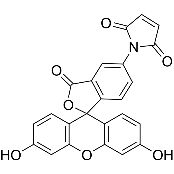
Fluorescein-5-maleimide (solution) (N-(5-Fluoresceinyl)maleimide (solution)) is a fluorescent dye. Fluorescein-5-maleimide can be used to detect the redox state of thiols in eukaryotic cells. Fluorescein-5-maleimide can label peptides and is used to detect negatively charged nanoparticles. Fluorescein-5-maleimide can also label actin to explore its interaction with cardiac myosin-binding protein C (cMyBP-C), which helps in developing small molecule modulators for heart failure. Fluorescein-5-maleimide can screen mutant proteins that contain cysteine residues. The excitation wavelength of Fluorescein-5-maleimide is 494 nm, and the emission wavelength is 519 nm.
Solvent and concentration: DMSO: 10 mM
For research use only. We do not sell to patients.
- CAS No.: 75350-46-8
- Formula: C24H13NO7
- Molecular Weight:427.36
-
Storage:
Please store the product under the recommended conditions in the Certificate of Analysis.


To place orders, for customer services and technical support, please contact: MedChemExpress USA
Tel: 609-228-6898 E-mail: [email protected] [email protected]


-
 Biological Activity
-
Chemical Information
- Purity & Documentation
- References
-
 Help & FAQs


-
HY-D0098
Fluorescein-5-maleimide -
HY-126793
2′,7′-Dichlorofluorescein diacetate -
HY-D0069
CPM -
HY-139109
IR-783 -
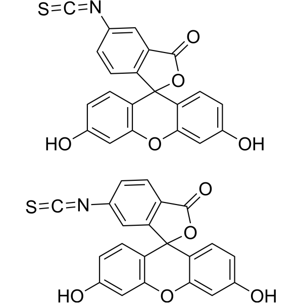
HY-15941
5(6)-FITC -
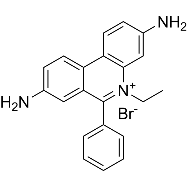
HY-D0021
EthD bromide -

HY-DY1050
Ethidium bromide (solution) -
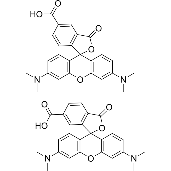
HY-15944
5(6)-TAMRA -

HY-DY1058
2′,7′-Dichlorofluorescein diacetate (solution) -
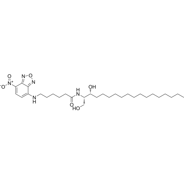
HY-141576
C6-NBD Sphinganine -
HY-141577
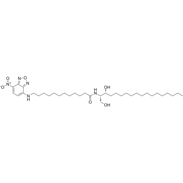
C12-NBD Sphinganine -

HY-DY1085
5(6)-TAMRA (solution)


Biological Activity
Description
Solvent and concentration: DMSO: 10 mM
In Vitro
Guide (The following is our recommended protocol. This protocol is only a guide and should be modified according to your specific needs).
1. Fluorescein-5-maleimide for detecting the redox status of thiols in cells
1.1 Prepare PBMCs from venous blood using Ficoll Paque density centrifugation and wash them in d-PBS.
1.2 Wash PBMCs (106cells in 100 μ L) labeled with anti human CD3-APC.
1.3 Stain with 0.1 μ M Fluorescein-5-maleimide and analyze by flow cytometry.
2. Preparation of Fluorescein-5-maleimide Fluorescent Probe[2]
2.1 Preparation of working fluid
Add the working solution (probe: peptide) in a 5:1 molar ratio to a peptide solution (4.06 mM) containing 20.3 mM N - (2-hydroxyethyl) piperazine-N '- ethanesulfonic acid KOH, 50 mM KCl, and 2 mM tris (2-carboxyethyl) phosphine buffer for peptide labeling.
2.2 Incubate the mixture (final volume of 125 μ L) in the dark at room temperature for 2 hours and gently shake.
2.3 Add excess DDT (20 mM) to quench the reaction.
2.4 Further purify the pure labeled peptide mixture by precipitation with acetone (1 mL).
2.5 Add 5 μ L of fluorescent probe to 15 μ L of nanoparticles and 130 μ L of buffer solution to detect their interaction.
2.6 Data analysis: Detecting nanoparticles-peptide interactions through changes in fluorescence intensity of labeled peptides and/or differential light scattering.
3. Fluorescein-5-maleimide is used to detect protein interactions (actin and cardiac myosin binding protein C (cMyBP-C))[3]
3.1 50 μ M G-actin was polymerized by adding 3 M KCl (final concentration of 100 mM) and 0.5 M MgCl2 (final concentration of 2 mM) under the conditions of 20 mM Tris pH 7.5, 0.2 mM CaCl2, and 0.2 mM ATP, and incubated at 23 ° C for 1 hour.
3.2 Add Fluorescein-5-maleimide (final concentration of 1 mM) and label at 23 ° C for 1 hour.
3.3 Add a five fold molar excess of DTT to stop labeling.
3.4 Incubate 0.25 μ M Fluorescein-5-maleimide actin and 0-20 μ M TMR-cC0-C2 (N-terminal fragment of cMyBP-C) together.
3.5 Data analysis (FLT-FRET assay): Firstly, excite Fluorescein-5-maleimide and detect its FLT to measure the changes in fluorescence resonance energy transfer (FRET) from Fluorescein-5-maleimide (donor) to TMR (acceptor), with a decrease in the FLT (and an increase in FRET) of Fluorescein-5-maleimide-actin indicating binding.
MedChemExpress (MCE) has not independently confirmed the accuracy of these methods. They are for reference only.


Chemical Information
-
CAS No. 75350-46-8
-
Appearance Liquid
-
Molecular Weight 427.36
-
Formula C24H13NO7
-
Color Light yellow to yellow
-
SMILES
O=C(C=C1)N(C2=CC3=C(C4(C5=C(OC6=C4C=CC(O)=C6)C=C(O)C=C5)OC3=O)C=C2)C1=O
-
Synonyms
N-(5-Fluoresceinyl)maleimide (solution)
-
Shipping
Room temperature in continental US; may vary elsewhere.
-
Storage
Please store the product under the recommended conditions in the Certificate of Analysis.


Purity & Documentation
-
Data Sheet (282 KB)
-
SDS (252 KB)
- English - EN (252 KB)
- Français - FR (252 KB)
- Deutsch - DE (252 KB)
- Norwegian - NO (252 KB)
- Español - ES (252 KB)
- Swedish - SV (252 KB)
- Italian - IT (252 KB)
- Korean - KR (252 KB)
- Portuguese - PT (252 KB)
-
Handling Instructions (2659 KB)
References
[1]. Wadley AJ, et al. Detecting intracellular thiol redox state in leukaemia and heterogeneous immune cell populations: An optimised protocol for digital flow cytometers. MethodsX. 2018 Nov 1;5:1473-1483. [Content Brief]
[2]. Akinloye O, et al. Peptide-based fluorescence biosensors for detection/measurement of nanoparticles. Anal Bioanal Chem. 2017 Feb;409(4):903-915. [Content Brief]
[3]. Bunch TA, et al. Drug discovery for heart failure targeting myosin-binding protein C. J Biol Chem. 2023 Dec;299(12):105369. [Content Brief]
[4]. McLachlin DT, et al. A method of screening for mutant proteins containing cysteine residues using fluorescein-5-maleimide. Protein Expr Purif. 1996 May;7(3):275-80. [Content Brief]

Calculators
Concentration (start) × Volume (start) = Concentration (final) × Volume (final)


Fluorescein-5-maleimide (solution) Related Classifications
HY-DY1088 Related Classifications
 Powered by Bioz