



IR-783
Based on 4 publication(s) in Google Scholar
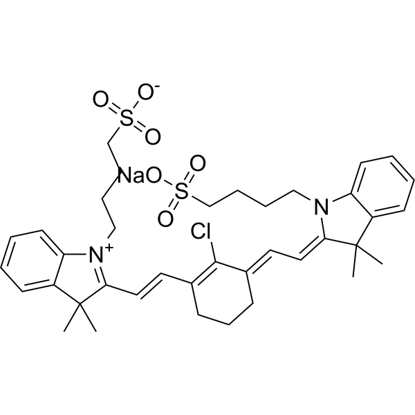
IR-783 (ADS 780WS) is a heptamethine cyanine dye. IR-783 induces Mitochondrial membrane potential loss, ATP depletion, mitochondrial permeability transition pore opening, Cytochrome c release and Apoptosis in breast cancer cells. IR-783 promotes the translocation of Drp1 from the cytosol to mitochondria. IR-783 increases the expression of mitochondrial fission proteins such as MFF and Fission-1. IR-783 possesses imaging, cancer-targeting and anticancer properties. IR-783 exerts anticancer effects against breast cancer. IR-783 can be used in breast cancer-related research.
Nur für Forschungszwecke. Wir verkaufen nicht an Patienten.
- Reinheit : 99.46%
- CAS. Nr.: 115970-66-6
- Formel: C38H46ClN2NaO6S2
- Molecular Weight:749.35
-
Speicherung:
4°C, sealed storage, away from moisture
* In solvent : -80°C, 6 months; -20°C, 1 month (sealed storage, away from moisture)

To place orders, for customer services and technical support, please contact: MedChemExpress USA
Tel: 609-228-6898 E-mail: [email protected] [email protected]


-
 Biologische Aktivität
-
Chemical Information
-
Lösungsmittel & Löslichkeit
- Protokoll
- Reinheit & Dokumentation
- Verweise
-
 Help & FAQs


-
HY-D1063
IR-780Fluorescent Probe -
HY-136886
IR-820Dye -

HY-W248590
IR-775 chlorideFlavonoid Dye -
HY-D0098
Fluorescein-5-maleimideFluorescent Dye -
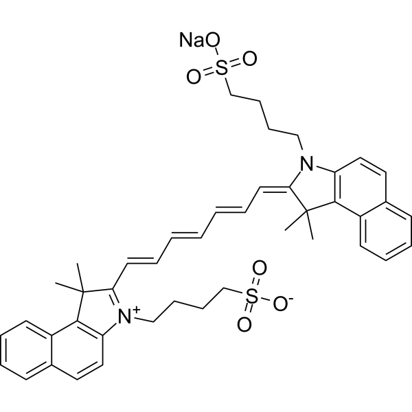
HY-D0711
Indocyanine greenFluorescent Agent -
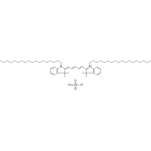
HY-D1028
DiD perchlorateDye -
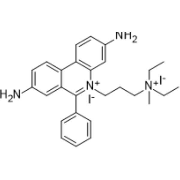
HY-D0815
Propidium IodideFluorochrome -
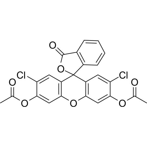
HY-126793
2′,7′-Dichlorofluorescein diacetateFluorescent Probe -
HY-15941
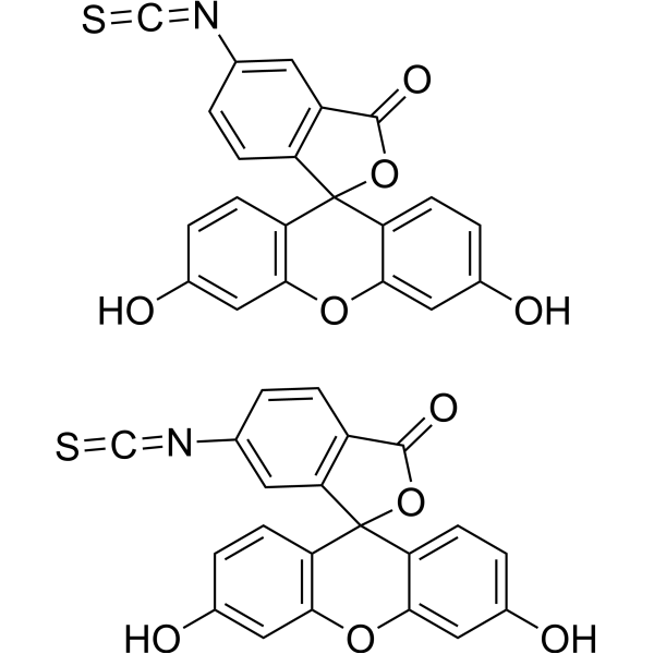
5(6)-FITCFluorescent Dye -
HY-123645
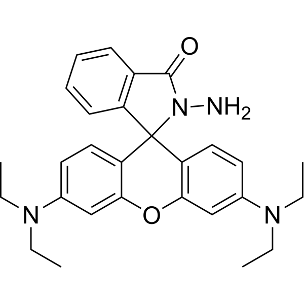
Rhodamine B hydrazideFluorescent Probe -
HY-101878
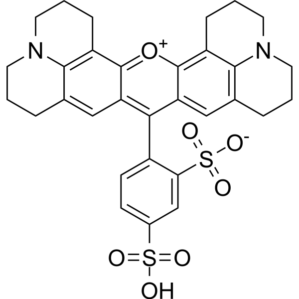
Texas RedRed Fluorescent Dye -
HY-15922
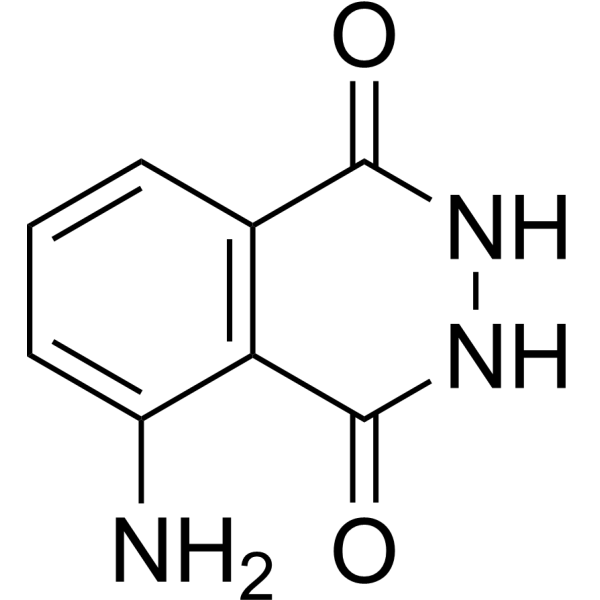
LuminolChemiluminescent Substance
Publications Citing Use of MedChemExpress (MCE) IR-783
More

Biologische Aktivität
Beschreibung
In Vitro
IR-783 (20-100 μM; 24 h for membrane potential assays) induces intracellular ATP depletion and loss of mitochondrial membrane potential in breast cancer MDA-MB-231 cells[1].
IR-783 (5-120 μM; 6-72 h) selectively inhibits the viability of breast cancer MDA-MB-231 cells in a dose- and time-dependent manner, but exerts no effect on normal hepatocyte LO2 cells[1].
IR-783 (20-100 μM; 24 h) induces apoptosis in breast cancer MDA-MB-231 cells in a dose-dependent manner[1].
MedChemExpress (MCE) has not independently confirmed the accuracy of these methods. They are for reference only.
-
Cell Line:human breast cancer MDA-MB-231 cells, human normal hepatocyte LO2 cells
-
Concentration:5-120 μM (24 h incubation); 40 μM (time-course incubation)
-
Incubation Time:6-72 h (40 μM concentration); 24 h (5-120 μM concentrations)
-
Result:Decreased MDA-MB-231 cell viability in a dose- and time-dependent manner, with significant reductions at all tested concentrations and time points relative to control.
Showed no apparent effect on LO2 cell viability even at 120 μM.
-
Cell Line:human breast cancer MDA-MB-231 cells
-
Concentration:20-100 μM
-
Incubation Time:24 h
-
Result:Increased the percentage of apoptotic MDA-MB-231 cells in a dose-dependent manner.
Increased cleaved-caspase-3 levels in a dose-dependent fashion.
Increased cleaved-poly ADP-ribose polymerase (C-PARP) levels relative to control.
In Vivo
MedChemExpress (MCE) has not independently confirmed the accuracy of these methods. They are for reference only.
-
Animal Model:Nude mice (female, 5 weeks old)[1]
-
Dosage:20 mg/kg
-
Administration:i.v.; daily; 4 weeks
-
Result:Inhibited tumour growth significantly after 3 weeks of exposure compared to controls (P < .05).
Showed no statistically significant changes in body weight compared to controls.
Induced no morphological differences in liver and kidney tissues, indicating low liver and kidney toxicity.
Increased numbers of TUNEL-positive cells in tumour tissue.
Increased immunoreactivity for cleaved-caspase-3 in tumour tissue.
Increased Drp1 expression in mitochondrial fractions of tumour tissue.
Increased colocalization of Drp1 and the mitochondrial marker TOM20 in tumour tissue.
- RNA extraction experimental
- Mitochondrial membrane-potential fluorescent assay
- Detection of Apoptosis
- TUNEL staining for apoptotic DNA fragmentation
- Annexin V plus membrane-impermeant dye apoptosis staining
- Fluorescent plasma-membrane potential dye assay
- Mitochondrial membrane-potential and mitochondrial mass staining
- Transepithelial/transendothelial electrical resistance assay
- Apoptosis Solutions
- Breast Cancer Modeling


Chemical Information
-
CAS. Nr. 115970-66-6
-
Appearance Solid
-
Molecular Weight 749.35
-
Formel C38H46ClN2NaO6S2
-
Color Green to dark green
-
SMILES
O=[S](CCCCN(/C1=C\C=C(CCC2)\C(Cl)=C2/C=C/C(C3(C)C)=[N+](C4=CC=CC=C34)CCCC[S](=O)([O-])=O)C5=CC=CC=C5C1(C)C)(O[Na])=O
-
Synonyms
ADS 780WS
-
Versand
Room temperature in continental US; may vary elsewhere.
-
Speicherung
4°C, sealed storage, away from moisture
* In solvent : -80°C, 6 months; -20°C, 1 month (sealed storage, away from moisture)


Publications (4)
-
Journal Impact Factor
-
Most Recent
-
Cell Rep Med
Targeted extracellular vesicle-photoimmunotherapy remodels stromal-immune microenvironment to boost chemo-immunotherapy in preclinical models. [Abstract]2026 Jun 3:102843. PMID: 42235518 -
ACS Appl Mater Interfaces
2026 Jan 25. PMID: 41582522 -
-
Eur J Pharm Biopharm
Spatiotemporally controlled systemic delivery reshapes the in vivo fate of a cationic antimicrobial peptide for lung-selective exposure and an improved therapeutic index. [Abstract]2026 Jul:224:115081. PMID: 42044857


Lösungsmittel & Löslichkeit
In Vitro:
DMSO : 116.67 mg/mL (155.69 mM; Need ultrasonic; Hygroscopic DMSO has a significant impact on the solubility of product, please use newly opened DMSO)
Please refer to the solubility information to select the appropriate solvent. Once prepared, please aliquot and store the solution to prevent product inactivation from repeated freeze-thaw cycles.
Storage method and period of stock solution: -80°C, 6 months; -20°C, 1 month (sealed storage, away from moisture). When stored at -80°C, please use it within 6 months. When stored at -20°C, please use it within 1 month.
Please refer to the solubility information to select the appropriate solvent. Once prepared, please aliquot and store the solution to prevent product inactivation from repeated freeze-thaw cycles.
Storage method and period of stock solution: -80°C, 6 months; -20°C, 1 month (sealed storage, away from moisture). When stored at -80°C, please use it within 6 months. When stored at -20°C, please use it within 1 month.
Konzentration (Stammlösung) × Volumen (Stammlösung) = Konzentration (Ziellösung) × Volumen (Ziellösung)


Reinheit & Dokumentation
-
Data Sheet (284 KB)
-
SDS (394 KB)
- English - EN (394 KB)
- Français - FR (394 KB)
- Deutsch - DE (394 KB)
- Norwegian - NO (394 KB)
- Español - ES (394 KB)
- Swedish - SV (394 KB)
- Italian - IT (394 KB)
- Korean - KR (394 KB)
- Portuguese - PT (394 KB)
-
Handling Instructions (2659 KB)
Verweise


Complete Stock Solution Preparation Table
Please refer to the solubility information to select the appropriate solvent. Once prepared, please aliquot and store the solution to prevent product inactivation from repeated freeze-thaw cycles.
Storage method and period of stock solution: -80°C, 6 months; -20°C, 1 month (sealed storage, away from moisture). When stored at -80°C, please use it within 6 months. When stored at -20°C, please use it within 1 month.
| Optional Solvent | Concentration Solvent Mass | 1 mg | 5 mg | 10 mg | 25 mg |
|---|---|---|---|---|---|
| DMSO | 1 mM | 1.3345 mL | 6.6724 mL | 13.3449 mL | 33.3622 mL |
| 5 mM | 0.2669 mL | 1.3345 mL | 2.6690 mL | 6.6724 mL | |
| 10 mM | 0.1334 mL | 0.6672 mL | 1.3345 mL | 3.3362 mL | |
| 15 mM | 0.0890 mL | 0.4448 mL | 0.8897 mL | 2.2241 mL | |
| 20 mM | 0.0667 mL | 0.3336 mL | 0.6672 mL | 1.6681 mL | |
| 25 mM | 0.0534 mL | 0.2669 mL | 0.5338 mL | 1.3345 mL | |
| 30 mM | 0.0445 mL | 0.2224 mL | 0.4448 mL | 1.1121 mL | |
| 40 mM | 0.0334 mL | 0.1668 mL | 0.3336 mL | 0.8341 mL | |
| 50 mM | 0.0267 mL | 0.1334 mL | 0.2669 mL | 0.6672 mL | |
| 60 mM | 0.0222 mL | 0.1112 mL | 0.2224 mL | 0.5560 mL | |
| 80 mM | 0.0167 mL | 0.0834 mL | 0.1668 mL | 0.4170 mL | |
| 100 mM | 0.0133 mL | 0.0667 mL | 0.1334 mL | 0.3336 mL |


IR-783 Related Classifications
HY-139109 Related Classifications
 Powered by Bioz