



2′,7′-Dichlorofluorescein diacetate (solution)
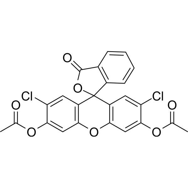
2′,7′-Dichlorofluorescein diacetate (DCFH2-DA) (solution) is a cell-permeable fluorescent probe. 2′,7′-Dichlorofluorescein diacetate can be used to detect the generation of reactive oxygen intermediates and for assessing the overall oxidative stress in toxicological phenomenon.
Solvent and concentration: DMSO: 10 mM
The 1 mL volume is defined as the base specification. All larger sizes correspond to incremental volumes of this base.
For research use only. We do not sell to patients.
- CAS No.: 2044-85-1
- Formula: C24H14Cl2O7
- Molecular Weight:485.27
-
Storage:
Please store the product under the recommended conditions in the Certificate of Analysis.


To place orders, for customer services and technical support, please contact: MedChemExpress USA
Tel: 609-228-6898 E-mail: [email protected] [email protected]


-
 Biological Activity
-
Chemical Information
- Purity & Documentation
- References
-
 Help & FAQs


-
HY-126793
2′,7′-Dichlorofluorescein diacetate -
HY-D0718
Nile Red -
HY-D0711
Indocyanine green -
HY-D0940
H2DCFDA -
HY-D0098
Fluorescein-5-maleimide -

HY-DY1008
Nile Red (solution) -
HY-W090090
BODIPY 493/503 -
HY-W040143
2',7'-Dichlorofluorescein -
HY-D1048
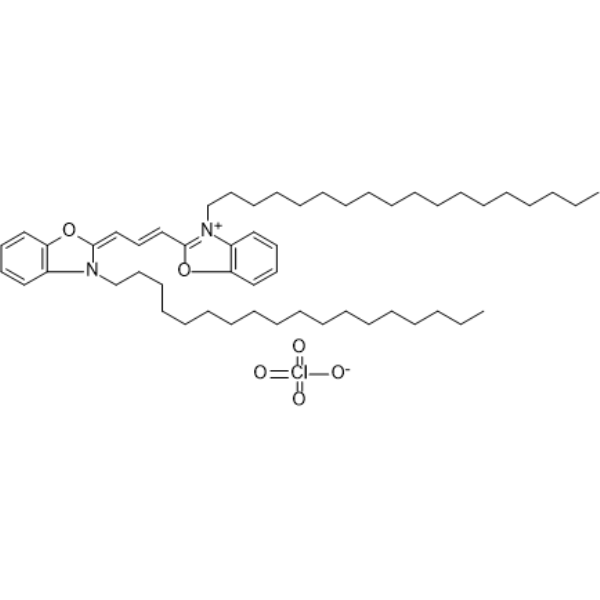
DiR -
HY-D0969
DiO -
HY-D0984A
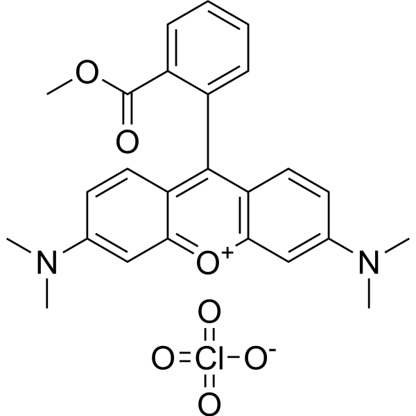
TMRM Perchlorate -
HY-101878
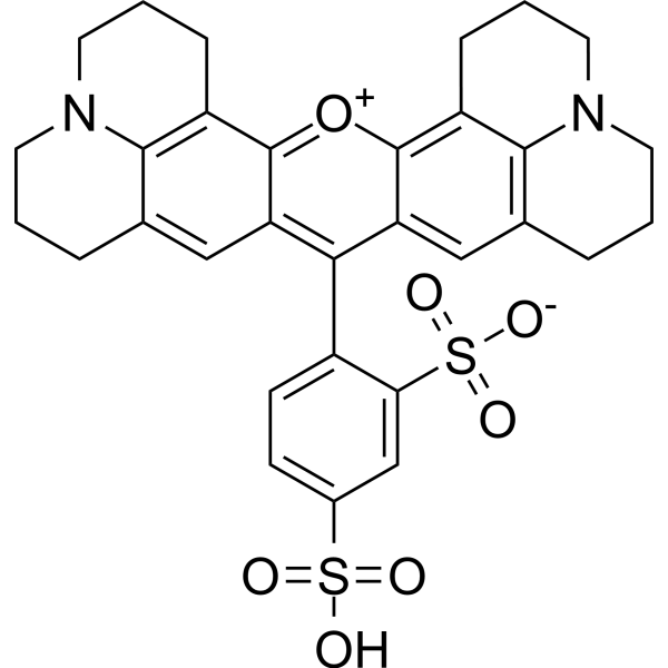
Texas Red


Biological Activity
Description
Solvent and concentration: DMSO: 10 mM
The 1 mL volume is defined as the base specification. All larger sizes correspond to incremental volumes of this base.
In Vitro
Guide (The following is our recommended protocol. This protocol is only a guide and should be modified according to your specific needs).
Using 2',7'-Dichlorofluorescein as a fluorescent probe to quantify intracellular ROS[2]
Experimental Reagents
20,70-dichlorofluorescein diacetate (DCFH2-DA): 100 nM.
CoCl2: 20 mM, used as an extracellular DCF fluorescence quencher.
Cell lines:
K562: Human erythromyelogenous leukemia cell line (drug-sensitive).
K562/adr: Drug-resistant subline of K562.
GLC4: Human small cell lung carcinoma cell line (drug-sensitive).
GLC4/adr: Drug-resistant subline of GLC4.
Culture medium: RPMI 1640, supplemented with 10% fetal bovine serum (FBS).
Experimental Procedure
1. Cell Culture:
(1) Seed K562, K562/adr, GLC4, and GLC4/adr cells into RPMI 1640 medium supplemented with 10% FBS, and culture in a 37°C, 5% CO2 incubator.
(2) The cell density should be 5×10⁵ cells/mL, and proceed to the next step after 24 hours.
2. ROS Measurement:
(1) Treat the cells: Add 20 μM CoCl2 (extracellular fluorescence quencher) and 100 nM DCHF-DA, and continue incubation for 30 minutes to ensure sufficient oxidation of DCHF by ROS.
(2) Measure fluorescence: Use a Perkin-Elmer LS-50B fluorimeter, set the excitation wavelength to 502 nm, and the emission wavelength to 523 nm, to monitor the fluorescence intensity of DCF.
(3) Data analysis:
Fit the kinetic model based on fluorescence intensity changes to calculate the deacetylation rate constant (ke) of DCHF-DA and the rate constant of ROS oxidation (k2[ROSi]). Use this model to quantitatively analyze ROS concentrations in different cell lines.
Experimental Results
1. ROS concentration: ROS concentrations vary between different cell lines. The average ROS concentration in K562 cells is 0.67 nM, 0.60 nM in K562/adr cells, 0.66 nM in GLC4 cells, and 0.81 nM in GLC4/adr cells.
2. Experimental validation: The use of CoCl2 as a quencher effectively removes external ROS signals, ensuring that the obtained DCF fluorescence signal accurately reflects intracellular ROS concentration.
Note: Drug-resistant cell lines (such as K562/adr and GLC4/adr) show higher ROS levels, suggesting that ROS accumulation may be involved in the mechanisms of drug resistance.
MedChemExpress (MCE) has not independently confirmed the accuracy of these methods. They are for reference only.


Chemical Information
-
CAS No. 2044-85-1
-
Appearance Liquid
-
Molecular Weight 485.27
-
Formula C24H14Cl2O7
-
Color Pink to red
-
SMILES
O=C1OC2(C3=C(OC4=C2C=C(Cl)C(OC(C)=O)=C4)C=C(OC(C)=O)C(Cl)=C3)C5=C1C=CC=C5
-
Shipping
Room temperature in continental US; may vary elsewhere.
-
Storage
Please store the product under the recommended conditions in the Certificate of Analysis.


Purity & Documentation
-
Data Sheet (277 KB)
-
SDS (252 KB)
- English - EN (252 KB)
- Français - FR (252 KB)
- Deutsch - DE (252 KB)
- Norwegian - NO (252 KB)
- Español - ES (252 KB)
- Swedish - SV (252 KB)
- Italian - IT (252 KB)
- Korean - KR (252 KB)
- Portuguese - PT (252 KB)
-
Handling Instructions (2659 KB)
References
[1]. Afri M, et al. Active oxygen chemistry within the liposomal bilayer. Part IV: Locating 2',7'-dichlorofluorescein (DCF), 2',7'-dichlorodihydrofluorescein (DCFH) and 2',7'-dichlorodihydrofluorescein diacetate (DCFH-DA) in the lipid bilayer. Chem Phys Lipids. 2004 Aug;131(1):123-33. [Content Brief]

Calculators
Concentration (start) × Volume (start) = Concentration (final) × Volume (final)


2′,7′-Dichlorofluorescein diacetate (solution) Related Classifications
HY-DY1058 Related Classifications
 Powered by Bioz