



PAA5
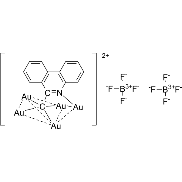
PAA5 is a methide carbon-centered polynuclear Au(I) cluster. PAA5 can release Au(I) causing Pro-oxidant response and accelerated ferroptosis. PAA5 increases the expression of pH2AX in a time-dependent manner. PAA5 has anticancer activity.
For research use only. We do not sell to patients.
- Formula: C14H8Au5B2F8N2-
- Molecular Weight:1348.66
-
Storage:
Please store the product under the recommended conditions in the Certificate of Analysis.

To place orders, for customer services and technical support, please contact: MedChemExpress USA
Tel: 609-228-6898 E-mail: [email protected] [email protected]


-
 Biological Activity
-
Chemical Information
- Protocols
- Purity & Documentation
- References
-
 Help & FAQs


All Endogenous Metabolite Isoforms
More

Biological Activity
Description
In Vitro
PAA5 (0-4 μM; 24 h) induces ferroptosis and increase of the ferroptosis marker gene prostaglandin-endoperoxide synthase 2 (PTGS2). PAA5 decreases cell viability in EJ cells with IC50 values of 1.0 μM and 2.7 μM for EJ and HUVEC cells, respectively[1].
PAA5 (1.5 μM; 4 h; EJ and HUVEC cells) releases active Au(I) metabolites within cells and increases GSH and ROS level[1].
PAA5 (4 μM; 1, 3 and 6 h, EJ cells) causes significant DNA damage as indicated by the increase of histone H2AX phosphorylation (pH2AX)[1].
MedChemExpress (MCE) has not independently confirmed the accuracy of these methods. They are for reference only.
-
Cell Line:HUVEC, EJ cells
-
Concentration:0, 0.5, 1, 1.5, 2, and 4 µM
-
Incubation Time:24 hours
-
Result:Decreased cell viability of EJ cells 57 and 55%, respectively, compare with HUVEC cells.
-
Cell Line:EJ cells
-
Concentration:4 µM
-
Incubation Time:1, 3, and 6 hours
-
Result:Increased the expression of pH2AX in a time dependent manner.
In Vivo
MedChemExpress (MCE) has not independently confirmed the accuracy of these methods. They are for reference only.
-
Animal Model:4-6 weeks, 18 g, female BALB/c nude mice[1]
-
Dosage:1.5 µM, 100 µL
-
Administration:Intravesical delivered into the bladder; 5 times every other day for 8 days
-
Result:Exhibited a good antitumor effect with the small average tumor volume of 564 ± 180 mm3 after 22 days and no significant body weight loss.


Chemical Information
-
Molecular Weight 1348.66
-
Formula C14H8Au5B2F8N2-
-
SMILES
[F-][B+3]([F-])([F-])[F-].[F-][B+3]([F-])([F-])[F-].C1(C(C234[Au@@]56[Au@]27[Au]358[Au]46)=[N]([Au]78)C9=C%10C=CC=C9)=C%10C=CC=C1.[2+]
-
Structure Classification
-
Initial Source
-
Shipping
Room temperature in continental US; may vary elsewhere.
-
Storage
Please store the product under the recommended conditions in the Certificate of Analysis.


Purity & Documentation
References

Calculators
Concentration (start) × Volume (start) = Concentration (final) × Volume (final)


PAA5 Related Classifications
HY-149036 Related Classifications
 Powered by Bioz